|
|
|
|
|
| Sample: |
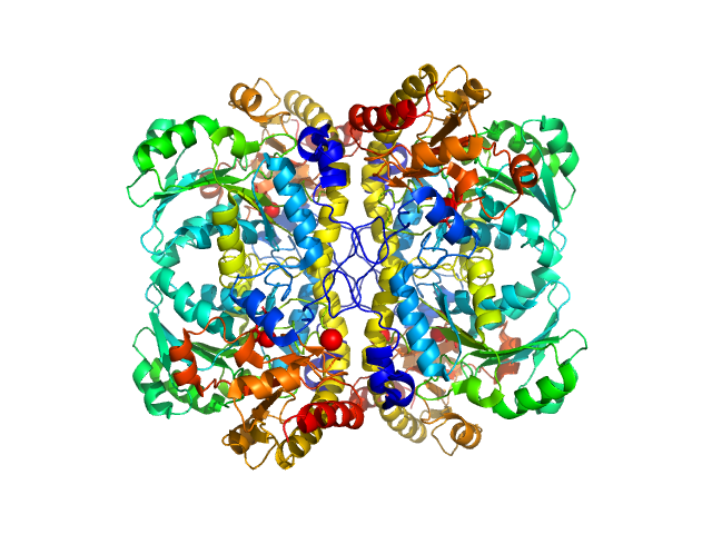
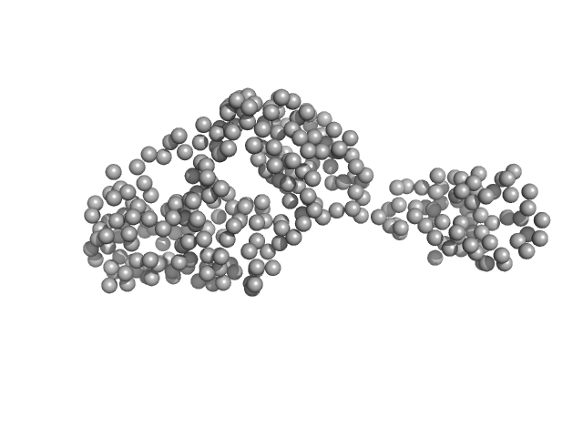
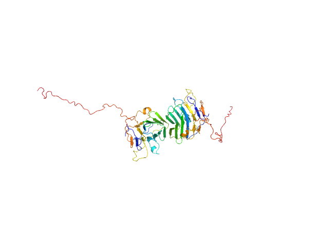
L-methionine gamma-lyase (K272S) tetramer, 174 kDa Clostridium sporogenes protein
|
| Buffer: |
PBS-D2O: 137 mM NaCl, 2.7 mM KCl, 10 mM Na2HPO4, 1.8 mM KH2PO4 (D2O buffer), pH: 7.4 |
| Experiment: |
SANS
data collected at YuMO SANS TOF spectrometer, IBR-2, Frank Laboratory of Neutron Physics, Joint Institute for Nuclear Research on 2019 May 19
|
Methionine gamma lyase fused with S3 domain VGF forms octamers and adheres to tumor cells via binding EGFR
Biochemical and Biophysical Research Communications :149319 (2023)
Bondarev N, Bagaeva D, Bazhenov S, Buben M, Bulushova N, Ryzhykau Y, Okhrimenko I, Zagryadskaya Y, Maslov I, Anisimova N, Sokolova D, Kuklin A, Pokrovsky V, Manukhov I
|
| RgGuinier |
3.7 |
nm |
| Dmax |
14.5 |
nm |
| VolumePorod |
211 |
nm3 |
|
|
|
|
|
|
|
| Sample: |
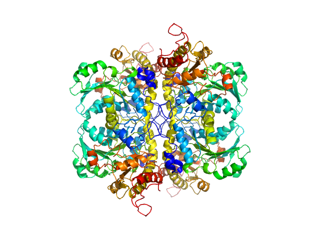
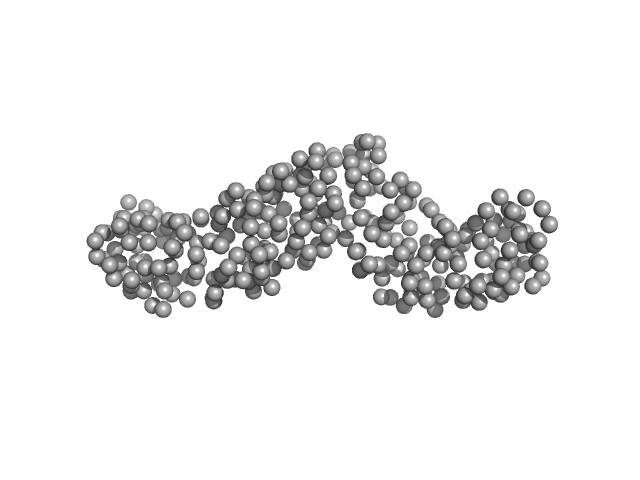
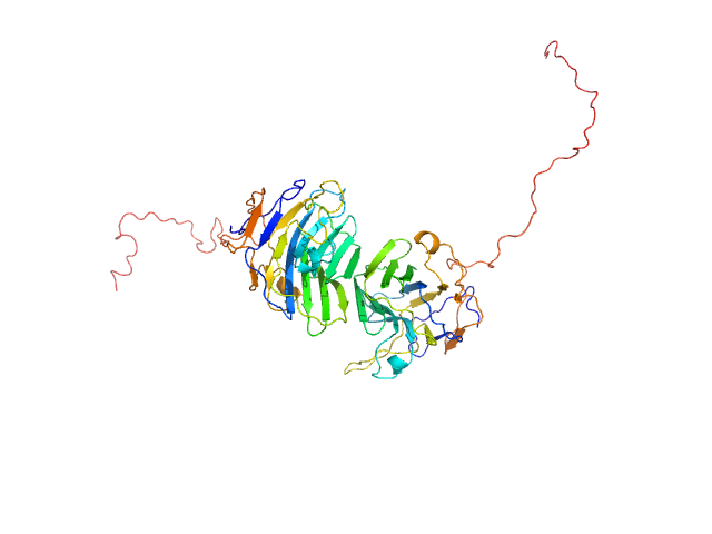
L-methionine gamma-lyase from Clostridium sporogenes fused with VGF S3 domain tetramer, 183 kDa Clostridium sporogenes protein
|
| Buffer: |
PBS-D2O: 137 mM NaCl, 2.7 mM KCl, 10 mM Na2HPO4, 1.8 mM KH2PO4 (D2O buffer), pH: 7.4 |
| Experiment: |
SANS
data collected at YuMO SANS TOF spectrometer, IBR-2, Frank Laboratory of Neutron Physics, Joint Institute for Nuclear Research on 2019 May 19
|
Methionine gamma lyase fused with S3 domain VGF forms octamers and adheres to tumor cells via binding EGFR
Biochemical and Biophysical Research Communications :149319 (2023)
Bondarev N, Bagaeva D, Bazhenov S, Buben M, Bulushova N, Ryzhykau Y, Okhrimenko I, Zagryadskaya Y, Maslov I, Anisimova N, Sokolova D, Kuklin A, Pokrovsky V, Manukhov I
|
| RgGuinier |
5.2 |
nm |
| Dmax |
17.3 |
nm |
| VolumePorod |
303 |
nm3 |
|
|
|
|
|
|
|
| Sample: |
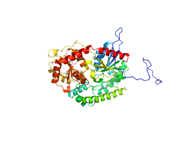
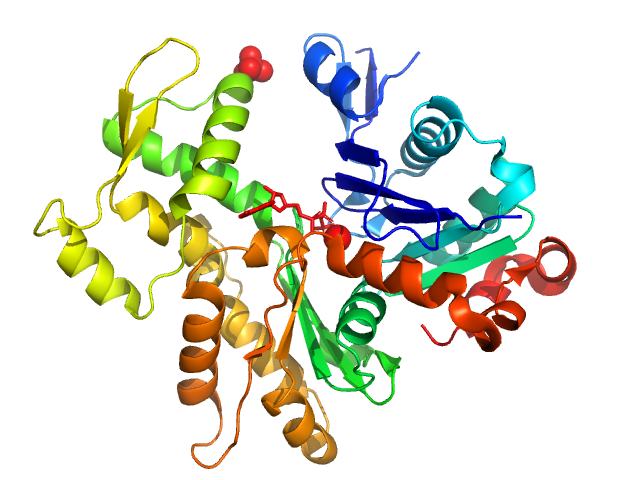
UDP-glycosyltransferase 202A2 monomer, 53 kDa Tetranychus urticae protein
|
| Buffer: |
20 mM sodium phosphate, 150 mM NaCl, pH: 7.8 |
| Experiment: |
SAXS
data collected at BioCAT 18ID, Advanced Photon Source (APS), Argonne National Laboratory on 2022 Jun 17
|
Structural and functional studies reveal the molecular basis of substrate promiscuity of a glycosyltransferase originating from a major agricultural pest
Journal of Biological Chemistry :105421 (2023)
Arriaza R, Abiskaroon B, Patel M, Daneshian L, Kluza A, Snoeck S, Watkins M, Hopkins J, Van Leeuwen T, Grbic M, Grbic V, Borowski T, Chruszcz M
|
| RgGuinier |
2.5 |
nm |
| Dmax |
9.5 |
nm |
| VolumePorod |
85 |
nm3 |
|
|
|
|
|
|
|
| Sample: |
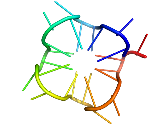
PolyGU RNA - (GU)12 monomer, 8 kDa synthetic RNA RNA
|
| Buffer: |
20 mM HEPES, 150 mM KCl, pH: 7 |
| Experiment: |
SAXS
data collected at 12-ID-B, Advanced Photon Source (APS), Argonne National Laboratory on 2022 Jun 10
|
Solution structure of poly(UG) RNA
Journal of Molecular Biology :168340 (2023)
Escobar C, Petersen R, Tonelli M, Fan L, Henzler-Wildman K, Butcher S
|
| RgGuinier |
1.3 |
nm |
| Dmax |
4.7 |
nm |
| VolumePorod |
8 |
nm3 |
|
|
|
|
|
|
|
| Sample: |
Actin, alpha skeletal muscle monomer, 42 kDa Oryctolagus cuniculus protein
|
| Buffer: |
5 mM Tris/Tris-HCl, 0.1 mM CaCl2, 1 mM NaN3, 0.2 mM ATP, pH: 8.1 |
| Experiment: |
SAXS
data collected at Rigaku MicroMax 007-HF, Moscow Institute of Physics and Technology (MIPT) on 2021 Jul 1
|
Small-angle X-ray scattering structural insights into alternative pathway of actin oligomerization associated with inactivated state
Biochemical and Biophysical Research Communications :149340 (2023)
Ryzhykau Y, Povarova O, Dronova E, Kuklina D, Antifeeva I, Ilyinsky N, Okhrimenko I, Semenov Y, Kuklin A, Ivanovich V, Fonin A, Uversky V, Turoverov K, Kuznetsova I
|
| RgGuinier |
2.9 |
nm |
| Dmax |
8.5 |
nm |
| VolumePorod |
58 |
nm3 |
|
|
|
|
|
|
|
| Sample: |
Heat-labile enterotoxin B chain monomer, 36 kDa Clostridium perfringens protein
|
| Buffer: |
10 mM HEPES, 100 mM NaCl, 2% glycerol, pH: 7.4 |
| Experiment: |
SAXS
data collected at BioCAT 18ID, Advanced Photon Source (APS), Argonne National Laboratory on 2020 Nov 18
|
Structural Basis of Clostridium perfringens Enterotoxin Activation and Oligomerization by Trypsin
Toxins 15(11):637 (2023)
Ogbu C, Kapoor S, Vecchio A
|
| RgGuinier |
2.7 |
nm |
| Dmax |
10.2 |
nm |
| VolumePorod |
46 |
nm3 |
|
|
|
|
|
|
|
| Sample: |
Heat-labile enterotoxin B chain monomer, 33 kDa Clostridium perfringens protein
|
| Buffer: |
10 mM HEPES, 100 mM NaCl, 2% glycerol, pH: 7.4 |
| Experiment: |
SAXS
data collected at BioCAT 18ID, Advanced Photon Source (APS), Argonne National Laboratory on 2020 Nov 18
|
Structural Basis of Clostridium perfringens Enterotoxin Activation and Oligomerization by Trypsin
Toxins 15(11):637 (2023)
Ogbu C, Kapoor S, Vecchio A
|
| RgGuinier |
2.6 |
nm |
| Dmax |
9.5 |
nm |
| VolumePorod |
40 |
nm3 |
|
|
|
|
|
|
|
| Sample: |
Isoform 1 of NAD-dependent protein deacetylase sirtuin-7 monomer, 45 kDa Homo sapiens protein
|
| Buffer: |
50 mM Tris-HCl, pH: 8 |
| Experiment: |
SAXS
data collected at EMBL P12, PETRA III on 2018 Dec 16
|
Substrates and Cyclic Peptide Inhibitors of the Oligonucleotide-Activated Sirtuin 7.
Angew Chem Int Ed Engl :e202314597 (2023)
Bolding JE, Nielsen AL, Jensen I, Hansen TN, Ryberg LA, Jameson ST, Harris P, Peters GHJ, Denu JM, Rogers JM, Olsen CA
|
| RgGuinier |
3.2 |
nm |
| Dmax |
11.5 |
nm |
| VolumePorod |
89 |
nm3 |
|
|
|
|
|
|
|
| Sample: |
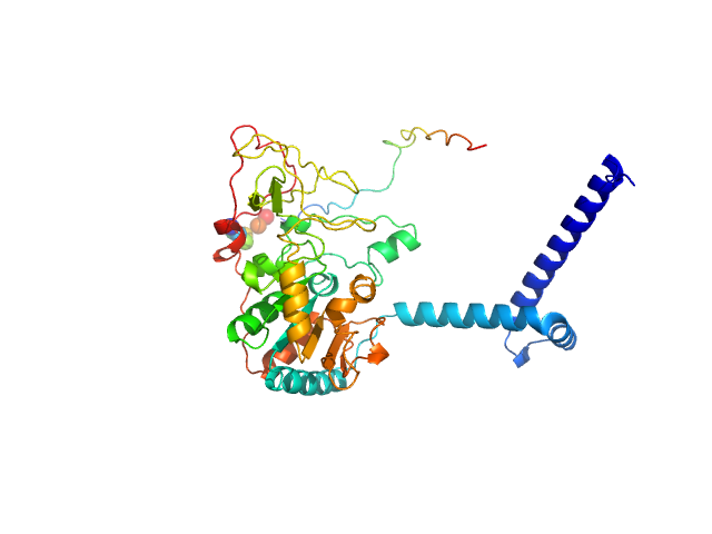
Adhesion G-protein coupled receptor G4 dimer, 55 kDa Homo sapiens protein
|
| Buffer: |
20 mM Tris-HCl, 150 mM NaCl, 5% (w/v) glycerol, pH: 8 |
| Experiment: |
SAXS
data collected at EMBL P12, PETRA III on 2019 Jun 21
|
The dimerized pentraxin-like domain of the adhesion G protein-coupled receptor 112 (ADGRG4) suggests function in sensing mechanical forces.
J Biol Chem :105356 (2023)
Kieslich B, Weiße RH, Brendler J, Ricken A, Schöneberg T, Sträter N
|
| RgGuinier |
2.7 |
nm |
| Dmax |
7.1 |
nm |
| VolumePorod |
67 |
nm3 |
|
|
|
|
|
|
|
| Sample: |
Adhesion G-protein coupled receptor G4 dimer, 55 kDa Homo sapiens protein
|
| Buffer: |
20 mM Tris-HCl, 150 mM NaCl, 5% (w/v) glycerol, pH: 8 |
| Experiment: |
SAXS
data collected at EMBL P12, PETRA III on 2019 Jun 21
|
The dimerized pentraxin-like domain of the adhesion G protein-coupled receptor 112 (ADGRG4) suggests function in sensing mechanical forces.
J Biol Chem :105356 (2023)
Kieslich B, Weiße RH, Brendler J, Ricken A, Schöneberg T, Sträter N
|
| RgGuinier |
2.8 |
nm |
| Dmax |
8.0 |
nm |
| VolumePorod |
65 |
nm3 |
|
|
 experimental SAS data")


12 experimental SAS data")


