|
|
|
|
|
| Sample: |
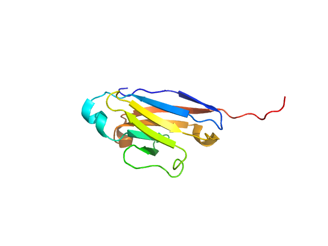
CMRF35-like molecule 8 monomer, 12 kDa Homo sapiens protein
|
| Buffer: |
MES buffer with 3mM DTT, pH: 5.5
|
| Experiment: |
SAXS
data collected at EMBL X33, DORIS III, DESY on 2006 Apr 6
|
Molecular analysis and solution structure from small-angle X-ray scattering of the human natural killer inhibitory receptor IRp60 (CD300a)
International Journal of Biological Macromolecules 40(3):193-200 (2007)
Dimasi N, Roessle M, Moran O, Candiano G, Svergun D, Biassoni R
|
| RgGuinier |
2.0 |
nm |
| Dmax |
7.0 |
nm |
| VolumePorod |
22 |
nm3 |
|
|
|
|
|
|
|
| Sample: |
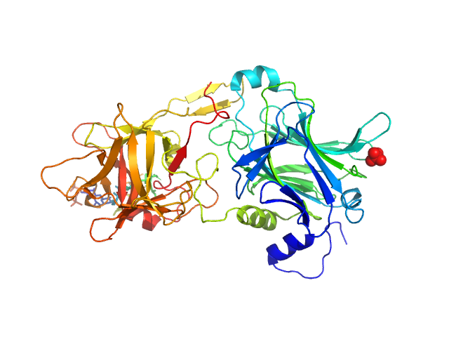
Hc fragment of Tetanus toxin dimer, 103 kDa Clostridium tetani (strain … protein
|
| Buffer: |
50 mM sodium phosphate buffer, 300 mM NaCl, pH: 7.8
|
| Experiment: |
SAXS
data collected at EMBL X33, DORIS III, DESY on 2004 May 14
|
The HC fragment of tetanus toxin forms stable, concentration-dependent dimers via an intermolecular disulphide bond.
J Mol Biol 365(1):123-34 (2007)
Qazi O, Bolgiano B, Crane D, Svergun DI, Konarev PV, Yao ZP, Robinson CV, Brown KA, Fairweather N
|
| RgGuinier |
4.0 |
nm |
| Dmax |
13.2 |
nm |
| VolumePorod |
150 |
nm3 |
|
|
|
|
|
|
|
| Sample: |
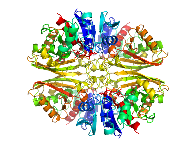
Glyceraldehyde-3-phosphate dehydrogenase 1 tetramer, 142 kDa Kluyveromyces marxianus protein
|
| Buffer: |
150 mM NaCl, 1 mM beta-mercaptoethanol, 1 mM EDTA, 10 mM TrisHCl, pH: 7.5
|
| Experiment: |
SAXS
data collected at EMBL X33, DORIS III, DESY on 2006 Mar 27
|
The Crystal and Solution Structures of Glyceraldehyde-3-phosphate Dehydrogenase Reveal Different Quaternary Structures
Journal of Biological Chemistry 281(44):33433-33440 (2006)
Ferreira-da-Silva F, Pereira P, Gales L, Roessle M, Svergun D, Moradas-Ferreira P, Damas A
|
| RgGuinier |
4.2 |
nm |
| Dmax |
12.0 |
nm |
| VolumePorod |
234 |
nm3 |
|
|
|
|
|
|

|
| Sample: |
Glyceraldehyde-3-phosphate dehydrogenase 1 bound to NAD+ tetramer, 142 kDa Kluyveromyces marxianus protein
|
| Buffer: |
150 mM NaCl, 1 mM beta-mercaptoethanol, 1 mM EDTA, 10 mM TrisHCl, pH: 7.5
|
| Experiment: |
SAXS
data collected at EMBL X33, DORIS III, DESY on 2006 Mar 27
|
The Crystal and Solution Structures of Glyceraldehyde-3-phosphate Dehydrogenase Reveal Different Quaternary Structures
Journal of Biological Chemistry 281(44):33433-33440 (2006)
Ferreira-da-Silva F, Pereira P, Gales L, Roessle M, Svergun D, Moradas-Ferreira P, Damas A
|
| RgGuinier |
3.7 |
nm |
| Dmax |
9.9 |
nm |
| VolumePorod |
202 |
nm3 |
|
|
|
|
|
|
|
| Sample: |
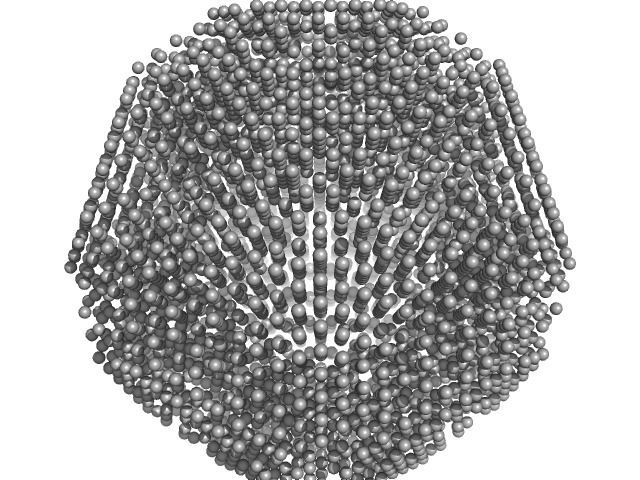
Lumazine Synthase , 960 kDa Bacillus subtilis protein
|
| Buffer: |
Borate buffer, pH: 7
|
| Experiment: |
SAXS
data collected at EMBL X33, DORIS III, DESY on 2004 Nov 25
|
Multiple assembly states of lumazine synthase: a model relating catalytic function and molecular assembly.
J Mol Biol 362(4):753-70 (2006)
Zhang X, Konarev PV, Petoukhov MV, Svergun DI, Xing L, Cheng RH, Haase I, Fischer M, Bacher A, Ladenstein R, Meining W
|
| RgGuinier |
6.2 |
nm |
| Dmax |
15.5 |
nm |
| VolumePorod |
1450 |
nm3 |
|
|
|
|
|
|

|
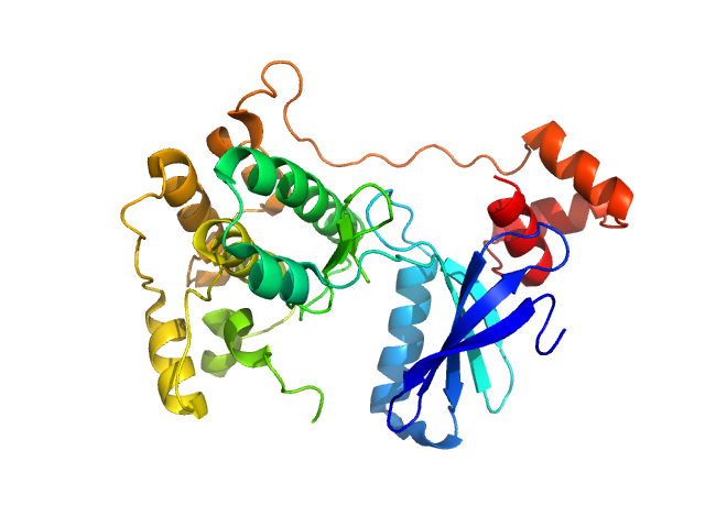
| Sample: |
Serine/threonine-protein kinase MARK2 monomer, 36 kDa Homo sapiens protein
|
| Buffer: |
0.1 M Bis-Tris, 0.2 M ammonium citrate, 1mM DTT, pH: 6.5
|
| Experiment: |
SAXS
data collected at EMBL X33, DORIS III, DESY on 2006 May 16
|
Structural Variations in the Catalytic and Ubiquitin-associated Domains of Microtubule-associated Protein/Microtubule Affinity Regulating Kinase (MARK) 1 and MARK2
Journal of Biological Chemistry 281(37):27586-27599 (2006)
Marx A, Nugoor C, Müller J, Panneerselvam S, Timm T, Bilang M, Mylonas E, Svergun D, Mandelkow E, Mandelkow E
|
| RgGuinier |
2.4 |
nm |
| Dmax |
8.0 |
nm |
| VolumePorod |
61 |
nm3 |
|
|
|
|
|
|
|
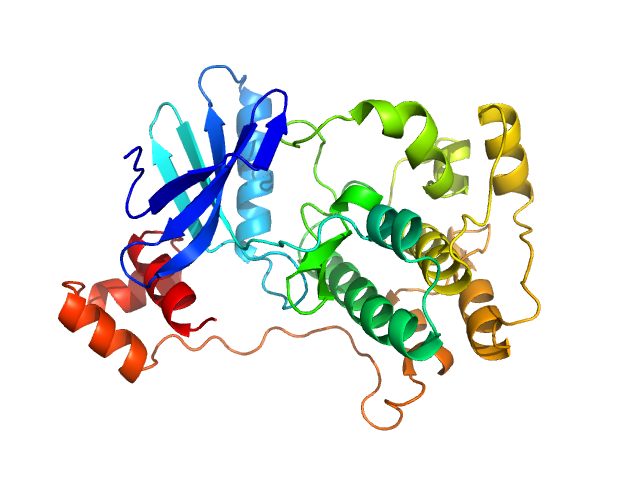
| Sample: |
Serine/threonine-protein kinase MARK2 monomer, 36 kDa Homo sapiens protein
|
| Buffer: |
0.1 M Bis-Tris, 0.2 M ammonium citrate, 1mM DTT, pH: 6.5
|
| Experiment: |
SAXS
data collected at EMBL X33, DORIS III, DESY on 2006 May 16
|
Structural Variations in the Catalytic and Ubiquitin-associated Domains of Microtubule-associated Protein/Microtubule Affinity Regulating Kinase (MARK) 1 and MARK2
Journal of Biological Chemistry 281(37):27586-27599 (2006)
Marx A, Nugoor C, Müller J, Panneerselvam S, Timm T, Bilang M, Mylonas E, Svergun D, Mandelkow E, Mandelkow E
|
| RgGuinier |
2.3 |
nm |
| Dmax |
8.0 |
nm |
| VolumePorod |
62 |
nm3 |
|
|
|
|
|
|
|
| Sample: |
Serine/threonine-protein kinase MARK1 monomer, 37 kDa Homo sapiens protein
|
| Buffer: |
0.1 M Bis-Tris, 0.2 M ammonium citrate, 1mM DTT, pH: 6.5
|
| Experiment: |
SAXS
data collected at EMBL X33, DORIS III, DESY on 2006 May 16
|
Structural Variations in the Catalytic and Ubiquitin-associated Domains of Microtubule-associated Protein/Microtubule Affinity Regulating Kinase (MARK) 1 and MARK2
Journal of Biological Chemistry 281(37):27586-27599 (2006)
Marx A, Nugoor C, Müller J, Panneerselvam S, Timm T, Bilang M, Mylonas E, Svergun D, Mandelkow E, Mandelkow E
|
| RgGuinier |
2.3 |
nm |
| Dmax |
8.0 |
nm |
| VolumePorod |
64 |
nm3 |
|
|
|
|
|
|
|
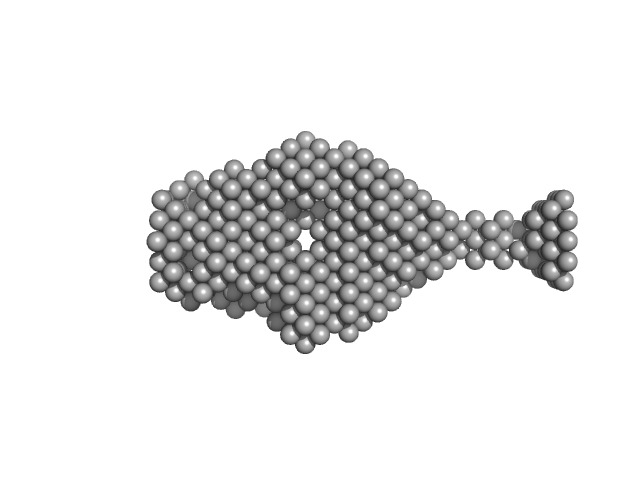
| Sample: |
Mouse ProNGF - pro-form of the NGF (nerve growth factor) dimer, 100 kDa Mus musculus protein
|
| Buffer: |
50 mM Na-phosphate, pH: 7
|
| Experiment: |
SAXS
data collected at EMBL X33, DORIS III, DESY on 2005 Jul 18
|
Structural and functional properties of mouse proNGF.
Biochem Soc Trans 34(Pt 4):605-6 (2006)
Paoletti F, Konarev PV, Covaceuszach S, Schwarz E, Cattaneo A, Lamba D, Svergun DI
|
| RgGuinier |
3.0 |
nm |
| Dmax |
11.0 |
nm |
| VolumePorod |
97 |
nm3 |
|
|
|
|
|
|
|
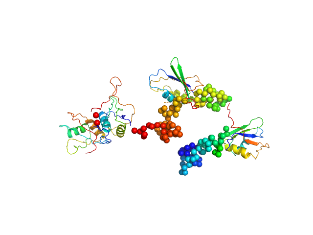
| Sample: |
Polypyrimidine tract-binding protein 1 monomer, 57 kDa Homo sapiens protein
|
| Buffer: |
25 mM Tris 100 mM NaCl 5.0 mM DTT, pH: 7.2
|
| Experiment: |
SAXS
data collected at EMBL X33, DORIS III, DESY on 2004 Feb 11
|
Conformation of polypyrimidine tract binding protein in solution.
Structure 14(6):1021-7 (2006)
Petoukhov MV, Monie TP, Allain FH, Matthews S, Curry S, Svergun DI
|
| RgGuinier |
4.0 |
nm |
| Dmax |
16.5 |
nm |
|
|


 experimental SAS data")
