Search

 
Advanced search  

25 hits found for Anderson

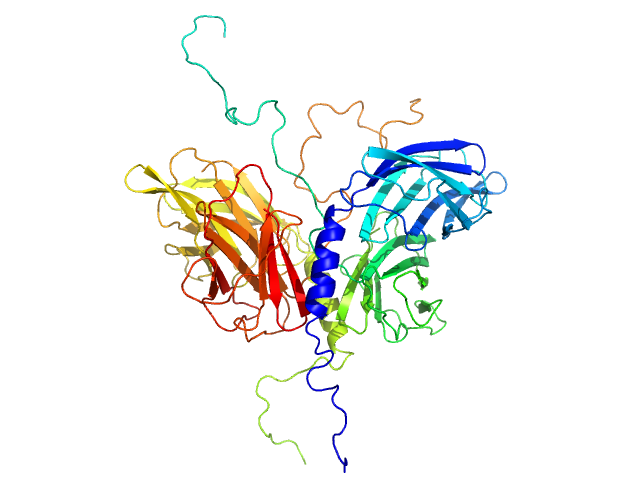
SASDEF3 – Paenibacillus xanthan lyase (PXL) at 4 °C

Paenibacillus xanthan lyase experimental SAS data
Paenibacillus xanthan lyase Kratky plot
Sample: Paenibacillus xanthan lyase monomer, 113 kDa Paenibacillus sp-62047 protein
Buffer: 20 mM Tris,, pH: 8.5
Experiment: SAXS data collected at BM29, ESRF on 2016 Dec 15
Structure and Dynamics of a Promiscuous Xanthan Lyase from Paenibacillus nanensis and the Design of Variants with Increased Stability and Activity. Cell Chem Biol 26(2):191-202.e6 (2019)
...Anderson L, Poulsen JN, Rasmussen KK, Agarwal S, Sainathan RK, Monrad RN, Svendsen A, Nielsen JE, Lo Leggio L, Rand KD
RgGuinier 3.7 nm
Dmax 13.1 nm
VolumePorod 137 nm3

SASDEG3 – Paenibacillus xanthan lyase (PXL) at 20 °C

Paenibacillus xanthan lyase experimental SAS data
Paenibacillus xanthan lyase Kratky plot
Sample: Paenibacillus xanthan lyase monomer, 113 kDa Paenibacillus sp-62047 protein
Buffer: 20 mM Tris,, pH: 8.5
Experiment: SAXS data collected at BM29, ESRF on 2016 Dec 15
Structure and Dynamics of a Promiscuous Xanthan Lyase from Paenibacillus nanensis and the Design of Variants with Increased Stability and Activity. Cell Chem Biol 26(2):191-202.e6 (2019)
...Anderson L, Poulsen JN, Rasmussen KK, Agarwal S, Sainathan RK, Monrad RN, Svendsen A, Nielsen JE, Lo Leggio L, Rand KD
RgGuinier 3.8 nm
Dmax 13.8 nm
VolumePorod 134 nm3

SASDQF5 – Zinc finger protein 410 (ZNF410 full length)

Zinc finger protein 410 experimental SAS data
BILBOMD model
Sample: Zinc finger protein 410 monomer, 52 kDa Homo sapiens protein
Buffer: 20 mM Tris, 250 mM NaCl, 0.1% v/v β-mercaptoethanol, pH: 7.5
Experiment: SAXS data collected at 12.3.1 (SIBYLS), Advanced Light Source (ALS) on 2020 Sep 30
Allosteric autoregulation of DNA binding via a DNA-mimicking protein domain: a biophysical study of ZNF410-DNA interaction using small angle X-ray scattering. Nucleic Acids Res (2023)
Kaur G, Ren R, Hammel M, Horton JR, Yang J, Cao Y, He C, Lan F, Lan X, Blobel GA, Blumenthal RM, Zhang X, Cheng X
RgGuinier 3.6 nm
Dmax 12.3 nm
VolumePorod 108 nm3

SASDQG5 – DNA (Zinc finger protein 410 recognition sequence)

DNA (Zinc finger protein 410 recognition sequence) experimental SAS data
DNA (Zinc finger protein 410 recognition sequence) Kratky plot
Sample: DNA (Zinc finger protein 410 recognition sequence) monomer, 11 kDa DNA
Buffer: 20 mM Tris, 250 mM NaCl, 0.1% v/v β-mercaptoethanol, pH: 7.5
Experiment: SAXS data collected at 12.3.1 (SIBYLS), Advanced Light Source (ALS) on 2020 Sep 30
Allosteric autoregulation of DNA binding via a DNA-mimicking protein domain: a biophysical study of ZNF410-DNA interaction using small angle X-ray scattering. Nucleic Acids Res (2023)
Kaur G, Ren R, Hammel M, Horton JR, Yang J, Cao Y, He C, Lan F, Lan X, Blobel GA, Blumenthal RM, Zhang X, Cheng X
RgGuinier 1.8 nm
Dmax 5.8 nm
VolumePorod 16 nm3

SASDQH5 – Zinc finger protein 410 (ZNF410 full length) bound to DNA

Zinc finger protein 410DNA (Zinc finger protein 410 recognition sequence) experimental SAS data
BILBOMD model
Sample: Zinc finger protein 410 monomer, 52 kDa Homo sapiens protein
DNA (Zinc finger protein 410 recognition sequence) monomer, 11 kDa DNA
Buffer: 20 mM Tris, 250 mM NaCl, 0.1% v/v β-mercaptoethanol, pH: 7.5
Experiment: SAXS data collected at 12.3.1 (SIBYLS), Advanced Light Source (ALS) on 2020 Sep 30
Allosteric autoregulation of DNA binding via a DNA-mimicking protein domain: a biophysical study of ZNF410-DNA interaction using small angle X-ray scattering. Nucleic Acids Res (2023)
Kaur G, Ren R, Hammel M, Horton JR, Yang J, Cao Y, He C, Lan F, Lan X, Blobel GA, Blumenthal RM, Zhang X, Cheng X
RgGuinier 4.4 nm
Dmax 14.3 nm
VolumePorod 76 nm3

SASDQJ5 – Zinc finger protein 410 (ZNF410): N-terminal region with 1-5 zinc fingers

Zinc finger protein 410 experimental SAS data
BILBOMD model
Sample: Zinc finger protein 410 monomer, 40 kDa Homo sapiens protein
Buffer: 20 mM Tris, 250 mM NaCl, 0.1% v/v β-mercaptoethanol, pH: 7.5
Experiment: SAXS data collected at 12.3.1 (SIBYLS), Advanced Light Source (ALS) on 2020 Sep 30
Allosteric autoregulation of DNA binding via a DNA-mimicking protein domain: a biophysical study of ZNF410-DNA interaction using small angle X-ray scattering. Nucleic Acids Res (2023)
Kaur G, Ren R, Hammel M, Horton JR, Yang J, Cao Y, He C, Lan F, Lan X, Blobel GA, Blumenthal RM, Zhang X, Cheng X
RgGuinier 3.2 nm
Dmax 10.7 nm
VolumePorod 78 nm3

SASDQL5 – Zinc finger protein 410 (ZNF410): C-terminal region with 1-5 zinc fingers

Zinc finger protein 410 experimental SAS data
BILBOMD model
Sample: Zinc finger protein 410 monomer, 29 kDa Homo sapiens protein
Buffer: 20 mM Tris, 250 mM NaCl, 0.1% v/v β-mercaptoethanol, pH: 7.5
Experiment: SAXS data collected at 12.3.1 (SIBYLS), Advanced Light Source (ALS) on 2020 Sep 30
Allosteric autoregulation of DNA binding via a DNA-mimicking protein domain: a biophysical study of ZNF410-DNA interaction using small angle X-ray scattering. Nucleic Acids Res (2023)
Kaur G, Ren R, Hammel M, Horton JR, Yang J, Cao Y, He C, Lan F, Lan X, Blobel GA, Blumenthal RM, Zhang X, Cheng X
RgGuinier 3.0 nm
Dmax 9.6 nm
VolumePorod 58 nm3

SASDQM5 – Zinc finger protein 410 (ZNF410): C-terminal region with 1-5 zinc fingers bound to DNA

DNA (Zinc finger protein 410 recognition sequence)Zinc finger protein 410 experimental SAS data
BILBOMD model
Sample: DNA (Zinc finger protein 410 recognition sequence) monomer, 11 kDa DNA
Zinc finger protein 410 monomer, 29 kDa Homo sapiens protein
Buffer: 20 mM Tris, 250 mM NaCl, 0.1% v/v β-mercaptoethanol, pH: 7.5
Experiment: SAXS data collected at 12.3.1 (SIBYLS), Advanced Light Source (ALS) on 2020 Sep 30
Allosteric autoregulation of DNA binding via a DNA-mimicking protein domain: a biophysical study of ZNF410-DNA interaction using small angle X-ray scattering. Nucleic Acids Res (2023)
Kaur G, Ren R, Hammel M, Horton JR, Yang J, Cao Y, He C, Lan F, Lan X, Blobel GA, Blumenthal RM, Zhang X, Cheng X
RgGuinier 3.1 nm
Dmax 11.9 nm
VolumePorod 52 nm3

SASDZS5 – Frozen human Phosphatidylinositol 3,4,5-trisphosphate-dependent Rac exchanger 2 DH/PH tandem (P-Rex2 DH/PH ) with sucrose as cryoprotectant

Phosphatidylinositol 3,4,5-trisphosphate-dependent Rac exchanger 2 DH/PH tandem experimental SAS data
BILBOMD model
Sample: Phosphatidylinositol 3,4,5-trisphosphate-dependent Rac exchanger 2 DH/PH tandem monomer, 44 kDa Homo sapiens protein
Buffer: 20 mM HEPES, 200 mM NaCl, 2 mM DTT, 1% glycerol, pH: 7
Experiment: SAXS data collected at 12.3.1 (SIBYLS), Advanced Light Source (ALS) on 2026 Feb 17
Freezing the P-Rex2 DH/PH tandem
Lauren Anderson
RgGuinier 2.6 nm
Dmax 8.4 nm
VolumePorod 48 nm3

SASDZT5 – Frozen human P-Rex2 DH/PH (Phosphatidylinositol 3,4,5-trisphosphate-dependent Rac exchanger 2 protein) without cryoprotectant

Phosphatidylinositol 3,4,5-trisphosphate-dependent Rac exchanger 2 DH/PH tandem experimental SAS data
BILBOMD model
Sample: Phosphatidylinositol 3,4,5-trisphosphate-dependent Rac exchanger 2 DH/PH tandem monomer, 44 kDa Homo sapiens protein
Buffer: 20 mM HEPES, 200 mM NaCl, 2 mM DTT, 1% glycerol, pH: 7
Experiment: SAXS data collected at 12.3.1 (SIBYLS), Advanced Light Source (ALS) on 2026 Feb 17
Freezing the P-Rex2 DH/PH tandem
Lauren Anderson
RgGuinier 2.6 nm
Dmax 8.5 nm
VolumePorod 55 nm3

SASDZU5 – Human Phosphatidylinositol 3,4,5-trisphosphate-dependent Rac exchanger 2 protein DH/PH domain and DEP1 module (P-Rex2 DH/PH-DEP1)

Phosphatidylinositol 3,4,5-trisphosphate-dependent Rac exchanger 2 DH/PH-DEP1 experimental SAS data
BILBOMD model
Sample: Phosphatidylinositol 3,4,5-trisphosphate-dependent Rac exchanger 2 DH/PH-DEP1 monomer, 55 kDa Homo sapiens protein
Buffer: 20 mM HEPES, 200 mM NaCl, 2 mM DTT, 1% glycerol, pH: 7
Experiment: SAXS data collected at 12.3.1 (SIBYLS), Advanced Light Source (ALS) on 2025 Jun 10
The Rho guanine-nucleotide exchange factor P-Rex2 exhibits structural and regulatory features distinct from the related RhoGEF P-Rex1 Journal of Biological Chemistry 302(7):113229 (2026)
Anderson L, Marde R, Muma G, Nayak V, Phan C, Li S, Cash J
RgGuinier 2.9 nm
Dmax 10.5 nm
VolumePorod 77 nm3

SASDZV5 – Human Phosphatidylinositol 3,4,5-trisphosphate-dependent Rac exchanger protein 2 DH/PH domain tandem (P-Rex2 DH/PH)

Phosphatidylinositol 3,4,5-trisphosphate-dependent Rac exchanger 2 DH/PH tandem experimental SAS data
BILBOMD model
Sample: Phosphatidylinositol 3,4,5-trisphosphate-dependent Rac exchanger 2 DH/PH tandem monomer, 44 kDa Homo sapiens protein
Buffer: 20 mM HEPES, 200 mM NaCl, 2 mM DTT, 1% glycerol, pH: 7
Experiment: SAXS data collected at 12.3.1 (SIBYLS), Advanced Light Source (ALS) on 2025 Jun 10
The Rho guanine-nucleotide exchange factor P-Rex2 exhibits structural and regulatory features distinct from the related RhoGEF P-Rex1 Journal of Biological Chemistry 302(7):113229 (2026)
Anderson L, Marde R, Muma G, Nayak V, Phan C, Li S, Cash J
RgGuinier 2.6 nm
Dmax 8.4 nm
VolumePorod 52 nm3

SASDNF8 – LARGE xylosyl- and glucuronyltransferase 1 (LARGE1dTM) dimer

Xylosyl- and glucuronyltransferase LARGE1 experimental SAS data
DAMMIF model
Sample: Xylosyl- and glucuronyltransferase LARGE1 dimer, 179 kDa Homo sapiens protein
Buffer: 20 mM HEPES, 150 mM NaCl, pH: 7.4
Experiment: SAXS data collected at BioCAT 18ID, Advanced Photon Source (APS), Argonne National Laboratory on 2022 Mar 18
LARGE1 processively polymerizes length-controlled matriglycan on prodystroglycan. Nat Commun 16(1):9028 (2025)
...Anderson ME, Venzke D, Campbell KP
RgGuinier 4.4 nm
Dmax 18.4 nm
VolumePorod 264 nm3

SASDNG8 – LARGE xylosyl- and glucuronyltransferase 1 (LARGE1dTM) dimer treated with PNGase F enzyme

Xylosyl- and glucuronyltransferase LARGE1 experimental SAS data
DAMMIF model
Sample: Xylosyl- and glucuronyltransferase LARGE1 dimer, 179 kDa Homo sapiens protein
Buffer: 20 mM HEPES, 150 mM NaCl, pH: 7.4
Experiment: SAXS data collected at BioCAT 18ID, Advanced Photon Source (APS), Argonne National Laboratory on 2022 Mar 18
LARGE1 processively polymerizes length-controlled matriglycan on prodystroglycan. Nat Commun 16(1):9028 (2025)
...Anderson ME, Venzke D, Campbell KP
RgGuinier 4.3 nm
Dmax 18.0 nm

SASDNH8 – Xylosyl- and glucuronyltransferase LARGE2 (LARGE2dTM) dimer

Xylosyl- and glucuronyltransferase LARGE2 experimental SAS data
DAMMIN model
Sample: Xylosyl- and glucuronyltransferase LARGE2 dimer, 168 kDa Mus musculus protein
Buffer: 20 mM HEPES, 150 mM NaCl, pH: 7.4
Experiment: SAXS data collected at BioCAT 18ID, Advanced Photon Source (APS), Argonne National Laboratory on 2022 Mar 18
LARGE1 processively polymerizes length-controlled matriglycan on prodystroglycan. Nat Commun 16(1):9028 (2025)
...Anderson ME, Venzke D, Campbell KP
RgGuinier 4.2 nm
Dmax 16.5 nm

SASDNJ8 – Xylosyl- and glucuronyltransferase LARGE2 (LARGE2dTM) dimer treated with PNGase F enzyme

Xylosyl- and glucuronyltransferase LARGE2 experimental SAS data
DAMMIN model
Sample: Xylosyl- and glucuronyltransferase LARGE2 dimer, 168 kDa Mus musculus protein
Buffer: 20 mM HEPES, 150 mM NaCl, pH: 7.4
Experiment: SAXS data collected at BioCAT 18ID, Advanced Photon Source (APS), Argonne National Laboratory on 2022 Mar 18
LARGE1 processively polymerizes length-controlled matriglycan on prodystroglycan. Nat Commun 16(1):9028 (2025)
...Anderson ME, Venzke D, Campbell KP
RgGuinier 4.1 nm
Dmax 15.9 nm
VolumePorod 231 nm3

SASDV47 – Peptide-linked fusion protein of Apoptosis-inducing factor 1 AIF(104-613) point mutant W196A and the N-terminal segment of Mitochondrial intermembrane space import and assembly protein 40 CHCHD4(1-45)

Mitochondrial intermembrane space import and assembly protein 40Apoptosis-inducing factor 1, mitochondrial experimental SAS data
Mitochondrial intermembrane space import and assembly protein 40 Apoptosis-inducing factor 1, mitochondrial Kratky plot
Sample: Mitochondrial intermembrane space import and assembly protein 40 dimer, 12 kDa Homo sapiens protein
Apoptosis-inducing factor 1, mitochondrial dimer, 113 kDa Homo sapiens protein
Buffer: 25 mM HEPES, 150 mM NaCl, 2 mM TCEP, pH: 7.5
Experiment: SAXS data collected at 12.3.1 (SIBYLS), Advanced Light Source (ALS) on 2018 Nov 28
NADH-bound AIF activates the mitochondrial CHCHD4/MIA40 chaperone by a substrate-mimicry mechanism. EMBO J (2025)
Brosey CA, Shen R, Tainer JA
RgGuinier 3.9 nm
Dmax 12.9 nm
VolumePorod 289 nm3

SASDEU7 – Stator protein FlaG soluble domain

Conserved flagellar protein FlaG soluble domain experimental SAS data
Conserved flagellar protein FlaG soluble domain Kratky plot
Sample: Conserved flagellar protein FlaG soluble domain monomer, 15 kDa Sulfolobus acidocaldarius protein
Buffer: 25 mM citric acid/sodium citrate, 150mM NaCl, 3% Glycerol, pH: 3
Experiment: SAXS data collected at 12.3.1 (SIBYLS), Advanced Light Source (ALS) on 2016 Nov 10
The structure of the periplasmic FlaG-FlaF complex and its essential role for archaellar swimming motility. Nat Microbiol (2019)
Tsai CL, Tripp P, Sivabalasarma S, Zhang C, Rodriguez-Franco M, Wipfler RL, Chaudhury P, Banerjee A, Beeby M, Whitaker RJ, Tainer JA, Albers SV
RgGuinier 3.7 nm
Dmax 18.0 nm
VolumePorod 133 nm3

SASDVY6 – Wild-type full-length Mitochondrial intermembrane space import and assembly protein 40 (CHCHD4)

Mitochondrial intermembrane space import and assembly protein 40 experimental SAS data
Mitochondrial intermembrane space import and assembly protein 40 Kratky plot
Sample: Mitochondrial intermembrane space import and assembly protein 40 monomer, 16 kDa Homo sapiens protein
Buffer: 25 mM HEPES, 150 mM NaCl, 2 mM TCEP, pH: 7.5
Experiment: SAXS data collected at 12.3.1 (SIBYLS), Advanced Light Source (ALS) on 2021 Oct 26
NADH-bound AIF activates the mitochondrial CHCHD4/MIA40 chaperone by a substrate-mimicry mechanism. EMBO J (2025)
Brosey CA, Shen R, Tainer JA
RgGuinier 2.9 nm
Dmax 9.2 nm
VolumePorod 56 nm3

SASDES7 – Stator protein complex FlaG/FlaF

Conserved flagellar protein FStator protein FlaG soluble domain experimental SAS data
MULTIFOXS model
Sample: Conserved flagellar protein F dimer, 32 kDa Sulfolobus acidocaldarius protein
Stator protein FlaG soluble domain dimer, 30 kDa Sulfolobus acidocaldarius protein
Buffer: 25 mM citric acid/sodium citrate, 150mM NaCl, 3% Glycerol, pH: 3
Experiment: SAXS data collected at 12.3.1 (SIBYLS), Advanced Light Source (ALS) on 2016 Nov 10
The structure of the periplasmic FlaG-FlaF complex and its essential role for archaellar swimming motility. Nat Microbiol (2019)
Tsai CL, Tripp P, Sivabalasarma S, Zhang C, Rodriguez-Franco M, Wipfler RL, Chaudhury P, Banerjee A, Beeby M, Whitaker RJ, Tainer JA, Albers SV
RgGuinier 3.2 nm
Dmax 12.5 nm
VolumePorod 109 nm3

SASDVZ6 – Mitochondrial intermembrane space import and assembly protein 40 (CHCHD4) triple mutant I12A/F14A/H20A (AIA-A)

Mitochondrial intermembrane space import and assembly protein 40 experimental SAS data
Mitochondrial intermembrane space import and assembly protein 40 Kratky plot
Sample: Mitochondrial intermembrane space import and assembly protein 40 monomer, 16 kDa Homo sapiens protein
Buffer: 25 mM HEPES, 150 mM NaCl, 2 mM TCEP, pH: 7.5
Experiment: SAXS data collected at 12.3.1 (SIBYLS), Advanced Light Source (ALS) on 2021 Oct 26
NADH-bound AIF activates the mitochondrial CHCHD4/MIA40 chaperone by a substrate-mimicry mechanism. EMBO J (2025)
Brosey CA, Shen R, Tainer JA
RgGuinier 3.1 nm
Dmax 10.5 nm
VolumePorod 62 nm3

SASDEV7 – Stator protein complex FlaG/FlaF-I96Y

Stator protein FlaG soluble domainConserved flagellar protein FlaF-I96Y soluble domain experimental SAS data
BILBOMD model
Sample: Stator protein FlaG soluble domain dimer, 30 kDa Sulfolobus acidocaldarius protein
Conserved flagellar protein FlaF-I96Y soluble domain dimer, 33 kDa Sulfolobus acidocaldarius protein
Buffer: 25 mM citric acid/sodium citrate, 150mM NaCl, 3% Glycerol, pH: 3
Experiment: SAXS data collected at 12.3.1 (SIBYLS), Advanced Light Source (ALS) on 2016 Nov 10
The structure of the periplasmic FlaG-FlaF complex and its essential role for archaellar swimming motility. Nat Microbiol (2019)
Tsai CL, Tripp P, Sivabalasarma S, Zhang C, Rodriguez-Franco M, Wipfler RL, Chaudhury P, Banerjee A, Beeby M, Whitaker RJ, Tainer JA, Albers SV
RgGuinier 2.7 nm
Dmax 8.2 nm
VolumePorod 90 nm3

SASDV27 – Mitochondrial intermembrane space import and assembly protein 40 (CHCHD4) point mutant H20D

Mitochondrial intermembrane space import and assembly protein 40 experimental SAS data
Mitochondrial intermembrane space import and assembly protein 40 Kratky plot
Sample: Mitochondrial intermembrane space import and assembly protein 40 monomer, 16 kDa Homo sapiens protein
Buffer: 25 mM HEPES, 150 mM NaCl, 2 mM TCEP, pH: 7.5
Experiment: SAXS data collected at 12.3.1 (SIBYLS), Advanced Light Source (ALS) on 2021 Oct 26
NADH-bound AIF activates the mitochondrial CHCHD4/MIA40 chaperone by a substrate-mimicry mechanism. EMBO J (2025)
Brosey CA, Shen R, Tainer JA
RgGuinier 3.2 nm
Dmax 11.2 nm
VolumePorod 66 nm3

SASDET7 – Stator protein complex FlaG-V118K/FlaF

Conserved flagellar protein FStator protein FlaG-V118K soluble domain experimental SAS data
MULTIFOXS model
Sample: Conserved flagellar protein F dimer, 32 kDa Sulfolobus acidocaldarius protein
Stator protein FlaG-V118K soluble domain dimer, 30 kDa Sulfolobus acidocaldarius protein
Buffer: 25 mM citric acid/sodium citrate, 150mM NaCl, 3% Glycerol, pH: 3
Experiment: SAXS data collected at 12.3.1 (SIBYLS), Advanced Light Source (ALS) on 2016 Nov 10
The structure of the periplasmic FlaG-FlaF complex and its essential role for archaellar swimming motility. Nat Microbiol (2019)
Tsai CL, Tripp P, Sivabalasarma S, Zhang C, Rodriguez-Franco M, Wipfler RL, Chaudhury P, Banerjee A, Beeby M, Whitaker RJ, Tainer JA, Albers SV
RgGuinier 3.2 nm
Dmax 12.5 nm
VolumePorod 108 nm3

SASDV37 – Mitochondrial intermembrane space import and assembly protein 40 (CHCHD4) point mutant L28D

Mitochondrial intermembrane space import and assembly protein 40 experimental SAS data
Mitochondrial intermembrane space import and assembly protein 40 Kratky plot
Sample: Mitochondrial intermembrane space import and assembly protein 40 monomer, 16 kDa Homo sapiens protein
Buffer: 25 mM HEPES, 150 mM NaCl, 2 mM TCEP, pH: 7.5
Experiment: SAXS data collected at 12.3.1 (SIBYLS), Advanced Light Source (ALS) on 2021 Oct 26
NADH-bound AIF activates the mitochondrial CHCHD4/MIA40 chaperone by a substrate-mimicry mechanism. EMBO J (2025)
Brosey CA, Shen R, Tainer JA
RgGuinier 2.8 nm
Dmax 9.2 nm
VolumePorod 52 nm3
