|
|
|
|
|
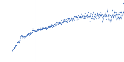
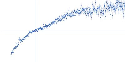
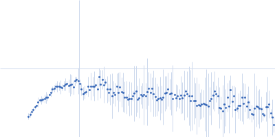
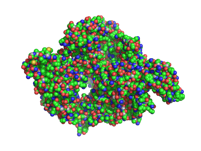
| Sample: |
Alpha-actinin-2 dimer, 208 kDa Homo sapiens protein
|
| Buffer: |
20 mM HEPES, 150 mM NaCl, pH: 8 |
| Experiment: |
SAXS
data collected at B21, Diamond Light Source on 2024 May 13
|
ACTN2 HCM-linked Missense Variants
Maya Noureddine
|
| RgGuinier |
13.0 |
nm |
| Dmax |
55.5 |
nm |
|
|
|
|
|
|
|
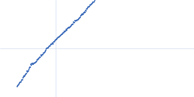
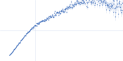
| Sample: |
Alpha-actinin-2 dimer, 208 kDa Homo sapiens protein
|
| Buffer: |
20 mM HEPES, 150 mM NaCl, pH: 8 |
| Experiment: |
SAXS
data collected at B21, Diamond Light Source on 2024 May 13
|
ACTN2 HCM-linked Missense Variants
Maya Noureddine
|
| RgGuinier |
11.6 |
nm |
| Dmax |
37.0 |
nm |
|
|
|
|
|
|
|
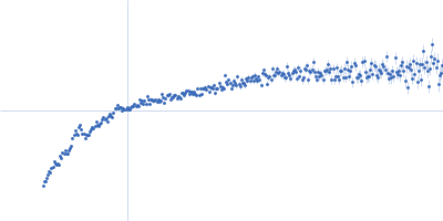
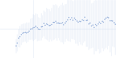
| Sample: |
Alpha-actinin-2 dimer, 208 kDa Homo sapiens protein
|
| Buffer: |
20 mM HEPES, 150 mM NaCl, pH: 8 |
| Experiment: |
SAXS
data collected at B21, Diamond Light Source on 2024 May 13
|
ACTN2 HCM-linked Missense Variants
Maya Noureddine
|
| RgGuinier |
12.9 |
nm |
| Dmax |
57.0 |
nm |
|
|
|
|
|
|
|
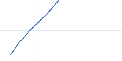
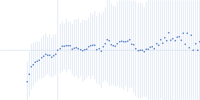
| Sample: |
Alpha-actinin-2 dimer, 208 kDa Homo sapiens protein
|
| Buffer: |
20 mM HEPES, 150 mM NaCl, pH: 8 |
| Experiment: |
SAXS
data collected at B21, Diamond Light Source on 2024 May 13
|
ACTN2 HCM-linked Missense Variants
Maya Noureddine
|
| RgGuinier |
11.5 |
nm |
| Dmax |
36.1 |
nm |
|
|
|
|
|
|
|
| Sample: |
Alpha-actinin-2 dimer, 208 kDa Homo sapiens protein
|
| Buffer: |
20 mM HEPES, 150 mM NaCl, pH: 8 |
| Experiment: |
SAXS
data collected at B21, Diamond Light Source on 2024 May 13
|
ACTN2 HCM-linked Missense Variants
Maya Noureddine
|
| RgGuinier |
11.6 |
nm |
| Dmax |
49.0 |
nm |
|
|
|
|
|
|
|
| Sample: |
Fc-hel8-L1-3 dimer, 109 kDa S.aureus protein
|
| Buffer: |
20 mM HEPES, 150 mM NaCl, pH 7.0, pH: 7 |
| Experiment: |
SAXS
data collected at EMBL P12, PETRA III on 2025 Apr 6
|
SEC-SAXS data of Fc-hel8-L1-3
Adriana Badarau
|
| RgGuinier |
6.4 |
nm |
| Dmax |
30.0 |
nm |
|
|
|
|
|
|
|
| Sample: |
Replicase polyprotein 1ab monomer, 9 kDa Severe acute respiratory … protein
Replicase polyprotein 1ab dimer, 44 kDa Severe acute respiratory … protein
ORF1ab polyprotein monomer, 106 kDa Human coronavirus OC43 protein
|
| Buffer: |
50 mM HEPES, 200 mM NaCl, pH: 8 |
| Experiment: |
SAXS
data collected at Anton Paar SAXSpace, CSIR-Central Drug Research Institute on 2025 Mar 24
|
RdRp
|
| RgGuinier |
5.2 |
nm |
| Dmax |
10.1 |
nm |
| VolumePorod |
213 |
nm3 |
|
|
|
|
|
|
|
| Sample: |
CD2-associated protein hexamer, 429 kDa Homo sapiens protein
|
| Buffer: |
10 mM potassium phosphate, pH: 7.6 |
| Experiment: |
SAXS
data collected at Anton Paar SAXSpace, CSIR-Central Drug Research Institute on 2025 May 16
|
CD2AP’s Structure and Oligomerization are compromised by the K301M mutation: implications for Nephrotic syndrome
Anil Kumar Pasupulati et .al
|
| RgGuinier |
10.7 |
nm |
| Dmax |
35.5 |
nm |
|
|
|
|
|
|
|
| Sample: |
CD2-associated protein trimer, 214 kDa Homo sapiens protein
|
| Buffer: |
10 mM potassium phosphate, pH: 7.6 |
| Experiment: |
SAXS
data collected at Anton Paar SAXSpace, CSIR-Central Drug Research Institute on 2025 May 18
|
CD2AP’s Structure and Oligomerization are compromised by the K301M mutation: implications for Nephrotic syndrome
Anil Kumar Pasupulati et .al
|
| RgGuinier |
9.6 |
nm |
| Dmax |
33.9 |
nm |
|
|
|
|
|
|
|
| Sample: |
Angiotensin Converting Enzyme2 dimer, 185 kDa Homo sapiens protein
|
| Buffer: |
50mM Tris, 200mM NaCl, pH: 8 |
| Experiment: |
SAXS
data collected at Anton Paar SAXSpace, CSIR-Central Drug Research Institute on 2021 Jul 29
|
ACE 2 protein
Raja Tripathi
|
| RgGuinier |
5.4 |
nm |
| Dmax |
27.5 |
nm |
| VolumePorod |
569 |
nm3 |
|
|