|
|
|
|
|
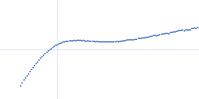
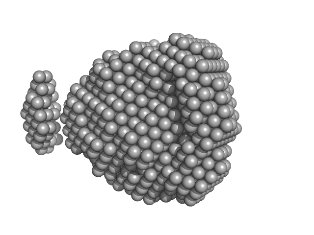
| Sample: |
Protein W monomer, 15 kDa Hendra virus (isolate … protein
|
| Buffer: |
50 mM sodium phosphate, 5 mM EDTA, pH: 6.5 |
| Experiment: |
SAXS
data collected at SWING, SOLEIL on 2021 Jun 12
|
Identification of a Region in the Common Amino-terminal Domain of Hendra Virus P, V, and W Proteins Responsible for Phase Transition and Amyloid Formation
Biomolecules 11(9):1324 (2021)
Salladini E, Gondelaud F, Nilsson J, Pesce G, Bignon C, Murrali M, Fabre R, Pierattelli R, Kajava A, Horvat B, Gerlier D, Mathieu C, Longhi S
|
| RgGuinier |
3.4 |
nm |
| Dmax |
15.5 |
nm |
| VolumePorod |
38 |
nm3 |
|
|
|
|
|
|
|
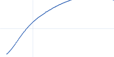
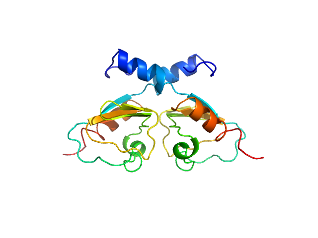
| Sample: |
Brain tumor protein monomer, 32 kDa Drosophila melanogaster protein
Maternal protein pumilio monomer, 38 kDa Drosophila melanogaster protein
Protein nanos monomer, 11 kDa Drosophila melanogaster protein
Hunchback mRNA Nanos Response Element 2 monomer, 7 kDa Drosophila melanogaster RNA
|
| Buffer: |
50 mM Tris, 150 mM NaCl, 1 mM DTT, 3% glycerol, pH: 7.4 |
| Experiment: |
SAXS
data collected at EMBL P12, PETRA III on 2019 Nov 12
|
Structure and dynamics of the quaternary hunchback mRNA translation repression complex.
Nucleic Acids Res 49(15):8866-8885 (2021)
Macošek J, Simon B, Linse JB, Jagtap PKA, Winter SL, Foot J, Lapouge K, Perez K, Rettel M, Ivanović MT, Masiewicz P, Murciano B, Savitski MM, Loedige I, Hub JS, Gabel F, Hennig J
|
| RgGuinier |
3.7 |
nm |
| Dmax |
12.7 |
nm |
| VolumePorod |
114 |
nm3 |
|
|
|
|
|
|
|
| Sample: |
YhbX/YhjW/YijP/YjdB family protein (L152F) monomer, 62 kDa Neisseria meningitidis serogroup … protein
|
| Buffer: |
50 mM HEPES, 100 mM NaCl, 0.14% Fos-Choline 12 (FC-12), pH: 7 |
| Experiment: |
SAXS
data collected at SAXS/WAXS, Australian Synchrotron on 2015 Nov 9
|
Conformational flexibility of EptA driven by an interdomain helix provides insights for enzyme-substrate recognition.
IUCrJ 8(Pt 5):732-746 (2021)
Anandan A, Dunstan NW, Ryan TM, Mertens HDT, Lim KYL, Evans GL, Kahler CM, Vrielink A
|
| RgGuinier |
4.6 |
nm |
| Dmax |
17.1 |
nm |
| VolumePorod |
186 |
nm3 |
|
|
|
|
|
|
|
| Sample: |
YhbX/YhjW/YijP/YjdB family protein (L152F) monomer, 62 kDa Neisseria meningitidis serogroup … protein
|
| Buffer: |
50 mM HEPES, 100 mM NaCl, 0.023% n-Dodecyl β-D-maltoside (DDM), pH: 7 |
| Experiment: |
SAXS
data collected at SAXS/WAXS, Australian Synchrotron on 2018 Nov 26
|
Conformational flexibility of EptA driven by an interdomain helix provides insights for enzyme-substrate recognition.
IUCrJ 8(Pt 5):732-746 (2021)
Anandan A, Dunstan NW, Ryan TM, Mertens HDT, Lim KYL, Evans GL, Kahler CM, Vrielink A
|
| RgGuinier |
4.2 |
nm |
| Dmax |
12.9 |
nm |
| VolumePorod |
304 |
nm3 |
|
|
|
|
|
|
|
| Sample: |
Fructokinase, PfkB monomer, 32 kDa Mycobacterium marinum (strain … protein
|
| Buffer: |
20 mM Tris-HCl, 100 mM NaCl, pH: 7.5 |
| Experiment: |
SAXS
data collected at BL19U2, Shanghai Synchrotron Radiation Facility (SSRF) on 2019 Dec 17
|
Structural analysis and functional study of phosphofructokinase B (PfkB) from Mycobacterium marinum
Biochemical and Biophysical Research Communications (2021)
Gao B, Ji R, Li Z, Su X, Li H, Sun Y, Ji C, Gan J, Li J
|
| RgGuinier |
2.0 |
nm |
| Dmax |
6.6 |
nm |
| VolumePorod |
58 |
nm3 |
|
|
|
|
|
|
|
| Sample: |
Phosphoprotein tetramer, 79 kDa Menangle virus protein
|
| Buffer: |
12.5 mM MOPS/KOH pH 7.0, 250 mM NaCl, pH: 7 |
| Experiment: |
SAXS
data collected at SAXS/WAXS, Australian Synchrotron on 2016 Nov 16
|
Structural Analysis of the Menangle Virus P Protein Reveals a Soft Boundary between Ordered and Disordered Regions
Viruses 13(9):1737 (2021)
Webby M, Herr N, Bulloch E, Schmitz M, Keown J, Goldstone D, Kingston R
|
| RgGuinier |
6.3 |
nm |
| Dmax |
23.4 |
nm |
| VolumePorod |
524 |
nm3 |
|
|
|
|
|
|
|
| Sample: |
Phosphoprotein monomer, 13 kDa Menangle virus protein
|
| Buffer: |
12.5 mM MOPS/KOH pH 7.0, 150 mM NaCl, pH: 7 |
| Experiment: |
SAXS
data collected at SAXS/WAXS, Australian Synchrotron on 2016 Aug 17
|
Structural Analysis of the Menangle Virus P Protein Reveals a Soft Boundary between Ordered and Disordered Regions
Viruses 13(9):1737 (2021)
Webby M, Herr N, Bulloch E, Schmitz M, Keown J, Goldstone D, Kingston R
|
| RgGuinier |
3.1 |
nm |
| Dmax |
12.2 |
nm |
| VolumePorod |
20 |
nm3 |
|
|
|
|
|
|
|
| Sample: |
Phosphoprotein monomer, 6 kDa Menangle virus protein
|
| Buffer: |
12.5 mM Tris/HCl pH 8.5, 150 mM NaCl, pH: 8.5 |
| Experiment: |
SAXS
data collected at SAXS/WAXS, Australian Synchrotron on 2017 Aug 15
|
Structural Analysis of the Menangle Virus P Protein Reveals a Soft Boundary between Ordered and Disordered Regions
Viruses 13(9):1737 (2021)
Webby M, Herr N, Bulloch E, Schmitz M, Keown J, Goldstone D, Kingston R
|
| RgGuinier |
2.5 |
nm |
| Dmax |
10.3 |
nm |
| VolumePorod |
12 |
nm3 |
|
|
|
|
|
|
|
| Sample: |
Transcriptional repressor BusR RCK_C domain dimer, 22 kDa Streptococcus agalactiae serotype … protein
|
| Buffer: |
100 mM NaCl, 30mM Hepes, pH: 7.5 |
| Experiment: |
SAXS
data collected at EMBL P12, PETRA III on 2019 Jul 2
|
BusR senses bipartite DNA binding motifs by a unique molecular ruler architecture.
Nucleic Acids Res (2021)
Bandera AM, Bartho J, Lammens K, Drexler DJ, Kleinschwärzer J, Hopfner KP, Witte G
|
| RgGuinier |
1.9 |
nm |
| Dmax |
6.4 |
nm |
| VolumePorod |
44 |
nm3 |
|
|
|
|
|
|
|
| Sample: |
Transcriptional repressor BusR tetramer, 95 kDa Streptococcus agalactiae protein
|
| Buffer: |
20mM HEPES, pH6.5, 100mM NaCl, 3% glycerol (v/v), pH: 6.5 |
| Experiment: |
SAXS
data collected at EMBL P12, PETRA III on 2019 Jul 2
|
BusR senses bipartite DNA binding motifs by a unique molecular ruler architecture.
Nucleic Acids Res (2021)
Bandera AM, Bartho J, Lammens K, Drexler DJ, Kleinschwärzer J, Hopfner KP, Witte G
|
| RgGuinier |
4.4 |
nm |
| Dmax |
13.9 |
nm |
| VolumePorod |
168 |
nm3 |
|
|

 Rg histogram")

 experimental SAS data")

 experimental SAS data")