Search

 
Advanced search  

26 hits found for Stransky

SASDV62 – Human Haemoglobin (Sigma-Aldrich) in 100mM Sodium Phosphate buffer, pH=7.0, SEC-SAXS

Hemoglobin subunit alphaHemoglobin subunit betaProtoporphyrin IX containing fe experimental SAS data
PDB (PROTEIN DATA BANK) model
Sample: Hemoglobin subunit alpha monomer, 15 kDa Homo sapiens protein
Hemoglobin subunit beta monomer, 16 kDa Homo sapiens protein
Protoporphyrin IX containing fe monomer, 1 kDa
Buffer: 100 mM Sodium Phosphate, pH: 7
Experiment: SAXS data collected at Anton Paar SAXSpoint 2.0, Institute of Biotechnology, Czech Academy of Sciences/Centre of Molecular Structure on 2022 May 24
Hemoglobin–PEG Interactions Probed by Small-Angle X-ray Scattering: Insights for Crystallization and Diagnostics Applications The Journal of Physical Chemistry B (2024)
...Stransky J, Andreasson J, Angelov B
RgGuinier 2.4 nm
Dmax 6.8 nm
VolumePorod 105 nm3

SASDV72 – Human Haemoglobin (Sigma-Aldrich) in 100mM Sodium Phosphate buffer, pH=7.0, c=5mg/ml, 1 day after SEC

Hemoglobin subunit alphaHemoglobin subunit betaProtoporphyrin IX containing fe experimental SAS data
Hemoglobin subunit alpha Hemoglobin subunit beta Protoporphyrin IX containing fe Kratky plot
Sample: Hemoglobin subunit alpha monomer, 15 kDa Homo sapiens protein
Hemoglobin subunit beta monomer, 16 kDa Homo sapiens protein
Protoporphyrin IX containing fe monomer, 1 kDa
Buffer: 100 mM Sodium Phosphate, pH: 7
Experiment: SAXS data collected at Anton Paar SAXSpoint 2.0, Institute of Biotechnology, Czech Academy of Sciences/Centre of Molecular Structure on 2022 Sep 30
Hemoglobin–PEG Interactions Probed by Small-Angle X-ray Scattering: Insights for Crystallization and Diagnostics Applications The Journal of Physical Chemistry B (2024)
...Stransky J, Andreasson J, Angelov B
RgGuinier 2.4 nm
Dmax 6.7 nm
VolumePorod 103 nm3

SASDV82 – Human Haemoglobin (Sigma-Aldrich) with 5% (w/v) PEG600 in 100mM Sodium Phosphate buffer pH=7.0, c=5 mg/ml

Hemoglobin subunit alphaHemoglobin subunit betaProtoporphyrin IX containing fe experimental SAS data
Hemoglobin subunit alpha Hemoglobin subunit beta Protoporphyrin IX containing fe Kratky plot
Sample: Hemoglobin subunit alpha monomer, 15 kDa Homo sapiens protein
Hemoglobin subunit beta monomer, 16 kDa Homo sapiens protein
Protoporphyrin IX containing fe monomer, 1 kDa
Buffer: 100mM Sodium Phosphate buffer with 5% (w/v) PEG600, pH: 7
Experiment: SAXS data collected at Anton Paar SAXSpoint 2.0, Institute of Biotechnology, Czech Academy of Sciences/Centre of Molecular Structure on 2022 Sep 30
Hemoglobin–PEG Interactions Probed by Small-Angle X-ray Scattering: Insights for Crystallization and Diagnostics Applications The Journal of Physical Chemistry B (2024)
...Stransky J, Andreasson J, Angelov B
RgGuinier 2.4 nm
Dmax 6.7 nm
VolumePorod 105 nm3

SASDV92 – Human Haemoglobin (Sigma-Aldrich) with 10% (w/v) PEG600 in 100mM Sodium Phosphate buffer pH=7.0, c=5 mg/ml

Hemoglobin subunit alphaHemoglobin subunit betaProtoporphyrin IX containing fe experimental SAS data
Hemoglobin subunit alpha Hemoglobin subunit beta Protoporphyrin IX containing fe Kratky plot
Sample: Hemoglobin subunit alpha monomer, 15 kDa Homo sapiens protein
Hemoglobin subunit beta monomer, 16 kDa Homo sapiens protein
Protoporphyrin IX containing fe monomer, 1 kDa
Buffer: 100mM Sodium Phosphate buffer with 10% (w/v) PEG600, pH: 7
Experiment: SAXS data collected at Anton Paar SAXSpoint 2.0, Institute of Biotechnology, Czech Academy of Sciences/Centre of Molecular Structure on 2022 Sep 30
Hemoglobin–PEG Interactions Probed by Small-Angle X-ray Scattering: Insights for Crystallization and Diagnostics Applications The Journal of Physical Chemistry B (2024)
...Stransky J, Andreasson J, Angelov B
RgGuinier 2.4 nm
Dmax 7.2 nm
VolumePorod 113 nm3

SASDVA2 – Human Haemoglobin (Sigma-Aldrich) with 20% (w/v) PEG600 in 100mM Sodium Phosphate buffer pH=7.0, c=5 mg/ml

Hemoglobin subunit alphaHemoglobin subunit betaProtoporphyrin IX containing fe experimental SAS data
Hemoglobin subunit alpha Hemoglobin subunit beta Protoporphyrin IX containing fe Kratky plot
Sample: Hemoglobin subunit alpha monomer, 15 kDa Homo sapiens protein
Hemoglobin subunit beta monomer, 16 kDa Homo sapiens protein
Protoporphyrin IX containing fe monomer, 1 kDa
Buffer: 100mM Sodium Phosphate buffer with 20% (w/v) PEG600, pH: 7
Experiment: SAXS data collected at Anton Paar SAXSpoint 2.0, Institute of Biotechnology, Czech Academy of Sciences/Centre of Molecular Structure on 2022 Sep 30
Hemoglobin–PEG Interactions Probed by Small-Angle X-ray Scattering: Insights for Crystallization and Diagnostics Applications The Journal of Physical Chemistry B (2024)
...Stransky J, Andreasson J, Angelov B
RgGuinier 2.6 nm
Dmax 7.6 nm
VolumePorod 113 nm3

SASDVB2 – Human Haemoglobin (Sigma-Aldrich) in 100mM Sodium Phosphate buffer, pH=7.0, c=5mg/ml, 1 week after SEC

Hemoglobin subunit alphaHemoglobin subunit betaProtoporphyrin IX containing fe experimental SAS data
Hemoglobin subunit alpha Hemoglobin subunit beta Protoporphyrin IX containing fe Kratky plot
Sample: Hemoglobin subunit alpha monomer, 15 kDa Homo sapiens protein
Hemoglobin subunit beta monomer, 16 kDa Homo sapiens protein
Protoporphyrin IX containing fe monomer, 1 kDa
Buffer: 100mM Sodium Phosphate buffer with 5% (w/v) PEG2000, pH: 7
Experiment: SAXS data collected at Anton Paar SAXSpoint 2.0, Institute of Biotechnology, Czech Academy of Sciences/Centre of Molecular Structure on 2022 Oct 6
Hemoglobin–PEG Interactions Probed by Small-Angle X-ray Scattering: Insights for Crystallization and Diagnostics Applications The Journal of Physical Chemistry B (2024)
...Stransky J, Andreasson J, Angelov B
RgGuinier 2.4 nm
Dmax 6.8 nm
VolumePorod 107 nm3

SASDVC2 – Human Haemoglobin (Sigma-Aldrich) with 5% PEG2000 in 100mM Sodium Phosphate buffer, pH=7.0, c=5mg/ml

Hemoglobin subunit betaHemoglobin subunit alphaProtoporphyrin IX containing fe experimental SAS data
Hemoglobin subunit beta Hemoglobin subunit alpha Protoporphyrin IX containing fe Kratky plot
Sample: Hemoglobin subunit beta monomer, 16 kDa Homo sapiens protein
Hemoglobin subunit alpha monomer, 15 kDa Homo sapiens protein
Protoporphyrin IX containing fe monomer, 1 kDa
Buffer: 100mM Sodium Phosphate buffer with 5% (w/v) PEG2000, pH: 7
Experiment: SAXS data collected at Anton Paar SAXSpoint 2.0, Institute of Biotechnology, Czech Academy of Sciences/Centre of Molecular Structure on 2022 Oct 6
Hemoglobin–PEG Interactions Probed by Small-Angle X-ray Scattering: Insights for Crystallization and Diagnostics Applications The Journal of Physical Chemistry B (2024)
...Stransky J, Andreasson J, Angelov B
RgGuinier 2.4 nm
Dmax 6.5 nm
VolumePorod 92 nm3

SASDVD2 – Human Haemoglobin (Sigma-Aldrich) with 5% (w/v) PEG4000 in 100mM Sodium Phosphate buffer, pH=7.0, c=5mg/ml

Hemoglobin subunit alphaHemoglobin subunit betaProtoporphyrin IX containing fe experimental SAS data
Hemoglobin subunit alpha Hemoglobin subunit beta Protoporphyrin IX containing fe Kratky plot
Sample: Hemoglobin subunit alpha monomer, 15 kDa Homo sapiens protein
Hemoglobin subunit beta monomer, 16 kDa Homo sapiens protein
Protoporphyrin IX containing fe monomer, 1 kDa
Buffer: 100mM Sodium Phosphate buffer with 5% (w/v) PEG4000, pH: 7
Experiment: SAXS data collected at Anton Paar SAXSpoint 2.0, Institute of Biotechnology, Czech Academy of Sciences/Centre of Molecular Structure on 2022 Oct 6
Hemoglobin–PEG Interactions Probed by Small-Angle X-ray Scattering: Insights for Crystallization and Diagnostics Applications The Journal of Physical Chemistry B (2024)
...Stransky J, Andreasson J, Angelov B
RgGuinier 2.4 nm
Dmax 6.1 nm
VolumePorod 91 nm3

SASDVE2 – Human Haemoglobin (Sigma-Aldrich) with 10% PEG2000 in 100mM Sodium Phosphate buffer, pH=7.0, c=5mg/ml

Hemoglobin subunit alphaHemoglobin subunit betaProtoporphyrin IX containing fe experimental SAS data
DAMMIF model
Sample: Hemoglobin subunit alpha monomer, 15 kDa Homo sapiens protein
Hemoglobin subunit beta monomer, 16 kDa Homo sapiens protein
Protoporphyrin IX containing fe monomer, 1 kDa
Buffer: 100mM Sodium Phosphate buffer with 10% (w/v) PEG2000, pH: 7
Experiment: SAXS data collected at Anton Paar SAXSpoint 2.0, Institute of Biotechnology, Czech Academy of Sciences/Centre of Molecular Structure on 2022 Oct 6
Hemoglobin–PEG Interactions Probed by Small-Angle X-ray Scattering: Insights for Crystallization and Diagnostics Applications The Journal of Physical Chemistry B (2024)
...Stransky J, Andreasson J, Angelov B
RgGuinier 2.8 nm
Dmax 11.3 nm
VolumePorod 131 nm3

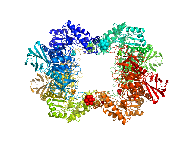
SASDKY7 – α-L-Fucosidase isoenzyme 2 from Paenibacillus thiaminolyticus - wild type

Alpha-L-fucosidase experimental SAS data
OTHER model
Sample: Alpha-L-fucosidase tetramer, 297 kDa Paenibacillus thiaminolyticus (Bacillus … protein
Buffer: 50 mM potassium phosphate, pH: 7.4
Experiment: SAXS data collected at Anton Paar SAXSpoint 2.0, Institute of Biotechnology, Czech Academy of Sciences/Centre of Molecular Structure on 2019 Jul 16
The first structure–function study of GH151 α‐ l ‐fucosidase uncovers new oligomerization pattern, active site complementation, and selective substrate specificity The FEBS Journal (2022)
Koval'ová T, Kovaľ T, Stránský J, Kolenko P, Dušková J, Švecová L, Vodičková P, Spiwok V, Benešová E, Lipovová P, Dohnálek J
RgGuinier 4.2 nm
Dmax 13.2 nm
VolumePorod 418 nm3

SASDKZ7 – α-L-Fucosidase isoenzyme 2 from Paenibacillus thiaminolyticus - mutant H503A

Alpha-L-fucosidase H503A experimental SAS data
PYMOL model
Sample: Alpha-L-fucosidase H503A tetramer, 297 kDa Paenibacillus thiaminolyticus (Bacillus … protein
Buffer: 50 mM potassium phosphate, pH: 7.4
Experiment: SAXS data collected at Anton Paar SAXSpoint 2.0, Institute of Biotechnology, Czech Academy of Sciences/Centre of Molecular Structure on 2019 Jul 16
The first structure–function study of GH151 α‐ l ‐fucosidase uncovers new oligomerization pattern, active site complementation, and selective substrate specificity The FEBS Journal (2022)
Koval'ová T, Kovaľ T, Stránský J, Kolenko P, Dušková J, Švecová L, Vodičková P, Spiwok V, Benešová E, Lipovová P, Dohnálek J
RgGuinier 4.5 nm
Dmax 13.5 nm
VolumePorod 433 nm3

SASDVL8 – Glycosidase/Rutinosidase from Mucor circinelloides - deglycosylated monomer

glucan 1,3-beta-glucosidase experimental SAS data
ALPHAFOLD model
Sample: glucan 1,3-beta-glucosidase monomer, 45 kDa Mucor circinelloides protein
Buffer: 50 mM Tris, 150 mM NaCl, pH: 7.1
Experiment: SAXS data collected at Anton Paar SAXSpoint 2.0, Institute of Biotechnology, Czech Academy of Sciences/Centre of Molecular Structure on 2020 Mar 26
Glucosidase/Rutinosidase from Mucor circinelloides, Penicillium chrysogenum and Aspergillus niger
Jan Stransky
RgGuinier 2.3 nm
Dmax 6.4 nm
VolumePorod 53 nm3

SASDVM8 – Glycosidase/Rutinosidase from Mucor circinelloides - deglycosylated dimer

glucan 1,3-beta-glucosidase experimental SAS data
Glycosidase/Rutinosidase from Mucor circinelloides - deglycosylated dimer Rg histogram
Sample: glucan 1,3-beta-glucosidase dimer, 92 kDa Mucor circinelloides protein
Buffer: 50 mM sodium citrate, 150 mM NaCl, pH: 4.5
Experiment: SAXS data collected at Anton Paar SAXSpoint 2.0, Institute of Biotechnology, Czech Academy of Sciences/Centre of Molecular Structure on 2022 Jun 27
Glucosidase/Rutinosidase from Mucor circinelloides, Penicillium chrysogenum and Aspergillus niger
Jan Stransky
RgGuinier 4.1 nm
Dmax 14.4 nm
VolumePorod 140 nm3

SASDVN8 – Glycosidase/Rutinosidase from Mucor circinelloides - glycosylated dimer

glucan 1,3-beta-glucosidase (short form, glycosylated) experimental SAS data
DAMMIN model
Sample: glucan 1,3-beta-glucosidase (short form, glycosylated) dimer, 89 kDa Mucor circinelloides protein
Buffer: 50 mM sodium citrate, 150 mM NaCl, pH: 4.5
Experiment: SAXS data collected at Anton Paar SAXSpoint 2.0, Institute of Biotechnology, Czech Academy of Sciences/Centre of Molecular Structure on 2022 Jun 28
Glucosidase/Rutinosidase from Mucor circinelloides, Penicillium chrysogenum and Aspergillus niger
Jan Stransky
RgGuinier 3.3 nm
Dmax 11.0 nm
VolumePorod 103 nm3

SASDVP8 – Glycosidase/Rutinosidase from Mucor circinelloides - glycosylated tetramer

glucan 1,3-beta-glucosidase (long form, glycosylated) experimental SAS data
Glycosidase/Rutinosidase from Mucor circinelloides - glycosylated tetramer Rg histogram
Sample: glucan 1,3-beta-glucosidase (long form, glycosylated) tetramer, 184 kDa Mucor circinelloides protein
Buffer: 50 mM sodium citrate, 150 mM NaCl, pH: 4.5
Experiment: SAXS data collected at Anton Paar SAXSpoint 2.0, Institute of Biotechnology, Czech Academy of Sciences/Centre of Molecular Structure on 2022 Jun 28
Glucosidase/Rutinosidase from Mucor circinelloides, Penicillium chrysogenum and Aspergillus niger
Jan Stransky
RgGuinier 4.8 nm
Dmax 15.1 nm
VolumePorod 171 nm3

SASDVQ8 – Glucosidase/Rutinosidase from Penicillium rubens - deglycosylated

glucan 1,3-beta-glucosidase experimental SAS data
ALPHAFOLD model
Sample: glucan 1,3-beta-glucosidase monomer, 42 kDa Penicillium rubens (strain … protein
Buffer: 50 mM sodium citrate, 150 mM NaCl, pH: 4.5
Experiment: SAXS data collected at Anton Paar SAXSpoint 2.0, Institute of Biotechnology, Czech Academy of Sciences/Centre of Molecular Structure on 2019 Nov 8
Glucosidase/Rutinosidase from Mucor circinelloides, Penicillium chrysogenum and Aspergillus niger
Jan Stransky
RgGuinier 2.1 nm
Dmax 5.5 nm
VolumePorod 65 nm3

SASDVR8 – Glucosidase/Rutinosidase from Penicillium rubens - glycosylated

glucan 1,3-beta-glucosidase experimental SAS data
ALPHAFOLD model
Sample: glucan 1,3-beta-glucosidase monomer, 42 kDa Penicillium rubens (strain … protein
Buffer: 50 mM sodium citrate, 150 mM NaCl, pH: 4.5
Experiment: SAXS data collected at Anton Paar SAXSpoint 2.0, Institute of Biotechnology, Czech Academy of Sciences/Centre of Molecular Structure on 2019 Nov 8
Glucosidase/Rutinosidase from Mucor circinelloides, Penicillium chrysogenum and Aspergillus niger
Jan Stransky
RgGuinier 2.2 nm
Dmax 7.6 nm
VolumePorod 68 nm3

SASDVS8 – Glucosidase/Rutinosidase from Aspergillus niger - deglycosylated

glucan 1,3-beta-glucosidase (W90L, N179H) experimental SAS data
PDB (PROTEIN DATA BANK) model
Sample: glucan 1,3-beta-glucosidase (W90L, N179H) monomer, 42 kDa Aspergillus niger (strain … protein
Buffer: McIlvain (Na2HPO4/citric acid) buffer, pH: 3.5
Experiment: SAXS data collected at Anton Paar SAXSpoint 2.0, Institute of Biotechnology, Czech Academy of Sciences/Centre of Molecular Structure on 2021 Jun 8
Glucosidase/Rutinosidase from Mucor circinelloides, Penicillium chrysogenum and Aspergillus niger
Jan Stransky
RgGuinier 2.2 nm
Dmax 6.1 nm
VolumePorod 55 nm3

SASDVT8 – Glucosidase/Rutinosidase from Aspergillus niger - glycosylated

glucan 1,3-beta-glucosidase (W90L, N179H) experimental SAS data
PDB (PROTEIN DATA BANK) model
Sample: glucan 1,3-beta-glucosidase (W90L, N179H) dimer, 83 kDa Aspergillus niger (strain … protein
Buffer: McIlvain (Na2HPO4/citric acid) buffer, pH: 3.5
Experiment: SAXS data collected at Anton Paar SAXSpoint 2.0, Institute of Biotechnology, Czech Academy of Sciences/Centre of Molecular Structure on 2021 Jun 8
Glucosidase/Rutinosidase from Mucor circinelloides, Penicillium chrysogenum and Aspergillus niger
Jan Stransky
RgGuinier 3.0 nm
Dmax 8.1 nm
VolumePorod 111 nm3

SASDPP3 – RTX719. Double-acylated truncated variant of B. pertussis adenylate cyclase toxin (CyaA)

Double-acylated variant of a genetic fusion comprising the C-terminal folding scaffold of RTX block V (residues 1562–1681 of CyaA) and the CyaA residues 719–1294 experimental SAS data
RTX719. Double-acylated truncated variant of B. pertussis adenylate cyclase toxin (CyaA) Rg histogram
Sample: Double-acylated variant of a genetic fusion comprising the C-terminal folding scaffold of RTX block V (residues 1562–1681 of CyaA) and the CyaA residues 719–1294 monomer, 78 kDa Bordetella pertussis protein
Buffer: 50 mM NaCl, 3 mM CaCl2, 20 mM Tris, pH: 8
Experiment: SAXS data collected at Anton Paar SAXSpoint 2.0, Institute of Biotechnology, Czech Academy of Sciences/Centre of Molecular Structure on 2022 Feb 15
Acyl chains stabilize the acylated domain and determine the receptor-mediated interaction of the Bordetella adenylate cyclase toxin with cell membrane. J Biol Chem :110392 (2025)
...Stransky J, Osicka R, Osickova A, Jurnecka D, Sebo P, Bumba L
RgGuinier 3.4 nm
Dmax 14.5 nm
VolumePorod 67 nm3

SASDV98 – Full-length N(5)-hydroxyornithine:cis-anhydromevalonyl coenzyme A-N(5)-transacylase sidF

N(5)-hydroxyornithine:cis-anhydromevalonyl coenzyme A-N(5)-transacylase sidF experimental SAS data
Full-length N(5)-hydroxyornithine:cis-anhydromevalonyl coenzyme A-N(5)-transacylase sidF Rg histogram
Sample: N(5)-hydroxyornithine:cis-anhydromevalonyl coenzyme A-N(5)-transacylase sidF tetramer, 223 kDa Aspergillus fumigatus (strain … protein
Buffer: 50 mM Tris, 200 mM NaCl, pH: 8
Experiment: SAXS data collected at Anton Paar SAXSpoint 2.0, Institute of Biotechnology, Czech Academy of Sciences/Centre of Molecular Structure on 2022 Jul 7
SidF, a dual substrate N5-acetyl-N5-hydroxy-L-ornithine transacetylase involved in Aspergillus fumigatus siderophore biosynthesis Journal of Structural Biology: X 11:100119 (2025)
...Stransky J, Demitri N, Haas H, Cianci M, Benini S
RgGuinier 4.0 nm
Dmax 19.0 nm
VolumePorod 412 nm3

SASDPQ3 – RTX719-K860R. Mono-acylated truncated variant of B. pertussis adenylate cyclase toxin (CyaA)

Mono-acylated variant of a genetic fusion comprising the C-terminal folding scaffold of RTX block V (residues 1562–1681 of CyaA) and the CyaA residues 719–1294 experimental SAS data
RTX719-K860R. Mono-acylated truncated variant of B. pertussis adenylate cyclase toxin (CyaA) Rg histogram
Sample: Mono-acylated variant of a genetic fusion comprising the C-terminal folding scaffold of RTX block V (residues 1562–1681 of CyaA) and the CyaA residues 719–1294 monomer, 78 kDa Bordetella pertussis protein
Buffer: 50 mM NaCl, 3 mM CaCl2, 20 mM Tris, pH: 8
Experiment: SAXS data collected at Anton Paar SAXSpoint 2.0, Institute of Biotechnology, Czech Academy of Sciences/Centre of Molecular Structure on 2022 Feb 16
Acyl chains stabilize the acylated domain and determine the receptor-mediated interaction of the Bordetella adenylate cyclase toxin with cell membrane. J Biol Chem :110392 (2025)
...Stransky J, Osicka R, Osickova A, Jurnecka D, Sebo P, Bumba L
RgGuinier 5.0 nm
Dmax 25.8 nm
VolumePorod 178 nm3

SASDVB8 – C-terminal truncation mutant of N(5)-hydroxyornithine:cis-anhydromevalonyl coenzyme A-N(5)-transacylase sidF

N(5)-hydroxyornithine:cis-anhydromevalonyl coenzyme A-N(5)-transacylase sidF (Δ444-462) experimental SAS data
CORAL model
Sample: N(5)-hydroxyornithine:cis-anhydromevalonyl coenzyme A-N(5)-transacylase sidF (Δ444-462) dimer, 107 kDa Aspergillus fumigatus (strain … protein
Buffer: 50 mM Tris, 200 mM NaCl, pH: 8
Experiment: SAXS data collected at Anton Paar SAXSpoint 2.0, Institute of Biotechnology, Czech Academy of Sciences/Centre of Molecular Structure on 2023 Jan 18
SidF, a dual substrate N5-acetyl-N5-hydroxy-L-ornithine transacetylase involved in Aspergillus fumigatus siderophore biosynthesis Journal of Structural Biology: X 11:100119 (2025)
...Stransky J, Demitri N, Haas H, Cianci M, Benini S
RgGuinier 3.5 nm
Dmax 15.0 nm
VolumePorod 189 nm3

SASDPR3 – RTX719-K983R. Mono-acylated truncated variant of B. pertussis adenylate cyclase toxin (CyaA)

Mono-acylated variant of a genetic fusion comprising the C-terminal folding scaffold of RTX block V (residues 1562–1681 of CyaA) and the CyaA residues 719–1294 experimental SAS data
RTX719-K983R. Mono-acylated truncated variant of B. pertussis adenylate cyclase toxin (CyaA) Rg histogram
Sample: Mono-acylated variant of a genetic fusion comprising the C-terminal folding scaffold of RTX block V (residues 1562–1681 of CyaA) and the CyaA residues 719–1294 monomer, 78 kDa Bordetella pertussis protein
Buffer: 50 mM NaCl, 3 mM CaCl2, 20 mM Tris, pH: 8
Experiment: SAXS data collected at Anton Paar SAXSpoint 2.0, Institute of Biotechnology, Czech Academy of Sciences/Centre of Molecular Structure on 2022 Feb 17
Acyl chains stabilize the acylated domain and determine the receptor-mediated interaction of the Bordetella adenylate cyclase toxin with cell membrane. J Biol Chem :110392 (2025)
...Stransky J, Osicka R, Osickova A, Jurnecka D, Sebo P, Bumba L
RgGuinier 5.3 nm
Dmax 26.3 nm
VolumePorod 187 nm3

SASDVA8 – The N-terminal domain of N(5)-hydroxyornithine:cis-anhydromevalonyl coenzyme A-N(5)-transacylase sidF

N(5)-hydroxyornithine:cis-anhydromevalonyl coenzyme A-N(5)-transacylase sidF experimental SAS data
The N-terminal domain of N(5)-hydroxyornithine:cis-anhydromevalonyl coenzyme A-N(5)-transacylase sidF Rg histogram
Sample: N(5)-hydroxyornithine:cis-anhydromevalonyl coenzyme A-N(5)-transacylase sidF monomer, 25 kDa Aspergillus fumigatus (strain … protein
Buffer: 50 mM Tris, 200 mM NaCl, pH: 8
Experiment: SAXS data collected at Anton Paar SAXSpoint 2.0, Institute of Biotechnology, Czech Academy of Sciences/Centre of Molecular Structure on 2023 Jan 18
SidF, a dual substrate N5-acetyl-N5-hydroxy-L-ornithine transacetylase involved in Aspergillus fumigatus siderophore biosynthesis Journal of Structural Biology: X 11:100119 (2025)
...Stransky J, Demitri N, Haas H, Cianci M, Benini S
RgGuinier 2.2 nm
Dmax 8.9 nm
VolumePorod 30 nm3

SASDPS3 – proRTX719. Non-acylated truncated variant of B. pertussis adenylate cyclase toxin (CyaA)

Non-acylated variant of a genetic fusion comprising the C-terminal folding scaffold of RTX block V (residues 1562–1681 of CyaA) and the CyaA residues 719–1294 experimental SAS data
proRTX719. Non-acylated truncated variant of B. pertussis adenylate cyclase toxin (CyaA) Rg histogram
Sample: Non-acylated variant of a genetic fusion comprising the C-terminal folding scaffold of RTX block V (residues 1562–1681 of CyaA) and the CyaA residues 719–1294 monomer, 78 kDa Bordetella pertussis protein
Buffer: 50 mM NaCl, 3 mM CaCl2, 20 mM Tris, pH: 8
Experiment: SAXS data collected at Anton Paar SAXSpoint 2.0, Institute of Biotechnology, Czech Academy of Sciences/Centre of Molecular Structure on 2022 Feb 22
Acyl chains stabilize the acylated domain and determine the receptor-mediated interaction of the Bordetella adenylate cyclase toxin with cell membrane. J Biol Chem :110392 (2025)
...Stransky J, Osicka R, Osickova A, Jurnecka D, Sebo P, Bumba L
RgGuinier 4.2 nm
Dmax 17.8 nm
VolumePorod 133 nm3