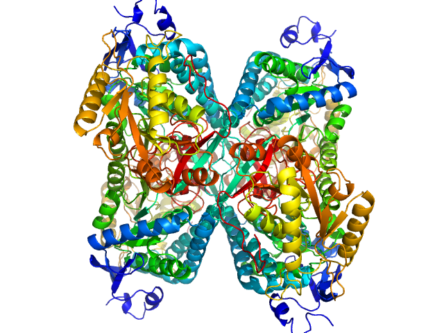
Search

 
Advanced search  

62 hits found for Tanner

SASDCH2 – Aldehyde dehydrogenase 7A1

Aldehyde dehydrogenase 7A1 (Alpha-aminoadipic semialdehyde dehydrogenase) experimental SAS data
NONE model
Sample: Aldehyde dehydrogenase 7A1 (Alpha-aminoadipic semialdehyde dehydrogenase) tetramer, 222 kDa Homo sapiens protein
Buffer: 50 mM Tris, 5% glycerol, 0.5 mM tris(3-hydroxypropyl)phosphine, 50 mM NaCl, pH: 7.8
Experiment: SAXS data collected at 12.3.1 (SIBYLS), Advanced Light Source (ALS) on 2014 Mar 9
Structural Basis of Substrate Recognition by Aldehyde Dehydrogenase 7A1. Biochemistry 54(35):5513-22 (2015)
...Tanner JJ
RgGuinier 3.8 nm
Dmax 11.5 nm
VolumePorod 270 nm3

SASDDJ2 – Calcium-bound polcalcin Phl p 7

polcalcin Phl p 7 experimental SAS data
MES-FOXS model
Sample: polcalcin Phl p 7 monomer, 9 kDa Phleum pratense protein
Buffer: 0.15M NaCl, 0.025M Hepes, pH 7.4, 100 uM Ca2+, pH: 7.4
Experiment: SAXS data collected at 12.3.1 (SIBYLS), Advanced Light Source (ALS) on 2011 Dec 8
Solution structures of polcalcin Phl p 7 in three ligation states: Apo-, hemi-Mg2+-bound, and fully Ca2+-bound. Proteins 81(2):300-15 (2013)
...Tanner JJ
RgGuinier 1.3 nm
Dmax 3.6 nm
VolumePorod 14 nm3

SASDDK2 – Aspergillus fumigatus UDP galactopyranose mutase

Aspergillus fumigatus UDP galactopyranose mutase experimental SAS data
MES-FOXS model
Sample: Aspergillus fumigatus UDP galactopyranose mutase tetramer, 228 kDa protein
Buffer: 20 mM HEPES, 45 mM NaCl, 0.5 mM Tris(hydroxypropyl)phosphine, pH: 7.5
Experiment: SAXS data collected at 12.3.1 (SIBYLS), Advanced Light Source (ALS) on 2010 Apr 19
Crystal structures and small-angle x-ray scattering analysis of UDP-galactopyranose mutase from the pathogenic fungus Aspergillus fumigatus. J Biol Chem 287(12):9041-51 (2012)
...Tanner JJ
RgGuinier 4.7 nm
Dmax 14.7 nm
VolumePorod 308 nm3

SASDDL2 – Sinorhizobium meliloti Proline Utilization A (PutA) lowest concentration, 1.00 mg/ml

Sinorhizobium meliloti (SmPutA) experimental SAS data
MES-FOXS model
Sample: Sinorhizobium meliloti (SmPutA) monomer, 132 kDa Sinorhizobium meliloti protein
Buffer: 50 mM Tris, 1% (v/v) glycerol, 0.5 mM THP, and 50 mM NaCl, pH: 7.8
Experiment: SAXS data collected at 12.3.1 (SIBYLS), Advanced Light Source (ALS) on 2014 Mar 27
Structures of Proline Utilization A (PutA) Reveal the Fold and Functions of the Aldehyde Dehydrogenase Superfamily Domain of Unknown Function. J Biol Chem 291(46):24065-24075 (2016)
...Tanner JJ
RgGuinier 3.4 nm
Dmax 11.0 nm
VolumePorod 171 nm3

SASDDM2 – Sinorhizobium meliloti Proline Utilization A (PutA) at 2.00 mg/ml

Sinorhizobium meliloti (SmPutA) experimental SAS data
MES-FOXS model
Sample: Sinorhizobium meliloti (SmPutA) monomer, 132 kDa Sinorhizobium meliloti protein
Buffer: 50 mM Tris, 1% (v/v) glycerol, 0.5 mM THP, and 50 mM NaCl, pH: 7.8
Experiment: SAXS data collected at 12.3.1 (SIBYLS), Advanced Light Source (ALS) on 2014 Mar 27
Structures of Proline Utilization A (PutA) Reveal the Fold and Functions of the Aldehyde Dehydrogenase Superfamily Domain of Unknown Function. J Biol Chem 291(46):24065-24075 (2016)
...Tanner JJ
RgGuinier 3.8 nm
Dmax 11.9 nm
VolumePorod 225 nm3

SASDDN2 – Sinorhizobium meliloti Proline Utilization A (PutA) at 3.00 mg/ml

Sinorhizobium meliloti (SmPutA) experimental SAS data
MES-FOXS model
Sample: Sinorhizobium meliloti (SmPutA) monomer, 132 kDa Sinorhizobium meliloti protein
Buffer: 50 mM Tris, 1% (v/v) glycerol, 0.5 mM THP, and 50 mM NaCl, pH: 7.8
Experiment: SAXS data collected at 12.3.1 (SIBYLS), Advanced Light Source (ALS) on 2014 Mar 27
Structures of Proline Utilization A (PutA) Reveal the Fold and Functions of the Aldehyde Dehydrogenase Superfamily Domain of Unknown Function. J Biol Chem 291(46):24065-24075 (2016)
...Tanner JJ
RgGuinier 3.8 nm
Dmax 11.8 nm
VolumePorod 248 nm3

SASDDP2 – Sinorhizobium meliloti Proline Utilization A (PutA) at high concentration, 4.00 mg/ml

Sinorhizobium meliloti (SmPutA) experimental SAS data
MES-FOXS model
Sample: Sinorhizobium meliloti (SmPutA) monomer, 132 kDa Sinorhizobium meliloti protein
Buffer: 50 mM Tris, 1% (v/v) glycerol, 0.5 mM THP, and 50 mM NaCl, pH: 7.8
Experiment: SAXS data collected at 12.3.1 (SIBYLS), Advanced Light Source (ALS) on 2014 Mar 27
Structures of Proline Utilization A (PutA) Reveal the Fold and Functions of the Aldehyde Dehydrogenase Superfamily Domain of Unknown Function. J Biol Chem 291(46):24065-24075 (2016)
...Tanner JJ
RgGuinier 3.9 nm
Dmax 11.9 nm
VolumePorod 277 nm3

SASDCP3 – Proline utilization A from Bdellovibrio bacteriovorus

Bifunctional protein PutA experimental SAS data
DAMMIF model
Sample: Bifunctional protein PutA dimer, 219 kDa Bdellovibrio bacteriovorus protein
Buffer: 50 mM Tris, 125 mM NaCl, 1 mM EDTA, and 1 mM tris(3-hydroxypropyl)phosphine (THP) at pH 7.5,, pH: 7.5
Experiment: SAXS data collected at 12.3.1 (SIBYLS), Advanced Light Source (ALS) on 2012 Jun 8
Biophysical investigation of type A PutAs reveals a conserved core oligomeric structure. FEBS J 284(18):3029-3049 (2017)
...Tanner JJ
RgGuinier 4.5 nm
Dmax 14.0 nm
VolumePorod 287 nm3

SASDDP3 – N-propargyl glycine-Inactivated Proline utilization A from Bradyrhizobium diazoefficiens (formerly Bradyrhizobium japonicum) collected by SEC-SAXS

Bifunctional protein PutA experimental SAS data
PDB (PROTEIN DATA BANK) model
Sample: Bifunctional protein PutA dimer, 215 kDa Bradyrhizobium diazoefficiens protein
Buffer: 50 mM Tris, 50 mM NaCl, 0.5 mM TCEP, 5% (v/v) glycerol, pH: 7.8
Experiment: SAXS data collected at BioCAT 18ID, Advanced Photon Source (APS), Argonne National Laboratory on 2017 Jul 16
Redox Modulation of Oligomeric State in Proline Utilization A. Biophys J 114(12):2833-2843 (2018)
...Tanner JJ
RgGuinier 4.6 nm
Dmax 14.4 nm
VolumePorod 324 nm3

SASDCQ3 – Proline utilization A from Desulfovibrio vulgaris 1.5 mg/mL

Bifunctional protein PutA experimental SAS data
Bifunctional protein PutA Kratky plot
Sample: Bifunctional protein PutA dimer, 229 kDa Desulfovibrio vulgaris protein
Buffer: 50 mM Tris-HCl, 50 mM NaCl, 0.5 mM EDTA, and 0.5 mM THP at pH 7.5., pH: 7.5
Experiment: SAXS data collected at 12.3.1 (SIBYLS), Advanced Light Source (ALS) on 2012 Jun 8
Biophysical investigation of type A PutAs reveals a conserved core oligomeric structure. FEBS J 284(18):3029-3049 (2017)
...Tanner JJ
RgGuinier 4.4 nm
Dmax 16.0 nm
VolumePorod 293 nm3

SASDDQ3 – Proline utilization A from Bradyrhizobium diazoefficiens (formerly Bradyrhizobium japonicum) collected by SEC-SAXS

Bifunctional protein PutA experimental SAS data
PDB (PROTEIN DATA BANK) model
Sample: Bifunctional protein PutA tetramer, 430 kDa Bradyrhizobium diazoefficiens protein
Buffer: 50 mM Tris, 50 mM NaCl, 0.5 mM TCEP, 5% (v/v) glycerol, pH: 7.8
Experiment: SAXS data collected at BioCAT 18ID, Advanced Photon Source (APS), Argonne National Laboratory on 2017 Jul 16
Redox Modulation of Oligomeric State in Proline Utilization A. Biophys J 114(12):2833-2843 (2018)
...Tanner JJ
RgGuinier 5.2 nm
Dmax 14.2 nm
VolumePorod 582 nm3

SASDCR3 – Proline utilization A from Legionella pneumophila 3 mg/mL

Bifunctional protein PutA experimental SAS data
Bifunctional protein PutA Kratky plot
Sample: Bifunctional protein PutA dimer, 238 kDa Legionella pneumophila subsp. … protein
Buffer: 50 mM Tris-HCl, 50 mM NaCl, 0.5 mM EDTA, and 0.5 mM THP at pH 7.5., pH: 7.5
Experiment: SAXS data collected at 12.3.1 (SIBYLS), Advanced Light Source (ALS) on 2010 Apr 20
Biophysical investigation of type A PutAs reveals a conserved core oligomeric structure. FEBS J 284(18):3029-3049 (2017)
...Tanner JJ
RgGuinier 4.6 nm
Dmax 16.0 nm
VolumePorod 291 nm3

SASDCS3 – Proline utilization A from Bradyrhizobium diazoefficiens (formerly Bradyrhizobium japonicum) 2.3 mg/mL

Proline dehydrogenase experimental SAS data
Proline dehydrogenase Kratky plot
Sample: Proline dehydrogenase tetramer, 430 kDa Bradyrhizobium diazoefficiens protein
Buffer: 50 mM Tris (pH 7.8), 50 mM NaCl, 0.5 mM Tris(2-carboxyethyl)phosphine, and 5% (v/v) glycerol, pH: 7.8
Experiment: SAXS data collected at 12.3.1 (SIBYLS), Advanced Light Source (ALS) on 2016 Dec 16
Biophysical investigation of type A PutAs reveals a conserved core oligomeric structure. FEBS J 284(18):3029-3049 (2017)
...Tanner JJ
RgGuinier 5.3 nm
Dmax 14.1 nm
VolumePorod 541 nm3

SASDCT3 – Proline utilization A from Bradyrhizobium diazoefficiens (formerly Bradyrhizobium japonicum) 4.7 mg/mL

Proline dehydrogenase experimental SAS data
Proline dehydrogenase Kratky plot
Sample: Proline dehydrogenase tetramer, 430 kDa Bradyrhizobium diazoefficiens protein
Buffer: 50 mM Tris (pH 7.8), 50 mM NaCl, 0.5 mM Tris(2-carboxyethyl)phosphine, and 5% (v/v) glycerol, pH: 7.8
Experiment: SAXS data collected at 12.3.1 (SIBYLS), Advanced Light Source (ALS) on 2016 Dec 12
Biophysical investigation of type A PutAs reveals a conserved core oligomeric structure. FEBS J 284(18):3029-3049 (2017)
...Tanner JJ
RgGuinier 5.2 nm
Dmax 14.6 nm
VolumePorod 553 nm3

SASDCU3 – Proline utilization A from Bradyrhizobium diazoefficiens (formerly Bradyrhizobium japonicum) 7.0 mg/mL

Proline dehydrogenase experimental SAS data
DAMMIF model
Sample: Proline dehydrogenase tetramer, 430 kDa Bradyrhizobium diazoefficiens protein
Buffer: 50 mM Tris (pH 7.8), 50 mM NaCl, 0.5 mM Tris(2-carboxyethyl)phosphine, and 5% (v/v) glycerol, pH: 7.8
Experiment: SAXS data collected at 12.3.1 (SIBYLS), Advanced Light Source (ALS) on 2016 Dec 12
Biophysical investigation of type A PutAs reveals a conserved core oligomeric structure. FEBS J 284(18):3029-3049 (2017)
...Tanner JJ
RgGuinier 5.2 nm
Dmax 13.7 nm
VolumePorod 560 nm3

SASDCV3 – Proline utilization A from Legionella pneumophila 5 mg/mL

Bifunctional protein PutA experimental SAS data
Bifunctional protein PutA Kratky plot
Sample: Bifunctional protein PutA dimer, 238 kDa Legionella pneumophila subsp. … protein
Buffer: 50 mM Tris-HCl, 50 mM NaCl, 0.5 mM EDTA, and 0.5 mM THP at pH 7.5., pH: 7.5
Experiment: SAXS data collected at 12.3.1 (SIBYLS), Advanced Light Source (ALS) on 2010 Apr 20
Biophysical investigation of type A PutAs reveals a conserved core oligomeric structure. FEBS J 284(18):3029-3049 (2017)
...Tanner JJ
RgGuinier 4.6 nm
Dmax 15.3 nm
VolumePorod 297 nm3

SASDCW3 – Proline utilization A from Legionella pneumophila 8 mg/mL

Bifunctional protein PutA experimental SAS data
DAMMIF model
Sample: Bifunctional protein PutA dimer, 238 kDa Legionella pneumophila subsp. … protein
Buffer: 50 mM Tris-HCl, 50 mM NaCl, 0.5 mM EDTA, and 0.5 mM THP at pH 7.5., pH: 7.5
Experiment: SAXS data collected at 12.3.1 (SIBYLS), Advanced Light Source (ALS) on 2010 Apr 20
Biophysical investigation of type A PutAs reveals a conserved core oligomeric structure. FEBS J 284(18):3029-3049 (2017)
...Tanner JJ
RgGuinier 4.6 nm
Dmax 15.5 nm
VolumePorod 295 nm3

SASDCX3 – Proline utilization A from Desulfovibrio vulgaris 3.0 mg/mL

Bifunctional protein PutA experimental SAS data
DAMMIF model
Sample: Bifunctional protein PutA dimer, 229 kDa Desulfovibrio vulgaris protein
Buffer: 50 mM Tris-HCl, 50 mM NaCl, 0.5 mM EDTA, and 0.5 mM THP at pH 7.5., pH: 7.5
Experiment: SAXS data collected at 12.3.1 (SIBYLS), Advanced Light Source (ALS) on 2012 Jun 8
Biophysical investigation of type A PutAs reveals a conserved core oligomeric structure. FEBS J 284(18):3029-3049 (2017)
...Tanner JJ
RgGuinier 4.4 nm
Dmax 16.0 nm
VolumePorod 295 nm3

SASDCY3 – Proline utilization A from Desulfovibrio vulgaris 4.5 mg/mL

Bifunctional protein PutA experimental SAS data
Bifunctional protein PutA Kratky plot
Sample: Bifunctional protein PutA dimer, 229 kDa Desulfovibrio vulgaris protein
Buffer: 50 mM Tris-HCl, 50 mM NaCl, 0.5 mM EDTA, and 0.5 mM THP at pH 7.5., pH: 7.5
Experiment: SAXS data collected at 12.3.1 (SIBYLS), Advanced Light Source (ALS) on 2012 Jun 8
Biophysical investigation of type A PutAs reveals a conserved core oligomeric structure. FEBS J 284(18):3029-3049 (2017)
...Tanner JJ
RgGuinier 4.4 nm
Dmax 16.0 nm
VolumePorod 294 nm3

SASDCZ3 – Proline utilization A from Bradyrhizobium diazoefficiens (formerly Bradyrhizobium japonicum) R51E mutant 2.3 mg/mL

Proline dehydrogenase experimental SAS data
Proline dehydrogenase Kratky plot
Sample: Proline dehydrogenase dimer, 215 kDa Bradyrhizobium diazoefficiens protein
Buffer: 50 mM Tris (pH 7.8), 50 mM NaCl, 0.5 mM Tris(2-carboxyethyl)phosphine, and 5% (v/v) glycerol, pH: 7.8
Experiment: SAXS data collected at 12.3.1 (SIBYLS), Advanced Light Source (ALS) on 2016 Dec 16
Biophysical investigation of type A PutAs reveals a conserved core oligomeric structure. FEBS J 284(18):3029-3049 (2017)
...Tanner JJ
RgGuinier 4.5 nm
Dmax 14.5 nm
VolumePorod 281 nm3

SASDC24 – Proline utilization A from Bradyrhizobium diazoefficiens (formerly Bradyrhizobium japonicum) R51E mutant 4.7 mg/mL

Proline dehydrogenase experimental SAS data
Proline dehydrogenase Kratky plot
Sample: Proline dehydrogenase dimer, 215 kDa Bradyrhizobium diazoefficiens protein
Buffer: 50 mM Tris (pH 7.8), 50 mM NaCl, 0.5 mM Tris(2-carboxyethyl)phosphine, and 5% (v/v) glycerol, pH: 7.8
Experiment: SAXS data collected at 12.3.1 (SIBYLS), Advanced Light Source (ALS) on 2016 Dec 16
Biophysical investigation of type A PutAs reveals a conserved core oligomeric structure. FEBS J 284(18):3029-3049 (2017)
...Tanner JJ
RgGuinier 4.5 nm
Dmax 13.9 nm
VolumePorod 283 nm3

SASDC34 – Proline utilization A from Bradyrhizobium diazoefficiens (formerly Bradyrhizobium japonicum) R51E mutant 7.0 mg/mL

Proline dehydrogenase experimental SAS data
DAMMIF model
Sample: Proline dehydrogenase dimer, 215 kDa Bradyrhizobium diazoefficiens protein
Buffer: 50 mM Tris (pH 7.8), 50 mM NaCl, 0.5 mM Tris(2-carboxyethyl)phosphine, and 5% (v/v) glycerol, pH: 7.8
Experiment: SAXS data collected at 12.3.1 (SIBYLS), Advanced Light Source (ALS) on 2016 Dec 16
Biophysical investigation of type A PutAs reveals a conserved core oligomeric structure. FEBS J 284(18):3029-3049 (2017)
...Tanner JJ
RgGuinier 4.5 nm
Dmax 14.6 nm
VolumePorod 289 nm3

SASDGH4 – Human alpha-aminoadipic semialdehyde dehydrogenase (ALDH)7A1 at 1.2 mg/mL

Alpha-aminoadipic semialdehyde dehydrogenase experimental SAS data
PDB (PROTEIN DATA BANK) model
Sample: Alpha-aminoadipic semialdehyde dehydrogenase tetramer, 222 kDa Homo sapiens protein
Buffer: 50 mM HEPES, 100 mM NaCl, 1 mM DTT, 10 mM NAD, 2% (v/v) glycerol, pH: 8
Experiment: SAXS data collected at 12.3.1 (SIBYLS), Advanced Light Source (ALS) on 2019 May 1
Structural Analysis of Pathogenic Mutations Targeting Glu427 of ALDH7A1, the Hot Spot Residue of Pyridoxine-Dependent Epilepsy. J Inherit Metab Dis (2019)
...Tanner JJ
RgGuinier 3.8 nm
Dmax 11.5 nm
VolumePorod 350 nm3

SASDGJ4 – Human alpha-aminoadipic semialdehyde dehydrogenase (ALDH)7A1 at 2.3 mg/mL

Alpha-aminoadipic semialdehyde dehydrogenase experimental SAS data
PDB (PROTEIN DATA BANK) model
Sample: Alpha-aminoadipic semialdehyde dehydrogenase tetramer, 222 kDa Homo sapiens protein
Buffer: 50 mM HEPES, 100 mM NaCl, 1 mM DTT, 10 mM NAD, 2% (v/v) glycerol, pH: 8
Experiment: SAXS data collected at 12.3.1 (SIBYLS), Advanced Light Source (ALS) on 2019 May 1
Structural Analysis of Pathogenic Mutations Targeting Glu427 of ALDH7A1, the Hot Spot Residue of Pyridoxine-Dependent Epilepsy. J Inherit Metab Dis (2019)
...Tanner JJ
RgGuinier 3.8 nm
Dmax 10.7 nm
VolumePorod 326 nm3

SASDGK4 – Human alpha-aminoadipic semialdehyde dehydrogenase (ALDH)7A1 at 4.7 mg/mL

Alpha-aminoadipic semialdehyde dehydrogenase experimental SAS data
PDB (PROTEIN DATA BANK) model
Sample: Alpha-aminoadipic semialdehyde dehydrogenase tetramer, 222 kDa Homo sapiens protein
Buffer: 50 mM HEPES, 100 mM NaCl, 1 mM DTT, 10 mM NAD, 2% (v/v) glycerol, pH: 8
Experiment: SAXS data collected at 12.3.1 (SIBYLS), Advanced Light Source (ALS) on 2019 May 1
Structural Analysis of Pathogenic Mutations Targeting Glu427 of ALDH7A1, the Hot Spot Residue of Pyridoxine-Dependent Epilepsy. J Inherit Metab Dis (2019)
...Tanner JJ
RgGuinier 3.8 nm
Dmax 10.7 nm
VolumePorod 315 nm3

SASDGL4 – Human alpha-aminoadipic semialdehyde dehydrogenase (ALDH)7A1 E399Q at 1.1 mg/mL

Alpha-aminoadipic semialdehyde dehydrogenase E399Q experimental SAS data
PDB (PROTEIN DATA BANK) model
Sample: Alpha-aminoadipic semialdehyde dehydrogenase E399Q , 56 kDa Homo sapiens protein
Buffer: 50 mM HEPES, 100 mM NaCl, 1 mM DTT, 10 mM NAD, 2% (v/v) glycerol, pH: 8
Experiment: SAXS data collected at 12.3.1 (SIBYLS), Advanced Light Source (ALS) on 2019 May 1
Structural Analysis of Pathogenic Mutations Targeting Glu427 of ALDH7A1, the Hot Spot Residue of Pyridoxine-Dependent Epilepsy. J Inherit Metab Dis (2019)
...Tanner JJ
RgGuinier 3.7 nm
Dmax 10.3 nm
VolumePorod 237 nm3

SASDGM4 – Human alpha-aminoadipic semialdehyde dehydrogenase (ALDH)7A1 E399Q at 2.1 mg/mL

Alpha-aminoadipic semialdehyde dehydrogenase E399Q experimental SAS data
PDB (PROTEIN DATA BANK) model
Sample: Alpha-aminoadipic semialdehyde dehydrogenase E399Q , 56 kDa Homo sapiens protein
Buffer: 50 mM HEPES, 100 mM NaCl, 1 mM DTT, 10 mM NAD, 2% (v/v) glycerol, pH: 8
Experiment: SAXS data collected at 12.3.1 (SIBYLS), Advanced Light Source (ALS) on 2019 May 1
Structural Analysis of Pathogenic Mutations Targeting Glu427 of ALDH7A1, the Hot Spot Residue of Pyridoxine-Dependent Epilepsy. J Inherit Metab Dis (2019)
...Tanner JJ
RgGuinier 3.8 nm
Dmax 10.8 nm
VolumePorod 238 nm3

SASDGN4 – Human alpha-aminoadipic semialdehyde dehydrogenase (ALDH)7A1 E399Q at 4.3 mg/mL

Alpha-aminoadipic semialdehyde dehydrogenase E399Q experimental SAS data
PDB (PROTEIN DATA BANK) model
Sample: Alpha-aminoadipic semialdehyde dehydrogenase E399Q , 56 kDa Homo sapiens protein
Buffer: 50 mM HEPES, 100 mM NaCl, 1 mM DTT, 10 mM NAD, 2% (v/v) glycerol, pH: 8
Experiment: SAXS data collected at 12.3.1 (SIBYLS), Advanced Light Source (ALS) on 2019 May 1
Structural Analysis of Pathogenic Mutations Targeting Glu427 of ALDH7A1, the Hot Spot Residue of Pyridoxine-Dependent Epilepsy. J Inherit Metab Dis (2019)
...Tanner JJ
RgGuinier 3.8 nm
Dmax 10.6 nm
VolumePorod 255 nm3

SASDGP4 – Human alpha-aminoadipic semialdehyde dehydrogenase (ALDH)7A1 E399D at 1.4 mg/mL

Alpha-aminoadipic semialdehyde dehydrogenase experimental SAS data
PDB (PROTEIN DATA BANK) model
Sample: Alpha-aminoadipic semialdehyde dehydrogenase , 56 kDa Homo sapiens protein
Buffer: 50 mM HEPES, 100 mM NaCl, 1 mM DTT, 10 mM NAD, 2% (v/v) glycerol, pH: 8
Experiment: SAXS data collected at 12.3.1 (SIBYLS), Advanced Light Source (ALS) on 2019 May 1
Structural Analysis of Pathogenic Mutations Targeting Glu427 of ALDH7A1, the Hot Spot Residue of Pyridoxine-Dependent Epilepsy. J Inherit Metab Dis (2019)
...Tanner JJ
RgGuinier 3.7 nm
Dmax 11.3 nm
VolumePorod 256 nm3

SASDGQ4 – Human alpha-aminoadipic semialdehyde dehydrogenase (ALDH)7A1 E399D at 2.9 mg/mL

Alpha-aminoadipic semialdehyde dehydrogenase experimental SAS data
PDB (PROTEIN DATA BANK) model
Sample: Alpha-aminoadipic semialdehyde dehydrogenase , 56 kDa Homo sapiens protein
Buffer: 50 mM HEPES, 100 mM NaCl, 1 mM DTT, 10 mM NAD, 2% (v/v) glycerol, pH: 8
Experiment: SAXS data collected at 12.3.1 (SIBYLS), Advanced Light Source (ALS) on 2019 May 1
Structural Analysis of Pathogenic Mutations Targeting Glu427 of ALDH7A1, the Hot Spot Residue of Pyridoxine-Dependent Epilepsy. J Inherit Metab Dis (2019)
...Tanner JJ
RgGuinier 3.8 nm
Dmax 11.4 nm
VolumePorod 260 nm3

SASDGR4 – Human alpha-aminoadipic semialdehyde dehydrogenase (ALDH)7A1 E399D at 5.7 mg/mL

Alpha-aminoadipic semialdehyde dehydrogenase E399D experimental SAS data
PDB (PROTEIN DATA BANK) model
Sample: Alpha-aminoadipic semialdehyde dehydrogenase E399D , 56 kDa Homo sapiens protein
Buffer: 50 mM HEPES, 100 mM NaCl, 1 mM DTT, 10 mM NAD, 2% (v/v) glycerol, pH: 8
Experiment: SAXS data collected at 12.3.1 (SIBYLS), Advanced Light Source (ALS) on 2019 May 1
Structural Analysis of Pathogenic Mutations Targeting Glu427 of ALDH7A1, the Hot Spot Residue of Pyridoxine-Dependent Epilepsy. J Inherit Metab Dis (2019)
...Tanner JJ
RgGuinier 3.9 nm
Dmax 10.6 nm
VolumePorod 272 nm3

SASDGS4 – Human alpha-aminoadipic semialdehyde dehydrogenase (ALDH)7A1 E399G at 1.6 mg/mL

Alpha-aminoadipic semialdehyde dehydrogenase E399G experimental SAS data
PDB (PROTEIN DATA BANK) model
Sample: Alpha-aminoadipic semialdehyde dehydrogenase E399G , 55 kDa Homo sapiens protein
Buffer: 50 mM HEPES, 100 mM NaCl, 1 mM DTT, 10 mM NAD, 2% (v/v) glycerol, pH: 8
Experiment: SAXS data collected at 12.3.1 (SIBYLS), Advanced Light Source (ALS) on 2019 May 1
Structural Analysis of Pathogenic Mutations Targeting Glu427 of ALDH7A1, the Hot Spot Residue of Pyridoxine-Dependent Epilepsy. J Inherit Metab Dis (2019)
...Tanner JJ
RgGuinier 3.8 nm
Dmax 11.3 nm
VolumePorod 290 nm3

SASDGT4 – Human alpha-aminoadipic semialdehyde dehydrogenase (ALDH)7A1 E399G at 3.2 mg/mL

Alpha-aminoadipic semialdehyde dehydrogenase E399G experimental SAS data
PDB (PROTEIN DATA BANK) model
Sample: Alpha-aminoadipic semialdehyde dehydrogenase E399G , 55 kDa Homo sapiens protein
Buffer: 50 mM HEPES, 100 mM NaCl, 1 mM DTT, 10 mM NAD, 2% (v/v) glycerol, pH: 8
Experiment: SAXS data collected at 12.3.1 (SIBYLS), Advanced Light Source (ALS) on 2019 May 1
Structural Analysis of Pathogenic Mutations Targeting Glu427 of ALDH7A1, the Hot Spot Residue of Pyridoxine-Dependent Epilepsy. J Inherit Metab Dis (2019)
...Tanner JJ
RgGuinier 3.7 nm
Dmax 10.6 nm
VolumePorod 250 nm3

SASDGU4 – Human alpha-aminoadipic semialdehyde dehydrogenase (ALDH)7A1 E399G at 6.5 mg/mL

Alpha-aminoadipic semialdehyde dehydrogenase E399G experimental SAS data
PDB (PROTEIN DATA BANK) model
Sample: Alpha-aminoadipic semialdehyde dehydrogenase E399G , 55 kDa Homo sapiens protein
Buffer: 50 mM HEPES, 100 mM NaCl, 1 mM DTT, 10 mM NAD, 2% (v/v) glycerol, pH: 8
Experiment: SAXS data collected at 12.3.1 (SIBYLS), Advanced Light Source (ALS) on 2019 May 1
Structural Analysis of Pathogenic Mutations Targeting Glu427 of ALDH7A1, the Hot Spot Residue of Pyridoxine-Dependent Epilepsy. J Inherit Metab Dis (2019)
...Tanner JJ
RgGuinier 3.8 nm
Dmax 10.0 nm
VolumePorod 270 nm3

SASDHV5 – Human 4-trimethylaminobutyraldehyde dehydrogenase (ALDH9A1) at 1.25 mg/mL

4-trimethylaminobutyraldehyde dehydrogenase experimental SAS data
OTHER model
Sample: 4-trimethylaminobutyraldehyde dehydrogenase tetramer, 215 kDa Homo sapiens protein
Buffer: 50 mM HEPES, 600 mM NaCl, 2% (v/v) glycerol, 1 mM DTT, 1 mM NAD+, pH: 8
Experiment: SAXS data collected at 12.3.1 (SIBYLS), Advanced Light Source (ALS) on 2019 May 28
Inhibition, crystal structures, and in-solution oligomeric structure of aldehyde dehydrogenase 9A1. Arch Biochem Biophys :108477 (2020)
...Tanner JJ
RgGuinier 3.8 nm
Dmax 10.9 nm
VolumePorod 252 nm3

SASDHW5 – Human 4-trimethylaminobutyraldehyde dehydrogenase (ALDH9A1) at 2.5 mg/mL

4-trimethylaminobutyraldehyde dehydrogenase experimental SAS data
OTHER model
Sample: 4-trimethylaminobutyraldehyde dehydrogenase tetramer, 215 kDa Homo sapiens protein
Buffer: 50 mM HEPES, 600 mM NaCl, 2% (v/v) glycerol, 1 mM DTT, 1 mM NAD+, pH: 8
Experiment: SAXS data collected at 12.3.1 (SIBYLS), Advanced Light Source (ALS) on 2019 May 28
Inhibition, crystal structures, and in-solution oligomeric structure of aldehyde dehydrogenase 9A1. Arch Biochem Biophys :108477 (2020)
...Tanner JJ
RgGuinier 3.8 nm
Dmax 10.8 nm
VolumePorod 240 nm3

SASDHX5 – Human 4-trimethylaminobutyraldehyde dehydrogenase (ALDH9A1) at 5 mg/mL

4-trimethylaminobutyraldehyde dehydrogenase experimental SAS data
OTHER model
Sample: 4-trimethylaminobutyraldehyde dehydrogenase tetramer, 215 kDa Homo sapiens protein
Buffer: 50 mM HEPES, 600 mM NaCl, 2% (v/v) glycerol, 1 mM DTT, 1 mM NAD+, pH: 8
Experiment: SAXS data collected at 12.3.1 (SIBYLS), Advanced Light Source (ALS) on 2019 May 28
Inhibition, crystal structures, and in-solution oligomeric structure of aldehyde dehydrogenase 9A1. Arch Biochem Biophys :108477 (2020)
...Tanner JJ
RgGuinier 3.8 nm
Dmax 10.5 nm
VolumePorod 240 nm3

SASDE96 – Aldehyde dehydrogenase 12 from Zea mays Extrapolated to Infinite Dilution

Aldehyde dehydrogenase 12 experimental SAS data
ALLOSMOD model
Sample: Aldehyde dehydrogenase 12 tetramer, 242 kDa Zea mays protein
Buffer: 50 mM Tris-HCl, 50 mM NaCl, 0.5 mM TCEP, and 5% (v/v) glycerol, pH: 7.8
Experiment: SAXS data collected at 12.3.1 (SIBYLS), Advanced Light Source (ALS) on 2016 Dec 6
Structural and Biochemical Characterization of Aldehyde Dehydrogenase 12, the Last Enzyme of Proline Catabolism in Plants. J Mol Biol (2018)
...Tanner JJ, Kopečný D
RgGuinier 4.1 nm
Dmax 14.4 nm
VolumePorod 351 nm3

SASDSX6 – Aquifex aeolicus domain of unknown function 507 at 1 mg/mL

DUF507 family protein experimental SAS data
CUSTOM IN-HOUSE model
Sample: DUF507 family protein monomer, 22 kDa Aquifex aeolicus (strain … protein
Buffer: 20 mM Hepes pH 7.2, 400 mM NaCl, pH: 7.2
Experiment: SAXS data collected at 12.3.1 (SIBYLS), Advanced Light Source (ALS) on 2021 Dec 7
Crystal structure of domain of unknown function 507 (DUF507) reveals a new protein fold. Sci Rep 13(1):13496 (2023)
...Tanner JJ
RgGuinier 2.3 nm
Dmax 9.0 nm
VolumePorod 39 nm3

SASDB27 – Chimeric EcRHH-RcPutA: The E.coli Proline utilization A RHH domain fused to R.capsulatus PutA

Proline utilization A experimental SAS data
Proline utilization A Kratky plot
Sample: Proline utilization A dimer, 251 kDa Escherchia coli, Rhodobacter … protein
Buffer: 50 mM Tris, 200 mM NaCl, 0.5 mM Tris(3-hydroxypropyl)phosphine, pH: 7.5
Experiment: SAXS data collected at 12.3.1 (SIBYLS), Advanced Light Source (ALS) on 2012 Oct 16
Engineering a trifunctional proline utilization A chimaera by fusing a DNA-binding domain to a bifunctional PutA. Biosci Rep 36(6) (2016)
...Tanner JJ, Becker DF
RgGuinier 5.2 nm
Dmax 18.3 nm
VolumePorod 308 nm3

SASDE47 – Aldehyde dehydrogenase 16 from Loktanella sp. (LsALDH16): 2 mg/ml

Aldehyde dehydrogenase 16 from Loktanella sp. experimental SAS data
PYMOL model
Sample: Aldehyde dehydrogenase 16 from Loktanella sp. dimer, 161 kDa Loktanella sp. 3ANDIMAR09 protein
Buffer: 20 mM Tris-HCl, 100 mM NaCl, 2.0% glycerol, 0.5 mM Tris(3-hydroxypropyl)phosphine, pH: 8
Experiment: SAXS data collected at 12.3.1 (SIBYLS), Advanced Light Source (ALS) on 2017 Dec 13
Crystal Structure of Aldehyde Dehydrogenase 16 Reveals Trans-Hierarchical Structural Similarity and a New Dimer. J Mol Biol (2018)
...Tanner JJ
RgGuinier 3.6 nm
Dmax 10.9 nm
VolumePorod 202 nm3

SASDE57 – Aldehyde dehydrogenase 16 from Loktanella sp. (LsALDH16): 4 mg/ml

Aldehyde dehydrogenase 16 from Loktanella sp. experimental SAS data
PYMOL model
Sample: Aldehyde dehydrogenase 16 from Loktanella sp. dimer, 161 kDa Loktanella sp. 3ANDIMAR09 protein
Buffer: 20 mM Tris-HCl, 100 mM NaCl, 2.0% glycerol, 0.5 mM Tris(3-hydroxypropyl)phosphine, pH: 8
Experiment: SAXS data collected at 12.3.1 (SIBYLS), Advanced Light Source (ALS) on 2017 Dec 13
Crystal Structure of Aldehyde Dehydrogenase 16 Reveals Trans-Hierarchical Structural Similarity and a New Dimer. J Mol Biol (2018)
...Tanner JJ
RgGuinier 3.6 nm
Dmax 11.2 nm
VolumePorod 204 nm3

SASDE67 – Aldehyde dehydrogenase 16 from Loktanella sp. (LsALDH16): 6 mg/ml

Aldehyde dehydrogenase 16 from Loktanella sp. experimental SAS data
PYMOL model
Sample: Aldehyde dehydrogenase 16 from Loktanella sp. dimer, 161 kDa Loktanella sp. 3ANDIMAR09 protein
Buffer: 20 mM Tris-HCl, 100 mM NaCl, 2.0% glycerol, 0.5 mM Tris(3-hydroxypropyl)phosphine, pH: 8
Experiment: SAXS data collected at 12.3.1 (SIBYLS), Advanced Light Source (ALS) on 2017 Dec 13
Crystal Structure of Aldehyde Dehydrogenase 16 Reveals Trans-Hierarchical Structural Similarity and a New Dimer. J Mol Biol (2018)
...Tanner JJ
RgGuinier 3.5 nm
Dmax 10.6 nm
VolumePorod 207 nm3

SASDE77 – Aldehyde dehydrogenase 16 from Loktanella sp. (LsALDH16): 8 mg/ml

Aldehyde dehydrogenase 16 from Loktanella sp. experimental SAS data
PYMOL model
Sample: Aldehyde dehydrogenase 16 from Loktanella sp. dimer, 161 kDa Loktanella sp. 3ANDIMAR09 protein
Buffer: 20 mM Tris-HCl, 100 mM NaCl, 2.0% glycerol, 0.5 mM Tris(3-hydroxypropyl)phosphine, pH: 8
Experiment: SAXS data collected at 12.3.1 (SIBYLS), Advanced Light Source (ALS) on 2017 Dec 13
Crystal Structure of Aldehyde Dehydrogenase 16 Reveals Trans-Hierarchical Structural Similarity and a New Dimer. J Mol Biol (2018)
...Tanner JJ
RgGuinier 3.6 nm
Dmax 10.8 nm
VolumePorod 205 nm3

SASDE87 – Aldehyde dehydrogenase family 16 member A1 from Homo sapiens (HsALDH16A1): 1 mg/ml

Aldehyde dehydrogenase family 16 member A1 from Homo sapiens experimental SAS data
PYMOL model
Sample: Aldehyde dehydrogenase family 16 member A1 from Homo sapiens dimer, 171 kDa Homo sapiens protein
Buffer: 20 mM Tris-HCl, 100 mM NaCl, 2.0% glycerol, 0.5 mM Tris(3-hydroxypropyl)phosphine, pH: 8
Experiment: SAXS data collected at 12.3.1 (SIBYLS), Advanced Light Source (ALS) on 2017 Dec 13
Crystal Structure of Aldehyde Dehydrogenase 16 Reveals Trans-Hierarchical Structural Similarity and a New Dimer. J Mol Biol (2018)
...Tanner JJ
RgGuinier 3.6 nm
Dmax 10.9 nm
VolumePorod 230 nm3

SASDE97 – Aldehyde dehydrogenase family 16 member A1 from Homo sapiens (HsALDH16A1): 1.6 mg/ml

Aldehyde dehydrogenase family 16 member A1 from Homo sapiens experimental SAS data
PYMOL model
Sample: Aldehyde dehydrogenase family 16 member A1 from Homo sapiens dimer, 171 kDa Homo sapiens protein
Buffer: 20 mM Tris-HCl, 100 mM NaCl, 2.0% glycerol, 0.5 mM Tris(3-hydroxypropyl)phosphine, pH: 8
Experiment: SAXS data collected at 12.3.1 (SIBYLS), Advanced Light Source (ALS) on 2017 Dec 13
Crystal Structure of Aldehyde Dehydrogenase 16 Reveals Trans-Hierarchical Structural Similarity and a New Dimer. J Mol Biol (2018)
...Tanner JJ
RgGuinier 3.8 nm
Dmax 11.2 nm
VolumePorod 236 nm3

SASDEA7 – Aldehyde dehydrogenase family 16 member A1 from Homo sapiens (HsALDH16A1): 3.2 mg/ml

Aldehyde dehydrogenase family 16 member A1 from Homo sapiens experimental SAS data
PYMOL model
Sample: Aldehyde dehydrogenase family 16 member A1 from Homo sapiens dimer, 171 kDa Homo sapiens protein
Buffer: 20 mM Tris-HCl, 100 mM NaCl, 2.0% glycerol, 0.5 mM Tris(3-hydroxypropyl)phosphine, pH: 8
Experiment: SAXS data collected at 12.3.1 (SIBYLS), Advanced Light Source (ALS) on 2017 Dec 13
Crystal Structure of Aldehyde Dehydrogenase 16 Reveals Trans-Hierarchical Structural Similarity and a New Dimer. J Mol Biol (2018)
...Tanner JJ
RgGuinier 3.8 nm
Dmax 11.5 nm
VolumePorod 237 nm3

SASDQ48 – Minimal proline dehydrogenase domain of proline utilization A (SmPutADeltaAlpha2) 1.1 mg/mL

Minimal proline dehydrogenase domain of proline utilization A (design #2) experimental SAS data
OTHER model
Sample: Minimal proline dehydrogenase domain of proline utilization A (design #2) dimer, 87 kDa Sinorhizobium meliloti protein
Buffer: 25 mM HEPES pH 7.6, 150 mM NaCl, and 1mM TCEP, pH: 7.6
Experiment: SAXS data collected at 12.3.1 (SIBYLS), Advanced Light Source (ALS) on 2022 Apr 12
Structure-based engineering of minimal Proline dehydrogenase domains for inhibitor discovery. Protein Eng Des Sel (2022)
...Tanner JJ
RgGuinier 2.7 nm
Dmax 9.5 nm
VolumePorod 102 nm3

SASDQ58 – Minimal proline dehydrogenase domain of proline utilization A (SmPutADeltaAlpha2) 2.3 mg/mL

Minimal proline dehydrogenase domain of proline utilization A (design #2) experimental SAS data
OTHER model
Sample: Minimal proline dehydrogenase domain of proline utilization A (design #2) dimer, 87 kDa Sinorhizobium meliloti protein
Buffer: 25 mM HEPES pH 7.6, 150 mM NaCl, and 1mM TCEP, pH: 7.6
Experiment: SAXS data collected at 12.3.1 (SIBYLS), Advanced Light Source (ALS) on 2022 Apr 12
Structure-based engineering of minimal Proline dehydrogenase domains for inhibitor discovery. Protein Eng Des Sel (2022)
...Tanner JJ
RgGuinier 2.9 nm
Dmax 9.7 nm
VolumePorod 102 nm3

SASDQ68 – Minimal proline dehydrogenase domain of proline utilization A (SmPutADeltaAlpha2) 3.4 mg/mL

Minimal proline dehydrogenase domain of proline utilization A (design #2) experimental SAS data
OTHER model
Sample: Minimal proline dehydrogenase domain of proline utilization A (design #2) dimer, 87 kDa Sinorhizobium meliloti protein
Buffer: 25 mM HEPES pH 7.6, 150 mM NaCl, and 1mM TCEP, pH: 7.6
Experiment: SAXS data collected at 12.3.1 (SIBYLS), Advanced Light Source (ALS) on 2022 Apr 12
Structure-based engineering of minimal Proline dehydrogenase domains for inhibitor discovery. Protein Eng Des Sel (2022)
...Tanner JJ
RgGuinier 3.0 nm
Dmax 9.8 nm
VolumePorod 108 nm3

SASDD29 – Low load concentration of apo alpha-aminoadipic semialdehyde dehydrogenase (ALDH7A1) collected by SEC-SAXS

Alpha-aminoadipic semialdehyde dehydrogenase experimental SAS data
PDB (PROTEIN DATA BANK) model
Sample: Alpha-aminoadipic semialdehyde dehydrogenase tetramer, 222 kDa Homo sapiens protein
Buffer: 50 mM Tris, 50 mM NaCl, 0.5 mM DTT, 5% (v/v) glycerol, pH: 7.8
Experiment: SAXS data collected at BioCAT 18ID, Advanced Photon Source (APS), Argonne National Laboratory on 2018 Feb 22
NAD+ Promotes Assembly of the Active Tetramer of Aldehyde Dehydrogenase 7A1. FEBS Lett (2018)
...Tanner JJ
RgGuinier 3.5 nm
VolumePorod 212 nm3

SASDD39 – Medium load concentration of apo alpha-aminoadipic semialdehyde dehydrogenase (ALDH7A1) collected by SEC-SAXS

Alpha-aminoadipic semialdehyde dehydrogenase experimental SAS data
PDB (PROTEIN DATA BANK) model
Sample: Alpha-aminoadipic semialdehyde dehydrogenase tetramer, 222 kDa Homo sapiens protein
Buffer: 50 mM Tris, 50 mM NaCl, 0.5 mM DTT, 5% (v/v) glycerol, pH: 7.8
Experiment: SAXS data collected at BioCAT 18ID, Advanced Photon Source (APS), Argonne National Laboratory on 2018 Feb 22
NAD+ Promotes Assembly of the Active Tetramer of Aldehyde Dehydrogenase 7A1. FEBS Lett (2018)
...Tanner JJ
RgGuinier 3.7 nm
VolumePorod 229 nm3

SASDD49 – High load concentration of apo alpha-aminoadipic semialdehyde dehydrogenase (ALDH7A1) collected by SEC-SAXS

Alpha-aminoadipic semialdehyde dehydrogenase experimental SAS data
PDB (PROTEIN DATA BANK) model
Sample: Alpha-aminoadipic semialdehyde dehydrogenase tetramer, 222 kDa Homo sapiens protein
Buffer: 50 mM Tris, 50 mM NaCl, 0.5 mM DTT, 5% (v/v) glycerol, pH: 7.8
Experiment: SAXS data collected at BioCAT 18ID, Advanced Photon Source (APS), Argonne National Laboratory on 2018 Feb 22
NAD+ Promotes Assembly of the Active Tetramer of Aldehyde Dehydrogenase 7A1. FEBS Lett (2018)
...Tanner JJ
RgGuinier 3.7 nm
VolumePorod 238 nm3

SASDD59 – Low load concentration of alpha-aminoadipic semialdehyde dehydrogenase (ALDH7A1) with nicotinamide adenine dinucleotide (NAD) collected by SEC-SAXS

Alpha-aminoadipic semialdehyde dehydrogenase experimental SAS data
PDB (PROTEIN DATA BANK) model
Sample: Alpha-aminoadipic semialdehyde dehydrogenase tetramer, 222 kDa Homo sapiens protein
Buffer: 50 mM Tris, 50 mM NaCl, 0.5 mM DTT, 5% (v/v) glycerol, 1 mM NAD, pH: 7.8
Experiment: SAXS data collected at BioCAT 18ID, Advanced Photon Source (APS), Argonne National Laboratory on 2018 Feb 22
NAD+ Promotes Assembly of the Active Tetramer of Aldehyde Dehydrogenase 7A1. FEBS Lett (2018)
...Tanner JJ
RgGuinier 3.7 nm
VolumePorod 277 nm3

SASDD69 – Medium load concentration of alpha-aminoadipic semialdehyde dehydrogenase (ALDH7A1) with nicotinamide adenine dinucleotide (NAD) collected by SEC-SAXS

Alpha-aminoadipic semialdehyde dehydrogenase experimental SAS data
PDB (PROTEIN DATA BANK) model
Sample: Alpha-aminoadipic semialdehyde dehydrogenase tetramer, 222 kDa Homo sapiens protein
Buffer: 50 mM Tris, 50 mM NaCl, 0.5 mM DTT, 5% (v/v) glycerol, 1 mM NAD, pH: 7.8
Experiment: SAXS data collected at BioCAT 18ID, Advanced Photon Source (APS), Argonne National Laboratory on 2018 Feb 22
NAD+ Promotes Assembly of the Active Tetramer of Aldehyde Dehydrogenase 7A1. FEBS Lett (2018)
...Tanner JJ
RgGuinier 3.8 nm
VolumePorod 275 nm3

SASDD79 – High load concentration of alpha-aminoadipic semialdehyde dehydrogenase ALDH7A1 with nicotinamide adenine dinucleotide (NAD) collected by SEC-SAXS

Alpha-aminoadipic semialdehyde dehydrogenase experimental SAS data
PDB (PROTEIN DATA BANK) model
Sample: Alpha-aminoadipic semialdehyde dehydrogenase tetramer, 222 kDa Homo sapiens protein
Buffer: 50 mM Tris, 50 mM NaCl, 0.5 mM DTT, 5% (v/v) glycerol, 1 mM NAD, pH: 7.8
Experiment: SAXS data collected at BioCAT 18ID, Advanced Photon Source (APS), Argonne National Laboratory on 2018 Feb 22
NAD+ Promotes Assembly of the Active Tetramer of Aldehyde Dehydrogenase 7A1. FEBS Lett (2018)
...Tanner JJ
RgGuinier 3.8 nm
VolumePorod 277 nm3

SASDNA9 – Acinetobacter baumannii putrescine N-hydroxylase, 1 mg/mL

L-lysine 6-monooxygenase (NADPH-requiring) experimental SAS data
ALLOSMOD model
Sample: L-lysine 6-monooxygenase (NADPH-requiring) tetramer, 214 kDa Acinetobacter baumannii MRSN … protein
Buffer: 25 mM HEPES pH 7.5, 150 mM NaCl, 1 mM TCEP, pH: 7.5
Experiment: SAXS data collected at 12.3.1 (SIBYLS), Advanced Light Source (ALS) on 2022 Apr 14
Kinetic and Structural Characterization of a Flavin-Dependent Putrescine N-Hydroxylase from Acinetobacter baumannii. Biochemistry (2022)
...Tanner JJ, Sobrado P
RgGuinier 4.1 nm
Dmax 15.0 nm
VolumePorod 394 nm3

SASDNB9 – Acinetobacter baumannii putrescine N-hydroxylase, 3 mg/mL

L-lysine 6-monooxygenase (NADPH-requiring) experimental SAS data
ALLOSMOD model
Sample: L-lysine 6-monooxygenase (NADPH-requiring) tetramer, 214 kDa Acinetobacter baumannii MRSN … protein
Buffer: 25 mM HEPES pH 7.5, 150 mM NaCl, 1 mM TCEP, pH: 7.5
Experiment: SAXS data collected at 12.3.1 (SIBYLS), Advanced Light Source (ALS) on 2022 Apr 14
Kinetic and Structural Characterization of a Flavin-Dependent Putrescine N-Hydroxylase from Acinetobacter baumannii. Biochemistry (2022)
...Tanner JJ, Sobrado P
RgGuinier 4.1 nm
Dmax 15.0 nm
VolumePorod 390 nm3

SASDNC9 – Acinetobacter baumannii putrescine N-hydroxylase, 5 mg/mL

L-lysine 6-monooxygenase (NADPH-requiring) experimental SAS data
ALLOSMOD model
Sample: L-lysine 6-monooxygenase (NADPH-requiring) tetramer, 214 kDa Acinetobacter baumannii MRSN … protein
Buffer: 25 mM HEPES pH 7.5, 150 mM NaCl, 1 mM TCEP, pH: 7.5
Experiment: SAXS data collected at 12.3.1 (SIBYLS), Advanced Light Source (ALS) on 2022 Apr 14
Kinetic and Structural Characterization of a Flavin-Dependent Putrescine N-Hydroxylase from Acinetobacter baumannii. Biochemistry (2022)
...Tanner JJ, Sobrado P
RgGuinier 4.2 nm
Dmax 15.0 nm
VolumePorod 390 nm3

SASDND9 – Acinetobacter baumannii putrescine N-hydroxylase, 6 mg/mL

L-lysine 6-monooxygenase (NADPH-requiring) experimental SAS data
ALLOSMOD model
Sample: L-lysine 6-monooxygenase (NADPH-requiring) tetramer, 214 kDa Acinetobacter baumannii MRSN … protein
Buffer: 25 mM HEPES pH 7.5, 150 mM NaCl, 1 mM TCEP, pH: 7.5
Experiment: SAXS data collected at 12.3.1 (SIBYLS), Advanced Light Source (ALS) on 2022 Apr 14
Kinetic and Structural Characterization of a Flavin-Dependent Putrescine N-Hydroxylase from Acinetobacter baumannii. Biochemistry (2022)
...Tanner JJ, Sobrado P
RgGuinier 4.2 nm
Dmax 15.4 nm
VolumePorod 390 nm3

SASDNE9 – Acinetobacter baumannii putrescine N-hydroxylase, 8 mg/mL

L-lysine 6-monooxygenase (NADPH-requiring) experimental SAS data
ALLOSMOD model
Sample: L-lysine 6-monooxygenase (NADPH-requiring) tetramer, 214 kDa Acinetobacter baumannii MRSN … protein
Buffer: 25 mM HEPES pH 7.5, 150 mM NaCl, 1 mM TCEP, pH: 7.5
Experiment: SAXS data collected at 12.3.1 (SIBYLS), Advanced Light Source (ALS) on 2022 Apr 14
Kinetic and Structural Characterization of a Flavin-Dependent Putrescine N-Hydroxylase from Acinetobacter baumannii. Biochemistry (2022)
...Tanner JJ, Sobrado P
RgGuinier 4.2 nm
Dmax 15.5 nm
VolumePorod 390 nm3

SASDNF9 – Acinetobacter baumannii putrescine N-hydroxylase, 9 mg/mL

L-lysine 6-monooxygenase (NADPH-requiring) experimental SAS data
ALLOSMOD model
Sample: L-lysine 6-monooxygenase (NADPH-requiring) tetramer, 214 kDa Acinetobacter baumannii MRSN … protein
Buffer: 25 mM HEPES pH 7.5, 150 mM NaCl, 1 mM TCEP, pH: 7.5
Experiment: SAXS data collected at 12.3.1 (SIBYLS), Advanced Light Source (ALS) on 2022 Apr 14
Kinetic and Structural Characterization of a Flavin-Dependent Putrescine N-Hydroxylase from Acinetobacter baumannii. Biochemistry (2022)
...Tanner JJ, Sobrado P
RgGuinier 4.2 nm
Dmax 15.0 nm
VolumePorod 396 nm3