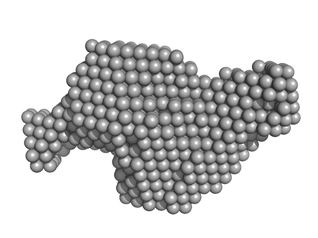
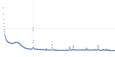
UniProt ID: Q94BT6 (29-190) Adagio protein 1 (Zeitlupe G46S:G80R)
|
|
|
|
| Sample: |
Adagio protein 1 (Zeitlupe G46S:G80R) dimer, 37 kDa Arabidopsis thaliana protein
|
| Buffer: |
50 mM HEPES, 100 mM NaCl, 2 mM TCEP, pH: 8 |
| Experiment: |
SAXS
data collected at G1, Cornell High Energy Synchrotron Source (CHESS) on 2017 Nov 2
|
Steric and Electronic Interactions at Gln154 in ZEITLUPE Induce Reorganization of the LOV Domain Dimer Interface.
Biochemistry (2020)
Pudasaini A, Green R, Song YH, Blumenfeld A, Karki N, Imaizumi T, Zoltowski BD
|
| RgGuinier |
2.2 |
nm |
| Dmax |
8.4 |
nm |
| VolumePorod |
41 |
nm3 |
|
|
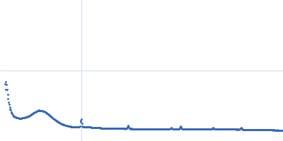
UniProt ID: P50098 (1-512) Inosine-5'-monophosphate dehydrogenase
|
|
|
|
| Sample: |
Inosine-5'-monophosphate dehydrogenase, 59 kDa Trypanosoma brucei brucei protein
|
| Buffer: |
20 mM Tris, 150 mM NaCl,, pH: 7 |
| Experiment: |
SAXS
data collected at EMBL P12, PETRA III on 2017 Nov 21
|
Rapid screening of in cellulo
grown protein crystals via a small-angle X-ray scattering/X-ray powder diffraction synergistic approach
Journal of Applied Crystallography 53(5) (2020)
Lahey-Rudolph J, Schönherr R, Jeffries C, Blanchet C, Boger J, Ferreira Ramos A, Riekehr W, Triandafillidis D, Valmas A, Margiolaki I, Svergun D, Redecke L
|
|
|
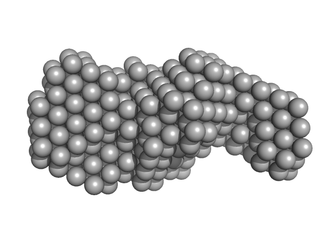
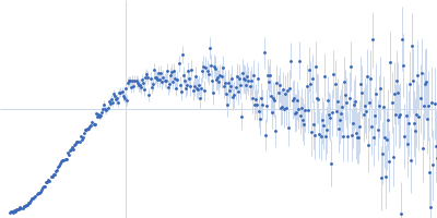
UniProt ID: P36056 (17-628) Probable S-adenosyl-L-methionine-dependent RNA methyltransferase RSM22, mitochondrial
|
|
|
|
| Sample: |
Probable S-adenosyl-L-methionine-dependent RNA methyltransferase RSM22, mitochondrial monomer, 70 kDa Saccharomyces cerevisiae protein
|
| Buffer: |
40 mM Tris pH 7.5, 500 mM NaCl, 5% glycerol, 2.5 mM DTT, pH: 7.5 |
| Experiment: |
SAXS
data collected at B21, Diamond Light Source on 2017 May 1
|
Probable S-adenosyl-L-methionine-dependent RNA methyltransferase RSM22
Jahangir Alam
|
| RgGuinier |
3.8 |
nm |
| Dmax |
13.9 |
nm |
| VolumePorod |
160 |
nm3 |
|
|
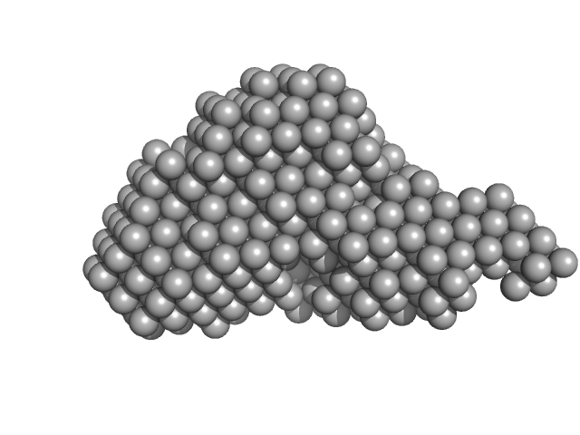
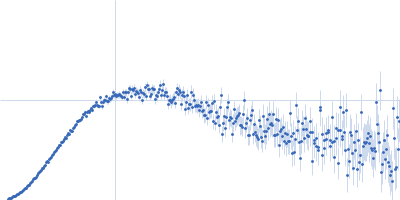
UniProt ID: P36056 (17-628) dimeric Probable S-adenosyl-L-methionine-dependent RNA methyltransferase RSM22, mitochondrial
|
|
|
|
| Sample: |
Dimeric Probable S-adenosyl-L-methionine-dependent RNA methyltransferase RSM22, mitochondrial dimer, 141 kDa Saccharomyces cerevisiae protein
|
| Buffer: |
40 mM Tris pH 7.5, 500 mM NaCl, 5% glycerol, 2.5 mM DTT, pH: 7.5 |
| Experiment: |
SAXS
data collected at B21, Diamond Light Source on 2017 May 1
|
Probable S-adenosyl-L-methionine-dependent RNA methyltransferase RSM22
Jahangir Alam
|
| RgGuinier |
5.0 |
nm |
| Dmax |
18.2 |
nm |
| VolumePorod |
516 |
nm3 |
|
|
UniProt ID: A4VAR6 (1-208) GATA-type iron responsive transcription factor Fep1 reconstituted
UniProt ID: None (None-None) 24-mer double strand DNA from the GATA promoter
|
|
|
|
| Sample: |
GATA-type iron responsive transcription factor Fep1 reconstituted monomer, 22 kDa Komagataella pastoris protein
24-mer double strand DNA from the GATA promoter dimer, 14 kDa synthetic oligonucleotide DNA
|
| Buffer: |
50 mM MOPS, 50 mM NaCl, pH: 7 |
| Experiment: |
SAXS
data collected at BM29, ESRF on 2018 Dec 5
|
Biophysical characterization of the complex between the iron-responsive transcription factor Fep1 and DNA.
Eur Biophys J (2021)
Miele AE, Cervoni L, Le Roy A, Cutone A, Musci G, Ebel C, Bonaccorsi di Patti MC
|
| RgGuinier |
3.5 |
nm |
| Dmax |
13.1 |
nm |
| VolumePorod |
73 |
nm3 |
|
|
UniProt ID: A4VAR6 (1-208) GATA-type iron responsive transcription factor Fep1
UniProt ID: None (None-None) 24-mer double strand DNA from the GATA promoter
|
|
|
|
| Sample: |
GATA-type iron responsive transcription factor Fep1 monomer, 22 kDa Komagataella pastoris protein
24-mer double strand DNA from the GATA promoter dimer, 14 kDa synthetic oligonucleotide DNA
|
| Buffer: |
50 mM MOPS, 50 mM NaCl, pH: 7 |
| Experiment: |
SAXS
data collected at BM29, ESRF on 2018 Dec 5
|
Biophysical characterization of the complex between the iron-responsive transcription factor Fep1 and DNA.
Eur Biophys J (2021)
Miele AE, Cervoni L, Le Roy A, Cutone A, Musci G, Ebel C, Bonaccorsi di Patti MC
|
| RgGuinier |
3.2 |
nm |
| Dmax |
12.0 |
nm |
| VolumePorod |
85 |
nm3 |
|
|
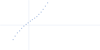
UniProt ID: A4VAR6 (1-208) GATA-type iron responsive transcription factor Fep1 reconstituted
|
|
|
|
| Sample: |
GATA-type iron responsive transcription factor Fep1 reconstituted monomer, 22 kDa Komagataella pastoris protein
|
| Buffer: |
50 mM MOPS, 50 mM NaCl, pH: 7 |
| Experiment: |
SAXS
data collected at BM29, ESRF on 2018 Dec 5
|
Biophysical characterization of the complex between the iron-responsive transcription factor Fep1 and DNA.
Eur Biophys J (2021)
Miele AE, Cervoni L, Le Roy A, Cutone A, Musci G, Ebel C, Bonaccorsi di Patti MC
|
| RgGuinier |
3.8 |
nm |
| Dmax |
16.5 |
nm |
| VolumePorod |
53 |
nm3 |
|
|
UniProt ID: A4VAR6 (1-208) GATA-type iron responsive transcription factor Fep1
|
|
|
|
| Sample: |
GATA-type iron responsive transcription factor Fep1 monomer, 22 kDa Komagataella pastoris protein
|
| Buffer: |
50 mM MOPS, 50 mM NaCl, pH: 7 |
| Experiment: |
SAXS
data collected at BM29, ESRF on 2018 Dec 5
|
Biophysical characterization of the complex between the iron-responsive transcription factor Fep1 and DNA.
Eur Biophys J (2021)
Miele AE, Cervoni L, Le Roy A, Cutone A, Musci G, Ebel C, Bonaccorsi di Patti MC
|
| RgGuinier |
3.5 |
nm |
| Dmax |
11.8 |
nm |
| VolumePorod |
41 |
nm3 |
|
|
UniProt ID: Q92133 (None-None) Histone H3
UniProt ID: P62799 (None-None) Histone H4
UniProt ID: P06897 (None-None) Histone H2A type 1
UniProt ID: Q92130 (None-None) Histone H2B
UniProt ID: None (None-None) Non-linker Ended Trinucleosome DNA
|
|
|
|
| Sample: |
Histone H3 monomer, 15 kDa Xenopus laevis protein
Histone H4 monomer, 11 kDa Xenopus laevis protein
Histone H2A type 1 monomer, 14 kDa Xenopus laevis protein
Histone H2B monomer, 14 kDa Xenopus laevis protein
Non-linker Ended Trinucleosome DNA monomer, 172 kDa DNA
|
| Buffer: |
20 mM Tris 150 mM NaCl 1 mM EDTA 1 mM DTT 50% w/v sucrose, pH: 7.5 |
| Experiment: |
SAXS
data collected at G1, Cornell High Energy Synchrotron Source (CHESS) on 2016 Mar 11
|
Solution structure(s) of trinucleosomes from contrast variation SAXS
Nucleic Acids Research (2021)
Mauney A, Muthurajan U, Luger K, Pollack L
|
| RgGuinier |
12.9 |
nm |
| Dmax |
41.8 |
nm |
|
|
 experimental SAS data")


![Fep1 wild type with [2Fe2S] cluster reconstituted Rg histogram](/media/fitting_files/extra/plots/SASDJ76_fit1_extra1_rghistogram_img.png "Fep1 wild type with [2Fe2S] cluster reconstituted Rg histogram")