|
|
|
|
|
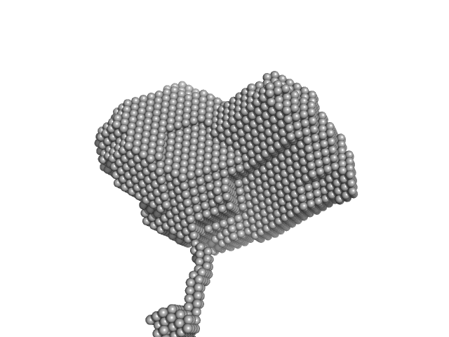
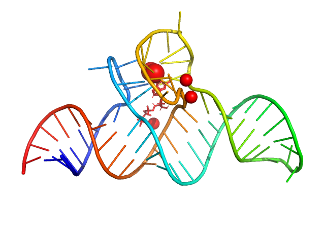
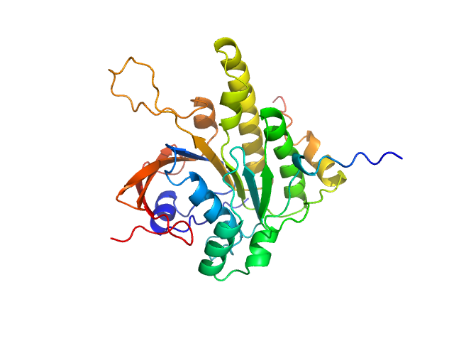
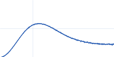
| Sample: |
Group IIC Intron Domain 1 monomer, 89 kDa Oceanobacillus iheyensis RNA
|
| Buffer: |
10 mM MgCl2, 5 mM Na-MES, pH: 6.5 |
| Experiment: |
SAXS
data collected at BM29, ESRF on 2021 Jul 7
|
Dynamic assembly of a large multidomain ribozyme visualized by cryo-electron microscopy.
Nat Commun 16(1):10195 (2025)
Jadhav S, Maiorca M, Manigrasso J, Saha S, Rakitch A, Muscat S, Mulvaney T, De Vivo M, Topf M, Marcia M
|
| RgGuinier |
3.8 |
nm |
| Dmax |
13.0 |
nm |
| VolumePorod |
188 |
nm3 |
|
|
|
|
|
|
|
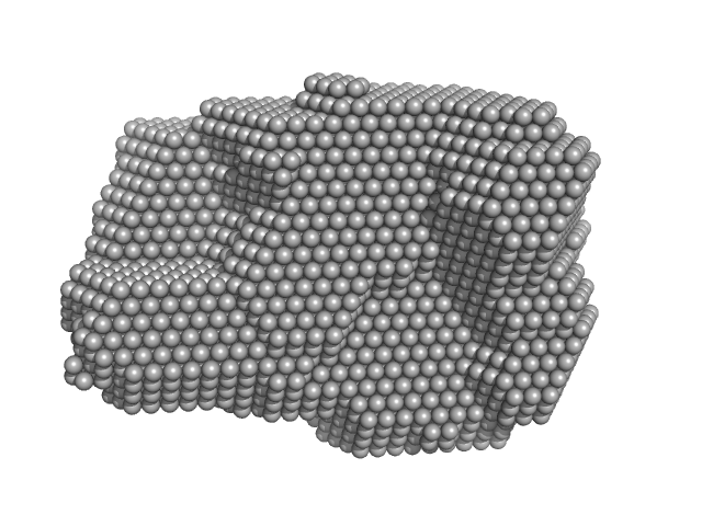
| Sample: |
Group IIC Intron Domain 1, 2 & 3 monomer, 103 kDa Oceanobacillus iheyensis RNA
|
| Buffer: |
10 mM MgCl2, 5 mM Na-MES, pH: 6.5 |
| Experiment: |
SAXS
data collected at BM29, ESRF on 2021 Sep 2
|
Dynamic assembly of a large multidomain ribozyme visualized by cryo-electron microscopy.
Nat Commun 16(1):10195 (2025)
Jadhav S, Maiorca M, Manigrasso J, Saha S, Rakitch A, Muscat S, Mulvaney T, De Vivo M, Topf M, Marcia M
|
| RgGuinier |
3.6 |
nm |
| Dmax |
12.0 |
nm |
| VolumePorod |
195 |
nm3 |
|
|
|
|
|
|
|
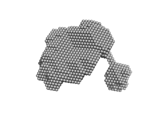
| Sample: |
Group IIC Intron Domain 1 & 2 monomer, 96 kDa Oceanobacillus iheyensis RNA
|
| Buffer: |
10 mM MgCl2, 5 mM Na-MES, pH: 6.5 |
| Experiment: |
SAXS
data collected at BM29, ESRF on 2021 Sep 2
|
Dynamic assembly of a large multidomain ribozyme visualized by cryo-electron microscopy.
Nat Commun 16(1):10195 (2025)
Jadhav S, Maiorca M, Manigrasso J, Saha S, Rakitch A, Muscat S, Mulvaney T, De Vivo M, Topf M, Marcia M
|
| RgGuinier |
3.6 |
nm |
| Dmax |
11.0 |
nm |
| VolumePorod |
179 |
nm3 |
|
|
|
|
|
|
|
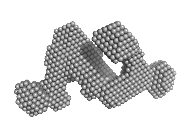
| Sample: |
Group IIC intron Domain 1, 2, 3 & 4 monomer, 125 kDa Oceanobacillus iheyensis RNA
|
| Buffer: |
10 mM MgCl2, 5 mM Na-MES, pH: 6.5 |
| Experiment: |
SAXS
data collected at BM29, ESRF on 2021 Sep 2
|
Dynamic assembly of a large multidomain ribozyme visualized by cryo-electron microscopy.
Nat Commun 16(1):10195 (2025)
Jadhav S, Maiorca M, Manigrasso J, Saha S, Rakitch A, Muscat S, Mulvaney T, De Vivo M, Topf M, Marcia M
|
| RgGuinier |
4.2 |
nm |
| Dmax |
12.6 |
nm |
| VolumePorod |
247 |
nm3 |
|
|
|
|
|
|
|
| Sample: |
Class V GTP aptamer apo, 22 kDa Escherichia coli RNA
|
| Buffer: |
50 mM Bis-Tris, 50 mM KCl, 2 mM MgCl2, pH: 6.5 |
| Experiment: |
SAXS
data collected at EMBL P12, PETRA III on 2024 Jul 19
|
Crystal structure of the class V GTP-binding RNA aptamer bound to its ligand: GTP recognition by a topologically complex intermolecular G-quadruplex.
Nucleic Acids Res 53(22) (2025)
Stafflinger H, Neißner K, Bartsch S, Pichler AK, Bartosik K, Dhamotharan K, Abele R, Duchardt-Ferner E, Micura R, Schindelin H, Wöhnert J
|
| RgGuinier |
3.3 |
nm |
| Dmax |
15.3 |
nm |
| VolumePorod |
99 |
nm3 |
|
|
|
|
|
|
|
| Sample: |
Class V GTP aptamer monomer, 22 kDa Escherichia coli RNA
Guanosine triphosphate monomer, 1 kDa
|
| Buffer: |
50 mM Bis-Tris, 50 mM KCl, 2 mM MgCl2, pH: 6.5 |
| Experiment: |
SAXS
data collected at EMBL P12, PETRA III on 2024 Jul 19
|
Crystal structure of the class V GTP-binding RNA aptamer bound to its ligand: GTP recognition by a topologically complex intermolecular G-quadruplex.
Nucleic Acids Res 53(22) (2025)
Stafflinger H, Neißner K, Bartsch S, Pichler AK, Bartosik K, Dhamotharan K, Abele R, Duchardt-Ferner E, Micura R, Schindelin H, Wöhnert J
|
| RgGuinier |
2.2 |
nm |
| Dmax |
7.6 |
nm |
| VolumePorod |
34 |
nm3 |
|
|
|
|
|
|
|
| Sample: |
Estrogen receptor alpha dimer, 55 kDa Homo sapiens protein
|
| Buffer: |
20 mM Tris-HCl pH 7.5, 150 mM NaCl, 5% glycerol, 2 mM TCEP, pH: 7.5 |
| Experiment: |
SAXS
data collected at BioSAXS, Australian Synchrotron on 2024 Nov 9
|
A ternary switch model governing ERα ligand binding domain conformation.
Nat Commun 16(1):10363 (2025)
McDougal DP, Pederick JL, Novick SJ, Jovcevski B, Warrender AK, Pascal BD, Griffin PR, Bruning JB
|
| RgGuinier |
2.4 |
nm |
| Dmax |
7.4 |
nm |
| VolumePorod |
74 |
nm3 |
|
|
|
|
|
|
|
| Sample: |
Estrogen receptor alpha dimer, 57 kDa Melanotaenia fluviatilis protein
|
| Buffer: |
20 mM Tris-HCl pH 7.5, 150 mM NaCl, 5% glycerol, 2 mM TCEP, pH: 7.5 |
| Experiment: |
SAXS
data collected at BioSAXS, Australian Synchrotron on 2024 Nov 9
|
A ternary switch model governing ERα ligand binding domain conformation.
Nat Commun 16(1):10363 (2025)
McDougal DP, Pederick JL, Novick SJ, Jovcevski B, Warrender AK, Pascal BD, Griffin PR, Bruning JB
|
| RgGuinier |
2.5 |
nm |
| Dmax |
9.2 |
nm |
| VolumePorod |
72 |
nm3 |
|
|
|
|
|
|
|
| Sample: |
EVH1 domain of Homer protein homolog 1 from mouse monomer, 14 kDa Mus musculus protein
|
| Buffer: |
20mM NaCl, 50 mM NaPi, 0.02% of sodium-azide, pH: 7.4 |
| Experiment: |
SAXS
data collected at Rigaku BioSAXS-2000, CEITEC on 2023 Nov 9
|
Structural Modeling and Dynamics of the Full-Length Homer1 Multimer.
Proteins (2025)
Kálmán ZE, Czajlik A, Maruzs B, Farkas F, Pap I, Homonnay C, Klumpler T, Batta G, Gáspári Z, Péterfia B
|
| RgGuinier |
1.6 |
nm |
| Dmax |
5.3 |
nm |
| VolumePorod |
23 |
nm3 |
|
|
|
|
|
|
|
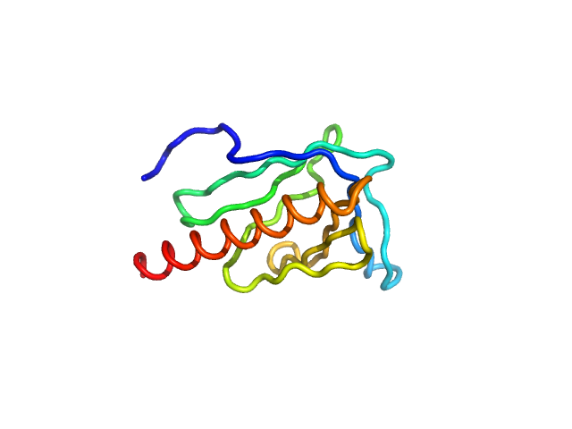
| Sample: |
Breast cancer type 2 susceptibility protein (BRC repeat 4) monomer, 4 kDa Homo sapiens protein
DNA repair protein RAD51 homolog 1 (Δ97) monomer, 26 kDa Homo sapiens protein
|
| Buffer: |
20 mM K₂HPO₄/KH₂PO₄, 100 mM NaCl, 200 mM Li₂SO₄, 1 mM DTT, 2% sucrose, pH: 8 |
| Experiment: |
SAXS
data collected at B21, Diamond Light Source on 2024 May 2
|
Breaking new ground into RAD51–BRC repeats interplay in Homologous Recombination
(2025)
Rinaldi F, Franco P, Veronesi M, Romeo E, Bresciani V, Varignani G, Catalano F, Bernetti M, Masetti M, Langer J, Girotto S, Cavalli A
|
| RgGuinier |
2.0 |
nm |
| Dmax |
6.6 |
nm |
| VolumePorod |
57 |
nm3 |
|
|


DNA repair protein RAD51 homolog 1 (Δ97) experimental SAS data")