|
|
|
|
|
| Sample: |
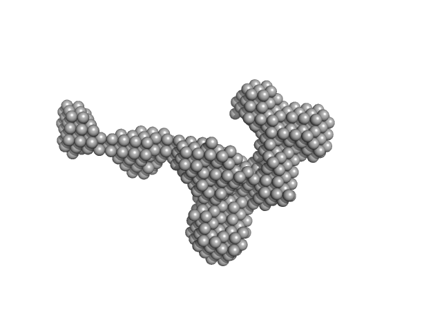
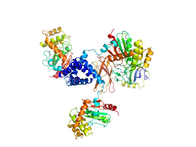
Stopper PN-Block (WA20) dimer, 25 kDa de novo protein protein
Extender PN-Block (FL4) monomer, 27 kDa de novo protein protein
|
| Buffer: |
20 mM HEPES, 100 mM NaCl, 200 mM ArgHCl, 10% glycerol, pH: 7.5 |
| Experiment: |
SAXS
data collected at BL-10C, Photon Factory (PF), High Energy Accelerator Research Organization (KEK) on 2014 Dec 19
|
Self-Assembling Supramolecular Nanostructures Constructed from de Novo Extender Protein Nanobuilding Blocks.
ACS Synth Biol 7(5):1381-1394 (2018)
Kobayashi N, Inano K, Sasahara K, Sato T, Miyazawa K, Fukuma T, Hecht MH, Song C, Murata K, Arai R
|
| RgGuinier |
3.3 |
nm |
| Dmax |
12.0 |
nm |
|
|
|
|
|
|
|
| Sample: |
Fluorescence recovery protein dimer, 23 kDa Synechocystis sp. PCC … protein
|
| Buffer: |
20 mM Tris-HCl, 150 mM NaCl, 0.1 mM EDTA, 2 mM dithiothreitol, 3 % v/v glycerol, pH: 7.6 |
| Experiment: |
SAXS
data collected at EMBL P12, PETRA III on 2017 Sep 1
|
Functional interaction of low-homology FRPs from different cyanobacteria with Synechocystis OCP.
Biochim Biophys Acta 1859(5):382-393 (2018)
Slonimskiy YB, Maksimov EG, Lukashev EP, Moldenhauer M, Jeffries CM, Svergun DI, Friedrich T, Sluchanko NN
|
| RgGuinier |
2.8 |
nm |
| Dmax |
10.5 |
nm |
| VolumePorod |
36 |
nm3 |
|
|
|
|
|
|
|
| Sample: |
Uncharacterized fluorescence recovery protein dimer, 24 kDa Arthrospira maxima CS-328 protein
|
| Buffer: |
20 mM Tris-HCl, 150 mM NaCl, 0.1 mM EDTA, 2 mM dithiothreitol, 3 % v/v glycerol, pH: 7.6 |
| Experiment: |
SAXS
data collected at EMBL P12, PETRA III on 2017 Sep 1
|
Functional interaction of low-homology FRPs from different cyanobacteria with Synechocystis OCP.
Biochim Biophys Acta 1859(5):382-393 (2018)
Slonimskiy YB, Maksimov EG, Lukashev EP, Moldenhauer M, Jeffries CM, Svergun DI, Friedrich T, Sluchanko NN
|
| RgGuinier |
2.7 |
nm |
| Dmax |
9.5 |
nm |
| VolumePorod |
35 |
nm3 |
|
|
|
|
|
|
|
| Sample: |
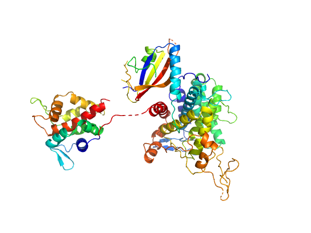
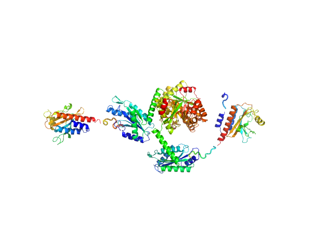
Ubiquitinating/deubiquitinating enzyme SdeA monomer, 72 kDa Legionella pneumophila subsp. … protein
|
| Buffer: |
10 mM HEPES 150 mM NaCl 1 mM TCEP, pH: 7.5 |
| Experiment: |
SAXS
data collected at EMBL P12, PETRA III on 2017 Nov 26
|
Insights into catalysis and function of phosphoribosyl-linked serine ubiquitination.
Nature 557(7707):734-738 (2018)
Kalayil S, Bhogaraju S, Bonn F, Shin D, Liu Y, Gan N, Basquin J, Grumati P, Luo ZQ, Dikic I
|
| RgGuinier |
3.5 |
nm |
| Dmax |
11.2 |
nm |
| VolumePorod |
128 |
nm3 |
|
|
|
|
|
|
|
| Sample: |
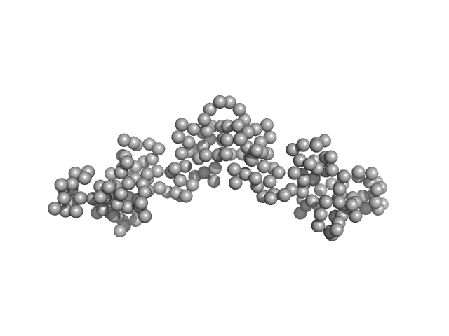
DsbA-like protein trimer, 74 kDa Proteus mirabilis protein
Putative metal resistance protein monomer, 30 kDa Proteus mirabilis protein
|
| Buffer: |
10mM HEPES, 150mM NaCl, pH: 7.5 |
| Experiment: |
SAXS
data collected at SAXS/WAXS, Australian Synchrotron on 2016 Nov 2
|
Disulfide isomerase activity of the dynamic, trimeric Proteus mirabilis ScsC protein is primed by the tandem immunoglobulin-fold domain of ScsB.
J Biol Chem 293(16):5793-5805 (2018)
Furlong EJ, Choudhury HG, Kurth F, Duff AP, Whitten AE, Martin JL
|
| RgGuinier |
3.9 |
nm |
| Dmax |
11.5 |
nm |
| VolumePorod |
145 |
nm3 |
|
|
|
|
|
|
|
| Sample: |
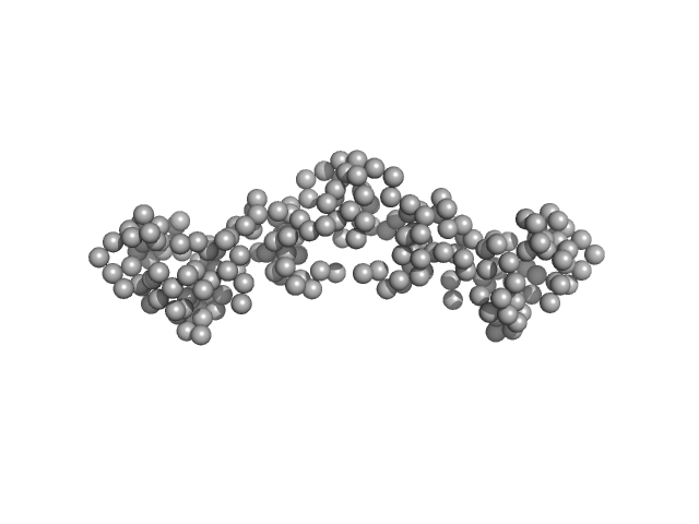
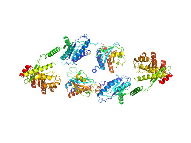
Sensory box/response regulator dimer, 136 kDa Mycobacterium smegmatis (strain … protein
|
| Buffer: |
20 mM HEPES, 100 mM NaCl, 5% glycerol, 2 mM MgCl2, pH: 7.5 |
| Experiment: |
SAXS
data collected at BL19U2, Shanghai Synchrotron Radiation Facility (SSRF) on 2016 Jun 26
|
The GDP-switched GAF domain of DcpA modulates the concerted synthesis/hydrolysis of c-di-GMP in Mycobacterium smegmatis.
Biochem J 475(7):1295-1308 (2018)
Chen HJ, Li N, Luo Y, Jiang YL, Zhou CZ, Chen Y, Li Q
|
| RgGuinier |
5.0 |
nm |
| Dmax |
20.0 |
nm |
| VolumePorod |
299 |
nm3 |
|
|
|
|
|
|
|
| Sample: |
Sensory box/response regulator dimer, 136 kDa Mycobacterium smegmatis (strain … protein
|
| Buffer: |
20 mM HEPES, 100 mM NaCl, 5% glycerol, 2 mM MgCl2, pH: 7.5 |
| Experiment: |
SAXS
data collected at BL19U2, Shanghai Synchrotron Radiation Facility (SSRF) on 2016 Jun 26
|
The GDP-switched GAF domain of DcpA modulates the concerted synthesis/hydrolysis of c-di-GMP in Mycobacterium smegmatis.
Biochem J 475(7):1295-1308 (2018)
Chen HJ, Li N, Luo Y, Jiang YL, Zhou CZ, Chen Y, Li Q
|
| RgGuinier |
4.8 |
nm |
| Dmax |
17.0 |
nm |
| VolumePorod |
271 |
nm3 |
|
|
|
|
|
|
|
| Sample: |
Calvin cycle protein CP12, chloroplastic monomer, 11 kDa Chlamydomonas reinhardtii protein
|
| Buffer: |
50 mM phosphate buffer, 50 mM NaCl, 20 mM oxidized DTT, pH: 6.5 |
| Experiment: |
SAXS
data collected at SWING, SOLEIL on 2015 Nov 3
|
Cryptic Disorder Out of Disorder: Encounter between Conditionally Disordered CP12 and Glyceraldehyde-3-Phosphate Dehydrogenase.
J Mol Biol 430(8):1218-1234 (2018)
Launay H, Barré P, Puppo C, Zhang Y, Maneville S, Gontero B, Receveur-Bréchot V
|
| RgGuinier |
2.3 |
nm |
| Dmax |
10.0 |
nm |
| VolumePorod |
22 |
nm3 |
|
|
|
|
|
|
|
| Sample: |
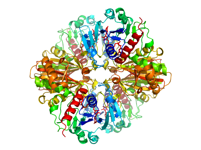
Glyceraldehyde-3-phosphate dehydrogenase tetramer, 148 kDa Chlamydomonas reinhardtii protein
|
| Buffer: |
30 mM Tris, 4 mM EDTA, 100 µM NAD, 5 mM free cysteine, pH: 7.9 |
| Experiment: |
SAXS
data collected at SWING, SOLEIL on 2011 Feb 7
|
Cryptic Disorder Out of Disorder: Encounter between Conditionally Disordered CP12 and Glyceraldehyde-3-Phosphate Dehydrogenase.
J Mol Biol 430(8):1218-1234 (2018)
Launay H, Barré P, Puppo C, Zhang Y, Maneville S, Gontero B, Receveur-Bréchot V
|
| RgGuinier |
3.2 |
nm |
| Dmax |
9.0 |
nm |
| VolumePorod |
206 |
nm3 |
|
|
|
|
|
|
|
| Sample: |
Calvin cycle protein CP12, chloroplastic monomer, 11 kDa Chlamydomonas reinhardtii protein
Glyceraldehyde-3-phosphate dehydrogenase tetramer, 148 kDa Chlamydomonas reinhardtii protein
|
| Buffer: |
30 mM Tris, 4 mM EDTA, 100 µM NAD, 5 mM free cysteine, pH: 7.9 |
| Experiment: |
SAXS
data collected at SWING, SOLEIL on 2010 Dec 8
|
Cryptic Disorder Out of Disorder: Encounter between Conditionally Disordered CP12 and Glyceraldehyde-3-Phosphate Dehydrogenase.
J Mol Biol 430(8):1218-1234 (2018)
Launay H, Barré P, Puppo C, Zhang Y, Maneville S, Gontero B, Receveur-Bréchot V
|
| RgGuinier |
3.8 |
nm |
| Dmax |
19.0 |
nm |
| VolumePorod |
251 |
nm3 |
|
|
extender PN-Block (FL4) experimental SAS data")


 Rg histogram")