Sign in
|
Register
Home
Browse
Submit data
About SASBDB
Help
Search
Advanced search
33
hits found for
Kursula
SASDB52
– Death associated protein kinase (wild-type)
Sample:
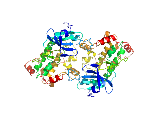
Death associated protein kinase wild-type , 37 kDa
Homo sapiens
protein
Buffer:
50 mM HEPES 250 mM NaCl 5mM CaCl2 0.25 mM TCEP 5% (v/v) glycerol, pH: 7.5
Experiment:
SAXS data collected at EMBL P12, PETRA III
on 2013 Dec 18
Death-Associated Protein Kinase Activity Is Regulated by Coupled Calcium/Calmodulin Binding to Two Distinct Sites.
Structure
24(6):851-61 (2016)
...
Kursula
P, Schultz C, McCarthy AA, Hart DJ, Wilmanns M
R
g
Guinier
2.7
nm
D
max
8.1
nm
Volume
Porod
101
nm
3
SASDB62
– Death associated protein kinase (D220K mutant)
Sample:
Death associated protein kinase (D220K mutant) , 37 kDa
Homo sapiens
protein
Buffer:
50 mM HEPES 250 mM NaCl 5mM CaCl2 0.25 mM TCEP 5% (v/v) glycerol, pH: 7.5
Experiment:
SAXS data collected at EMBL P12, PETRA III
on 2013 Dec 18
Death-Associated Protein Kinase Activity Is Regulated by Coupled Calcium/Calmodulin Binding to Two Distinct Sites.
Structure
24(6):851-61 (2016)
...
Kursula
P, Schultz C, McCarthy AA, Hart DJ, Wilmanns M
R
g
Guinier
2.5
nm
D
max
9.0
nm
Volume
Porod
89
nm
3
SASDB72
– Death associated protein kinase (Basic Loop mutant)
Sample:
Death associated protein kinase (Basic Loop mutant) , 37 kDa
Homo sapiens
protein
Buffer:
50 mM HEPES 250 mM NaCl 5mM CaCl2 0.25 mM TCEP 5% (v/v) glycerol, pH: 7.5
Experiment:
SAXS data collected at EMBL P12, PETRA III
on 2013 Dec 18
Death-Associated Protein Kinase Activity Is Regulated by Coupled Calcium/Calmodulin Binding to Two Distinct Sites.
Structure
24(6):851-61 (2016)
...
Kursula
P, Schultz C, McCarthy AA, Hart DJ, Wilmanns M
R
g
Guinier
2.4
nm
D
max
7.5
nm
Volume
Porod
78
nm
3
SASDAN4
– Calmodulin:peptide complex
Sample:
Calmodulin monomer, 17 kDa
Homo sapiens
protein
C-terminal region of human myelin basic protein monomer, 2 kDa
Homo sapiens
protein
Buffer:
25 mM Tris75 200 mM NaCl, pH: 7.5
Experiment:
SAXS data collected at EMBL X33, DORIS III, DESY
on 2006 Nov 28
Interaction between the C-terminal region of human myelin basic protein and calmodulin: analysis of complex formation and solution structure.
BMC Struct Biol
8:10 (2008)
...
Kursula
P
R
g
Guinier
2.1
nm
D
max
7.0
nm
Volume
Porod
36
nm
3
SASDBZ4
– Tyrosine hydroxylase (Isoform 1, Homo sapiens)
Sample:
Tyrosine hydroxylase, isoform 1 tetramer, 222 kDa
Homo sapiens
protein
Buffer:
20 mM Na-HEPES 200 mM NaCl, pH: 7
Experiment:
SAXS data collected at EMBL P12, PETRA III
on 2015 Jun 14
Stable preparations of tyrosine hydroxylase provide the solution structure of the full-length enzyme.
Sci Rep
6:30390 (2016)
...
Kursula
P, Martinez A, Flydal MI
R
g
Guinier
4.7
nm
D
max
20.0
nm
Volume
Porod
520
nm
3
SASDLA5
– Wild-type human myelin protein P2
Sample:
Myelin P2 protein monomer, 15 kDa
Homo sapiens
protein
Buffer:
20 mM HEPES, 300 mM NaCl, 1 mM DTT, pH: 7.5
Experiment:
SAXS data collected at B21, Diamond Light Source
on 2020 Jul 22
Human myelin protein P2: from crystallography to time-lapse membrane imaging and neuropathy-associated variants.
FEBS J
(2021)
...
Kursula
P
R
g
Guinier
1.5
nm
D
max
4.2
nm
Volume
Porod
17
nm
3
SASDLB5
– CMT mutant i50del of human myelin protein P2
Sample:
Myelin P2 disease mutant I50del monomer, 15 kDa protein
Buffer:
20 mM HEPES, 300 mM NaCl, 1 mM DTT, pH: 7.5
Experiment:
SAXS data collected at B21, Diamond Light Source
on 2020 Jul 22
Human myelin protein P2: from crystallography to time-lapse membrane imaging and neuropathy-associated variants.
FEBS J
(2021)
...
Kursula
P
R
g
Guinier
1.5
nm
D
max
4.0
nm
Volume
Porod
17
nm
3
SASDLC5
– CMT mutant M114T of human myelin protein P2
Sample:
Human P2 M114T mutant monomer, 15 kDa protein
Buffer:
20 mM HEPES, 300 mM NaCl, 1 mM DTT, pH: 7.5
Experiment:
SAXS data collected at B21, Diamond Light Source
on 2020 Jul 22
Human myelin protein P2: from crystallography to time-lapse membrane imaging and neuropathy-associated variants.
FEBS J
(2021)
...
Kursula
P
R
g
Guinier
1.5
nm
D
max
4.0
nm
Volume
Porod
16
nm
3
SASDLD5
– CMT mutant V115A of human myelin protein P2
Sample:
Human P2 V115A mutant monomer, 15 kDa protein
Buffer:
20 mM HEPES, 300 mM NaCl, 1 mM DTT, pH: 7.5
Experiment:
SAXS data collected at B21, Diamond Light Source
on 2020 Jul 22
Human myelin protein P2: from crystallography to time-lapse membrane imaging and neuropathy-associated variants.
FEBS J
(2021)
...
Kursula
P
R
g
Guinier
1.5
nm
D
max
4.0
nm
Volume
Porod
17
nm
3
SASDND6
– Human ganglioside-induced differentiation-associated protein 1 (GDAP1) missense mutant H123R, construct GDAP1∆302-358
Sample:
Ganglioside-induced differentiation-associated protein 1 (H123R) dimer, 70 kDa
Homo sapiens
protein
Buffer:
25 mM HEPES, 300 mM NaCl, pH: 7.5
Experiment:
SAXS data collected at SWING, SOLEIL
on 2020 Oct 8
Structural insights into Charcot-Marie-Tooth disease-linked mutations in human GDAP1.
FEBS Open Bio
(2022)
...
Kursula
P
R
g
Guinier
3.1
nm
D
max
9.9
nm
Volume
Porod
107
nm
3
SASDNE6
– Human ganglioside-induced differentiation-associated protein 1 (GDAP1) missense mutant R120W, construct GDAP1∆302-358
Sample:
Ganglioside-induced differentiation-associated protein 1 (R120W) dimer, 70 kDa
Homo sapiens
protein
Buffer:
25 mM HEPES, 300 mM NaCl, pH: 7.5
Experiment:
SAXS data collected at SWING, SOLEIL
on 2020 Jul 1
Structural insights into Charcot-Marie-Tooth disease-linked mutations in human GDAP1.
FEBS Open Bio
(2022)
...
Kursula
P
R
g
Guinier
3.1
nm
D
max
10.0
nm
Volume
Porod
105
nm
3
SASDJR7
– Mouse cysteine sulphinic acid decarboxylase (CSAD)
Sample:
Cysteine sulfinic acid decarboxylase dimer, 117 kDa
Mus musculus
protein
Buffer:
20 mM HEPES, 200 mM NaCl, pH: 7.5
Experiment:
SAXS data collected at SWING, SOLEIL
on 2018 Feb 4
Structure and substrate specificity determinants of the taurine biosynthetic enzyme cysteine sulphinic acid decarboxylase.
J Struct Biol
:107674 (2020)
...
Kursula
P
R
g
Guinier
3.4
nm
D
max
15.0
nm
Volume
Porod
198
nm
3
SASDRZ7
– Human wild-type nicotinamide phosphoribosyltransferase (NAMPT)
Sample:
Nicotinamide phosphoribosyltransferase dimer, 114 kDa
Homo sapiens
protein
Buffer:
20 mM Tris-HCl, 500 mM NaCl, 6 mM MgCl2, 5% (v/v) glycerol, pH: 8
Experiment:
SAXS data collected at EMBL P12, PETRA III
on 2019 May 20
Identification of structural determinants of nicotinamide phosphoribosyl transferase (NAMPT) activity and substrate selectivity.
J Struct Biol
:108004 (2023)
...
Kursula
P, Ziegler M
R
g
Guinier
3.3
nm
D
max
12.9
nm
Volume
Porod
181
nm
3
SASDR28
– Human wild-type nicotinamide phosphoribosyltransferase (NAMPT) in the presence of nicotinamide
Sample:
Nicotinamide phosphoribosyltransferase dimer, 114 kDa
Homo sapiens
protein
Buffer:
20 mM Tris-HCl, 500 mM NaCl, 6 mM MgCl2, 5% (v/v) glycerol, 1 mM nicotinamide, pH: 8
Experiment:
SAXS data collected at EMBL P12, PETRA III
on 2019 May 20
Identification of structural determinants of nicotinamide phosphoribosyl transferase (NAMPT) activity and substrate selectivity.
J Struct Biol
:108004 (2023)
...
Kursula
P, Ziegler M
R
g
Guinier
3.2
nm
D
max
12.8
nm
Volume
Porod
166
nm
3
SASDR38
– Human wild-type nicotinamide phosphoribosyltransferase (NAMPT) in the presence of nicotinic acid
Sample:
Nicotinamide phosphoribosyltransferase dimer, 114 kDa
Homo sapiens
protein
Buffer:
20 mM Tris-HCl, 500 mM NaCl, 6 mM MgCl2, 5% (v/v) glycerol, 1 mM nicotinic acid, pH: 8
Experiment:
SAXS data collected at EMBL P12, PETRA III
on 2019 May 20
Identification of structural determinants of nicotinamide phosphoribosyl transferase (NAMPT) activity and substrate selectivity.
J Struct Biol
:108004 (2023)
...
Kursula
P, Ziegler M
R
g
Guinier
3.3
nm
D
max
12.9
nm
Volume
Porod
180
nm
3
SASDR48
– Human wild-type nicotinamide phosphoribosyltransferase (NAMPT) in the presence of phosphoribosyl pyrophosphate
Sample:
Nicotinamide phosphoribosyltransferase dimer, 114 kDa
Homo sapiens
protein
Buffer:
20 mM Tris-HCl, 500 mM NaCl, 6 mM MgCl2, 5% (v/v) glycerol, 1 mM phosphoribosyl pyrophosphate, pH: 8
Experiment:
SAXS data collected at EMBL P12, PETRA III
on 2019 May 20
Identification of structural determinants of nicotinamide phosphoribosyl transferase (NAMPT) activity and substrate selectivity.
J Struct Biol
:108004 (2023)
...
Kursula
P, Ziegler M
R
g
Guinier
3.3
nm
D
max
12.9
nm
Volume
Porod
173
nm
3
SASDR58
– Human nicotinamide phosphoribosyltransferase Δ42-51 loop mutant (NAMPT Δ42-51)
Sample:
Nicotinamide phosphoribosyltransferase Δ42-51 dimer, 111 kDa
Homo sapiens
protein
Buffer:
20 mM Tris-HCl, 500 mM NaCl, 6 mM MgCl2, 5% (v/v) glycerol, pH: 8
Experiment:
SAXS data collected at EMBL P12, PETRA III
on 2019 May 20
Identification of structural determinants of nicotinamide phosphoribosyl transferase (NAMPT) activity and substrate selectivity.
J Struct Biol
:108004 (2023)
...
Kursula
P, Ziegler M
R
g
Guinier
3.2
nm
D
max
11.0
nm
Volume
Porod
159
nm
3
SASDR68
– Human nicotinamide phosphoribosyltransferase Δ42-51 loop mutant (NAMPT Δ42-51) in the presence of nicotinamide
Sample:
Nicotinamide phosphoribosyltransferase Δ42-51 dimer, 111 kDa
Homo sapiens
protein
Buffer:
20 mM Tris-HCl, 500 mM NaCl, 6 mM MgCl2, 5% (v/v) glycerol, 1 mM nicotinamide, pH: 8
Experiment:
SAXS data collected at EMBL P12, PETRA III
on 2019 May 20
Identification of structural determinants of nicotinamide phosphoribosyl transferase (NAMPT) activity and substrate selectivity.
J Struct Biol
:108004 (2023)
...
Kursula
P, Ziegler M
R
g
Guinier
3.2
nm
D
max
10.9
nm
Volume
Porod
151
nm
3
SASDR78
– Human nicotinamide phosphoribosyltransferase Δ42-51 loop mutant (NAMPT Δ42-51) in the presence of nicotinic acid
Sample:
Nicotinamide phosphoribosyltransferase Δ42-51 dimer, 111 kDa
Homo sapiens
protein
Buffer:
20 mM Tris-HCl, 500 mM NaCl, 6 mM MgCl2, 5% (v/v) glycerol, 1 mM nicotinic acid, pH: 8
Experiment:
SAXS data collected at EMBL P12, PETRA III
on 2019 May 20
Identification of structural determinants of nicotinamide phosphoribosyl transferase (NAMPT) activity and substrate selectivity.
J Struct Biol
:108004 (2023)
...
Kursula
P, Ziegler M
R
g
Guinier
3.3
nm
D
max
11.2
nm
Volume
Porod
152
nm
3
SASDR88
– Human nicotinamide phosphoribosyltransferase Δ42-51 loop mutant (NAMPT Δ42-51) in the presence of phosphoribosyl pyrophosphate
Sample:
Nicotinamide phosphoribosyltransferase Δ42-51 dimer, 111 kDa
Homo sapiens
protein
Buffer:
20 mM Tris-HCl, 500 mM NaCl, 6 mM MgCl2, 5% (v/v) glycerol, 1 mM phosphoribosyl pyrophosphate, pH: 8
Experiment:
SAXS data collected at EMBL P12, PETRA III
on 2019 May 20
Identification of structural determinants of nicotinamide phosphoribosyl transferase (NAMPT) activity and substrate selectivity.
J Struct Biol
:108004 (2023)
...
Kursula
P, Ziegler M
R
g
Guinier
3.2
nm
D
max
11.4
nm
Volume
Porod
154
nm
3
SASDJR8
– Dimeric human ganglioside-induced differentiation-associated protein 1, construct GDAP1∆295-358
Sample:
Ganglioside-induced differentiation-associated protein 1, construct GDAP1∆295-358 dimer, 68 kDa
Homo sapiens
protein
Buffer:
25 mM HEPES, 300 mM NaCl, pH: 7.5
Experiment:
SAXS data collected at EMBL P12, PETRA III
on 2019 May 21
Structure of the Complete Dimeric Human GDAP1 Core Domain Provides Insights into Ligand Binding and Clustering of Disease Mutations
Frontiers in Molecular Biosciences
7 (2021)
...
Kursula
P
R
g
Guinier
3.1
nm
D
max
101.6
nm
Volume
Porod
111
nm
3
SASDJS8
– Dimeric human ganglioside-induced differentiation-associated protein 1, construct GDAP1∆295-358 with hexadecanedioic acid
Sample:
Ganglioside-induced differentiation-associated protein 1, construct GDAP1∆295-358 dimer, 68 kDa
Homo sapiens
protein
Buffer:
25 mM HEPES, 300 mM NaCl, pH: 7.5
Experiment:
SAXS data collected at SWING, SOLEIL
on 2019 Jul 11
Structure of the Complete Dimeric Human GDAP1 Core Domain Provides Insights into Ligand Binding and Clustering of Disease Mutations
Frontiers in Molecular Biosciences
7 (2021)
...
Kursula
P
R
g
Guinier
3.1
nm
D
max
9.2
nm
Volume
Porod
112
nm
3
SASDJT8
– Monomeric human ganglioside-induced differentiation-associated protein 1, construct GDAP1∆295-358
Sample:
Ganglioside-induced differentiation-associated protein 1, construct GDAP1∆295-358 monomer, 34 kDa
Homo sapiens
protein
Buffer:
25 mM HEPES, 300 mM NaCl, pH: 7.5
Experiment:
SAXS data collected at EMBL P12, PETRA III
on 2019 May 21
Structure of the Complete Dimeric Human GDAP1 Core Domain Provides Insights into Ligand Binding and Clustering of Disease Mutations
Frontiers in Molecular Biosciences
7 (2021)
...
Kursula
P
R
g
Guinier
2.7
nm
D
max
92.3
nm
Volume
Porod
71
nm
3
SASDJU8
– Monomeric human ganglioside-induced differentiation-associated protein 1, construct GDAP1∆295-358 with hexadecanedioic acid
Sample:
Ganglioside-induced differentiation-associated protein 1, construct GDAP1∆295-358 monomer, 34 kDa
Homo sapiens
protein
Buffer:
25 mM HEPES, 300 mM NaCl, pH: 7.5
Experiment:
SAXS data collected at SWING, SOLEIL
on 2019 Jul 11
Structure of the Complete Dimeric Human GDAP1 Core Domain Provides Insights into Ligand Binding and Clustering of Disease Mutations
Frontiers in Molecular Biosciences
7 (2021)
...
Kursula
P
R
g
Guinier
2.5
nm
D
max
8.9
nm
Volume
Porod
63
nm
3
SASDJV8
– Dimeric human ganglioside-induced differentiation-associated protein 1, construct GDAP1∆303-358
Sample:
Ganglioside-induced differentiation-associated protein 1, construct GDAP1∆303-358 dimer, 70 kDa
Homo sapiens
protein
Buffer:
25 mM HEPES, 300 mM NaCl, pH: 7.5
Experiment:
SAXS data collected at SWING, SOLEIL
on 2020 Jun 28
Structure of the Complete Dimeric Human GDAP1 Core Domain Provides Insights into Ligand Binding and Clustering of Disease Mutations
Frontiers in Molecular Biosciences
7 (2021)
...
Kursula
P
R
g
Guinier
3.1
nm
D
max
9.9
nm
Volume
Porod
106
nm
3
SASDJW8
– Monomeric human ganglioside-induced differentiation-associated protein 1, construct GDAP1∆303-358, mutant Y29E/C88A
Sample:
Ganglioside-induced differentiation-associated protein 1, construct GDAP1∆303-358, mutant Y29E/C88A monomer, 35 kDa
Homo sapiens
protein
Buffer:
25 mM HEPES, 300 mM NaCl, pH: 7.5
Experiment:
SAXS data collected at SWING, SOLEIL
on 2020 Jun 28
Structure of the Complete Dimeric Human GDAP1 Core Domain Provides Insights into Ligand Binding and Clustering of Disease Mutations
Frontiers in Molecular Biosciences
7 (2021)
...
Kursula
P
R
g
Guinier
2.5
nm
D
max
8.7
nm
Volume
Porod
59
nm
3
SASDJX8
– Dimeric human ganglioside-induced differentiation-associated protein 1, construct GDAP1∆319-358
Sample:
Ganglioside-induced differentiation-associated protein 1, construct GDAP1∆319-358 dimer, 74 kDa
Homo sapiens
protein
Buffer:
25 mM HEPES, 300 mM NaCl, pH: 7.5
Experiment:
SAXS data collected at SWING, SOLEIL
on 2019 Jul 11
Structure of the Complete Dimeric Human GDAP1 Core Domain Provides Insights into Ligand Binding and Clustering of Disease Mutations
Frontiers in Molecular Biosciences
7 (2021)
...
Kursula
P
R
g
Guinier
3.5
nm
D
max
10.8
nm
Volume
Porod
130
nm
3
SASDJY8
– Dimeric human ganglioside-induced differentiation-associated protein 1, construct GDAP1∆319-358 with hexadecanedioic acid
Sample:
Ganglioside-induced differentiation-associated protein 1, construct GDAP1∆319-358 dimer, 74 kDa
Homo sapiens
protein
Buffer:
25 mM HEPES, 300 mM NaCl, pH: 7.5
Experiment:
SAXS data collected at SWING, SOLEIL
on 2019 Jul 11
Structure of the Complete Dimeric Human GDAP1 Core Domain Provides Insights into Ligand Binding and Clustering of Disease Mutations
Frontiers in Molecular Biosciences
7 (2021)
...
Kursula
P
R
g
Guinier
3.1
nm
D
max
9.6
nm
Volume
Porod
121
nm
3
SASDJZ8
– Monomeric human ganglioside-induced differentiation-associated protein 1, construct GDAP1∆319-358
Sample:
Ganglioside-induced differentiation-associated protein 1, construct GDAP1∆319-358 monomer, 37 kDa
Homo sapiens
protein
Buffer:
25 mM HEPES, 300 mM NaCl, pH: 7.5
Experiment:
SAXS data collected at SWING, SOLEIL
on 2019 Jul 11
Structure of the Complete Dimeric Human GDAP1 Core Domain Provides Insights into Ligand Binding and Clustering of Disease Mutations
Frontiers in Molecular Biosciences
7 (2021)
...
Kursula
P
R
g
Guinier
2.7
nm
D
max
9.0
nm
Volume
Porod
69
nm
3
SASDSZ8
– The DNA binding domain (DBD) of Hepatocyte nuclear factor 1-alpha (HNF-1A)
Sample:
Hepatocyte nuclear factor 1-alpha monomer, 23 kDa
Homo sapiens
protein
Buffer:
20 mM HEPES, 500 mM NaCl, 1 mM TCEP,, pH: 8
Experiment:
SAXS data collected at EMBL P12, PETRA III
on 2020 Jun 25
Structural properties of the HNF-1A transactivation domain
Frontiers in Molecular Biosciences
10 (2023)
...
Kursula
P
R
g
Guinier
2.7
nm
D
max
10.2
nm
Volume
Porod
22
nm
3
SASDJ29
– Monomeric human ganglioside-induced differentiation-associated protein 1, construct GDAP1∆319-358 with hexadecanedioic acid
Sample:
Ganglioside-induced differentiation-associated protein 1, construct GDAP1∆319-358 monomer, 37 kDa
Homo sapiens
protein
Buffer:
25 mM HEPES, 300 mM NaCl, pH: 7.5
Experiment:
SAXS data collected at SWING, SOLEIL
on 2019 Jul 11
Structure of the Complete Dimeric Human GDAP1 Core Domain Provides Insights into Ligand Binding and Clustering of Disease Mutations
Frontiers in Molecular Biosciences
7 (2021)
...
Kursula
P
R
g
Guinier
2.5
nm
D
max
8.6
nm
Volume
Porod
65
nm
3
SASDS29
– The DNA binding and transactivation domains (DBD-TAD) of Hepatocyte nuclear factor 1-alpha (HNF-1A)
Sample:
Hepatocyte nuclear factor 1-alpha monomer, 62 kDa
Homo sapiens
protein
Buffer:
4 mM Tris, 100 mM NaCl, 1 mM TCEP,, pH: 8.5
Experiment:
SAXS data collected at CoSAXS, MAX IV
on 2022 Apr 2
Structural properties of the HNF-1A transactivation domain
Frontiers in Molecular Biosciences
10 (2023)
...
Kursula
P
R
g
Guinier
4.9
nm
D
max
17.0
nm
Volume
Porod
109
nm
3
SASDJ39
– Monomeric human ganglioside-induced differentiation-associated protein 1-like 1, GDAP1L1
Sample:
Ganglioside-induced differentiation-associated protein 1-like 1 (∆125-143 isoform) monomer, 44 kDa
Homo sapiens
protein
Buffer:
20 mM TRIS pH 7.5, 150 mM NaCl, 1 mM TCEP, pH: 7.5
Experiment:
SAXS data collected at B21, Diamond Light Source
on 2018 Dec 10
Structure of the Complete Dimeric Human GDAP1 Core Domain Provides Insights into Ligand Binding and Clustering of Disease Mutations
Frontiers in Molecular Biosciences
7 (2021)
...
Kursula
P
R
g
Guinier
2.7
nm
D
max
10.0
nm
Volume
Porod
73
nm
3