|
|
|
|
|
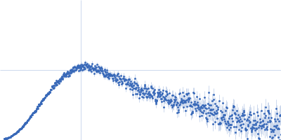
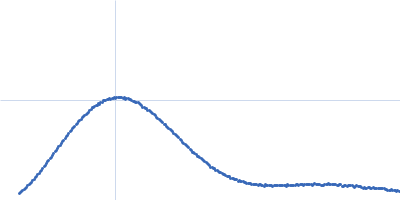
| Sample: |
Pro-matrix metalloproteinase-1 (Interstitial collagenase) monomer, 52 kDa Homo sapiens protein
|
| Buffer: |
50 mM Tris-HCl, 150 mM Sodium chloride, 10 mM Calcium chloride, pH: 7.4 |
| Experiment: |
SAXS
data collected at B21, Diamond Light Source on 2015 May 21
|
Conformationally constrained mutant of human matrix metalloproteinase-1
Rob Holland
|
| RgGuinier |
2.7 |
nm |
| Dmax |
9.0 |
nm |
| VolumePorod |
73 |
nm3 |
|
|
|
|
|
|
|
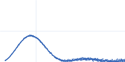
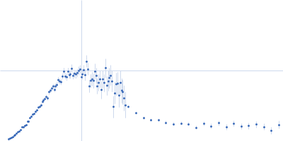
| Sample: |
Pro-matrix metalloproteinase-1 (Interstitial collagenase) (proMMP-1 S243C, S318C) monomer, 52 kDa Homo sapiens protein
|
| Buffer: |
50 mM Tris-HCl, 150 mM Sodium chloride, 10 mM Calcium chloride, pH: 7.4 |
| Experiment: |
SAXS
data collected at B21, Diamond Light Source on 2015 May 21
|
Conformationally constrained mutant of human matrix metalloproteinase-1
Rob Holland
|
| RgGuinier |
2.7 |
nm |
| Dmax |
8.9 |
nm |
| VolumePorod |
61 |
nm3 |
|
|
|
|
|
|
|
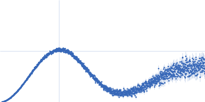
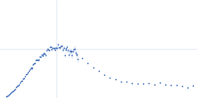
| Sample: |
Xylose isomerase tetramer, 173 kDa Streptomyces rubiginosus protein
|
| Buffer: |
100 mM HEPES, 1 mM MgCl2, pH: 7 |
| Experiment: |
SAXS
data collected at EMBL P12, PETRA III on 2016 Dec 21
|
A high flux setup for millisecond-scale small-angle X-ray scattering studies on macromolecular solutions
Clement Blanchet
|
|
|
|
|
|
|
|
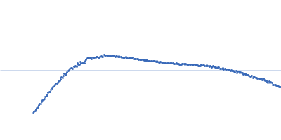
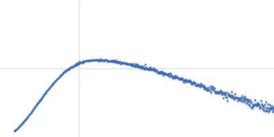
| Sample: |
Cytochrome C monomer, 12 kDa Bos taurus protein
|
| Buffer: |
TRIS 50mM, pH: 7.4 |
| Experiment: |
SAXS
data collected at EMBL P12, PETRA III on 2016 Dec 21
|
A high flux setup for millisecond-scale small-angle X-ray scattering studies on macromolecular solutions
Clement Blanchet
|
|
|
|
|
|
|
|
| Sample: |
PAS fold family dimer, 78 kDa Coleofasciculus chthonoplastes PCC … protein
|
| Buffer: |
20 mM HEPES, 150 mM NaCl, 5 mM MgCl2, 5 % w/v Glycerol, pH: 7.5 |
| Experiment: |
SAXS
data collected at cSAXS, Swiss Light Source on 2015 Mar 11
|
MPAC Delta132
Robert Lindner
|
| RgGuinier |
4.4 |
nm |
| Dmax |
15.9 |
nm |
| VolumePorod |
132 |
nm3 |
|
|
|
|
|
|
|
| Sample: |
Glycine decarboxylase dimer, 219 kDa Homo sapiens protein
|
| Buffer: |
10 mM TRIS pH 7.5, 100 mM NaCl, 1 mM DTT , 100 mM glycine, pH: 7.5 |
| Experiment: |
SAXS
data collected at BM29, ESRF on 2017 Nov 12
|
Glycine decarboxylase (P-protein of glycine cleavage system)
Bart Van Laer
|
| RgGuinier |
4.0 |
nm |
| VolumePorod |
315 |
nm3 |
|
|
|
|
|
|
|
| Sample: |
Glycine decarboxylase dimer, 219 kDa Homo sapiens neanderthalensis protein
|
| Buffer: |
10 mM TRIS pH 7.5, 100 mM NaCl, 1 mM DTT, pH: 7.5 |
| Experiment: |
SAXS
data collected at BM29, ESRF on 2017 Nov 12
|
Glycine decarboxylase (P-protein of glycine cleavage system)
Bart Van Laer
|
| RgGuinier |
4.0 |
nm |
| VolumePorod |
309 |
nm3 |
|
|
|
|
|
|
|
| Sample: |
Proton-gated ion channel pentamer, 183 kDa Gloeobacter violaceus (strain … protein
|
| Buffer: |
D2O, 20 mM Tris, 150 mM NaCl, 0.5 mM matched-out deuterated DDM,, pH: 7.5 |
| Experiment: |
SANS
data collected at D22, Institut Laue-Langevin (ILL) on 2019 Jun 20
|
Probing solution structure of the pentameric ligand-gated ion channel GLIC by small-angle neutron scattering
Proceedings of the National Academy of Sciences 118(37):e2108006118 (2021)
Lycksell M, Rovšnik U, Bergh C, Johansen N, Martel A, Porcar L, Arleth L, Howard R, Lindahl E
|
| RgGuinier |
3.8 |
nm |
| Dmax |
12.0 |
nm |
| VolumePorod |
235 |
nm3 |
|
|
|
|
|
|
|
| Sample: |
Proton-gated ion channel pentamer, 183 kDa Gloeobacter violaceus (strain … protein
|
| Buffer: |
D2O, 20 mM Tris, 150 mM NaCl, 0.5 mM matched-out deuterated DDM,, pH: 7.5 |
| Experiment: |
SANS
data collected at D22, Institut Laue-Langevin (ILL) on 2019 Jun 21
|
Probing solution structure of the pentameric ligand-gated ion channel GLIC by small-angle neutron scattering
Proceedings of the National Academy of Sciences 118(37):e2108006118 (2021)
Lycksell M, Rovšnik U, Bergh C, Johansen N, Martel A, Porcar L, Arleth L, Howard R, Lindahl E
|
| RgGuinier |
4.0 |
nm |
| Dmax |
17.7 |
nm |
| VolumePorod |
225 |
nm3 |
|
|
|
|
|
|
|
| Sample: |
Brain tumor protein monomer, 32 kDa Drosophila melanogaster protein
Maternal protein pumilio monomer, 38 kDa Drosophila melanogaster protein
Protein nanos monomer, 11 kDa Drosophila melanogaster protein
Hunchback mRNA Nanos Response Element 2 monomer, 7 kDa Drosophila melanogaster RNA
|
| Buffer: |
50 mM Tris, 150 mM NaCl, 1 mM DTT, 3% glycerol, pH: 7.4 |
| Experiment: |
SAXS
data collected at EMBL P12, PETRA III on 2019 Nov 12
|
Structure and dynamics of the quaternary hunchback mRNA translation repression complex.
Nucleic Acids Res 49(15):8866-8885 (2021)
Macošek J, Simon B, Linse JB, Jagtap PKA, Winter SL, Foot J, Lapouge K, Perez K, Rettel M, Ivanović MT, Masiewicz P, Murciano B, Savitski MM, Loedige I, Hub JS, Gabel F, Hennig J
|
| RgGuinier |
3.7 |
nm |
| Dmax |
12.7 |
nm |
| VolumePorod |
114 |
nm3 |
|
|
 experimental SAS data")
 (proMMP-1 S243C, S318C) experimental SAS data")