Search

 
Advanced search  

61 hits found for Santos

SASDFZ9 – RuvB-like 1 and 2, sperm-associated antigen 1 complex (His-RuvBL1/RuvBL2/SPAG1)

RuvB-like 1RuvB-like 2Sperm-associated antigen 1 experimental SAS data
DAMMIF model
Sample: RuvB-like 1 hexamer, 311 kDa Homo sapiens protein
RuvB-like 2 hexamer, 311 kDa Homo sapiens protein
Sperm-associated antigen 1 monomer, 104 kDa Homo sapiens protein
Buffer: 20 mM HEPES, 150 mM NaCl, 1% glycerol, 5 mM TCEP, pH: 7.5
Experiment: SAXS data collected at SWING, SOLEIL on 2019 Mar 21
R2SP
Raphael ...Santos Morais
RgGuinier 6.8 nm
Dmax 34.8 nm
VolumePorod 1800 nm3

SASDG22 – RuvB-like 1 and 2 dodecamer (His-RuvBL1/RuvBL2)

RuvB-like 1RuvB-like 2 experimental SAS data
DAMMIF model
Sample: RuvB-like 1 hexamer, 311 kDa Homo sapiens protein
RuvB-like 2 hexamer, 311 kDa Homo sapiens protein
Buffer: 20 mM HEPES, 150 mM NaCl, 1% glycerol, 5 mM TCEP, pH: 7.5
Experiment: SAXS data collected at SWING, SOLEIL on 2019 Mar 3
R2SP
Raphael ...Santos Morais
RgGuinier 6.3 nm
Dmax 23.4 nm
VolumePorod 1550 nm3

SASDG32 – Sperm-associated antigen 1, PIH1 domain-containing protein 2 heterodimer complex (SPAG1/PIH1D2)

Sperm-associated antigen 1PIH1 domain-containing protein 2 experimental SAS data
DAMMIF model
Sample: Sperm-associated antigen 1 monomer, 104 kDa Homo sapiens protein
PIH1 domain-containing protein 2 monomer, 36 kDa Homo sapiens protein
Buffer: 20 mM HEPES, 150 mM NaCl, 1% glycerol, 5 mM TCEP, pH: 7.5
Experiment: SAXS data collected at SWING, SOLEIL on 2019 Mar 21
R2SP
Raphael ...Santos Morais
RgGuinier 7.0 nm
Dmax 30.9 nm
VolumePorod 664 nm3

SASDG42 – Truncated sperm-associated antigen 1 (622-926), PIH1 domain-containing protein 2 heterodimer complex (SPAG1(622-926)/PIH1D2)

PIH1 domain-containing protein 2Sperm-associated antigen 1 experimental SAS data
DAMMIF model
Sample: PIH1 domain-containing protein 2 monomer, 36 kDa Homo sapiens protein
Sperm-associated antigen 1 monomer, 35 kDa Homo sapiens protein
Buffer: 20 mM HEPES, 150 mM NaCl, 1% glycerol, 5 mM TCEP, pH: 7.5
Experiment: SAXS data collected at SWING, SOLEIL on 2019 Mar 21
R2SP
Raphael ...Santos Morais
RgGuinier 5.0 nm
Dmax 19.3 nm
VolumePorod 123 nm3

SASDJE2 – Beta-ketoacyl synthase (Bamb_5925) C-terminal docking domain

Beta-ketoacyl synthase Bamb_5925 experimental SAS data
DAMMIN model
Sample: Beta-ketoacyl synthase Bamb_5925 monomer, 4 kDa Burkholderia ambifaria protein
Buffer: 20 mM Tris-HCl, 200 mM NaCl, 5% glycerol, pH: 7.5
Experiment: SAXS data collected at SWING, SOLEIL on 2019 Jan 23
Towards improved understanding of intersubunit interactions in modular polyketide biosynthesis: docking in the enacyloxin IIa polyketide synthase. J Struct Biol :107581 (2020)
...Santos-Morais R, Gruez A, Chagot B, Weissman KJ
RgGuinier 1.6 nm
Dmax 6.2 nm
VolumePorod 3 nm3

SASDJF2 – Beta-ketoacyl synthase (Bamb_5924) N-terminal docking domain

Beta-ketoacyl synthase Bamb_5924 experimental SAS data
DAMFILT model
Sample: Beta-ketoacyl synthase Bamb_5924 dimer, 10 kDa Burkholderia ambifaria protein
Buffer: 20 mM Tris-HCl, 200 mM NaCl, 5% glycerol, pH: 7.5
Experiment: SAXS data collected at SWING, SOLEIL on 2017 Oct 6
Towards improved understanding of intersubunit interactions in modular polyketide biosynthesis: docking in the enacyloxin IIa polyketide synthase. J Struct Biol :107581 (2020)
...Santos-Morais R, Gruez A, Chagot B, Weissman KJ
RgGuinier 1.9 nm
Dmax 7.5 nm
VolumePorod 22 nm3

SASDJG2 – Beta-ketoacyl synthase (Bamb_5920) C-terminal docking domain

Beta-ketoacyl synthase Bamb_5920 experimental SAS data
DAMMIN model
Sample: Beta-ketoacyl synthase Bamb_5920 monomer, 2 kDa Burkholderia ambifaria protein
Buffer: 20 mM Tris-HCl, 200 mM NaCl, 5% glycerol, pH: 7.5
Experiment: SAXS data collected at SWING, SOLEIL on 2019 Jan 23
Towards improved understanding of intersubunit interactions in modular polyketide biosynthesis: docking in the enacyloxin IIa polyketide synthase. J Struct Biol :107581 (2020)
...Santos-Morais R, Gruez A, Chagot B, Weissman KJ
RgGuinier 1.2 nm
Dmax 4.9 nm
VolumePorod 3 nm3

SASDJH2 – Beta-ketoacyl synthase (Bamb_5924) N-terminal docking domain mutant K32A

Beta-ketoacyl synthase Bamb_5924, K32A mutant experimental SAS data
Beta-ketoacyl synthase Bamb_5924, K32A mutant Kratky plot
Sample: Beta-ketoacyl synthase Bamb_5924, K32A mutant dimer, 10 kDa Burkholderia ambifaria protein
Buffer: 20 mM Tris-HCl, 200 mM NaCl, 5% glycerol, pH: 7.5
Experiment: SAXS data collected at SWING, SOLEIL on 2019 Jan 23
Towards improved understanding of intersubunit interactions in modular polyketide biosynthesis: docking in the enacyloxin IIa polyketide synthase. J Struct Biol :107581 (2020)
...Santos-Morais R, Gruez A, Chagot B, Weissman KJ
RgGuinier 1.8 nm
Dmax 8.0 nm
VolumePorod 22 nm3

SASDJJ2 – Beta-ketoacyl synthase (Bamb_5924) N-terminal docking domain mutant R17A

Beta-ketoacyl synthase Bamb_5924, R17A mutant experimental SAS data
Beta-ketoacyl synthase Bamb_5924, R17A mutant Kratky plot
Sample: Beta-ketoacyl synthase Bamb_5924, R17A mutant dimer, 10 kDa Burkholderia ambifaria protein
Buffer: 20 mM Tris-HCl, 200 mM NaCl, 5% glycerol, pH: 7.5
Experiment: SAXS data collected at SWING, SOLEIL on 2019 Jan 23
Towards improved understanding of intersubunit interactions in modular polyketide biosynthesis: docking in the enacyloxin IIa polyketide synthase. J Struct Biol :107581 (2020)
...Santos-Morais R, Gruez A, Chagot B, Weissman KJ
RgGuinier 1.8 nm
Dmax 8.2 nm
VolumePorod 20 nm3

SASDJK2 – Artificial fusion protein of the beta-ketoacyl synthase Bamb_5920 C-terminal docking domain and the beta-ketoacyl synthase Bamb_5919 N-terminal docking domain

Beta-ketoacyl synthase Bamb_5920 CDD/Bamb_5919 NDD artificial protein fusion experimental SAS data
DAMFILT model
Sample: Beta-ketoacyl synthase Bamb_5920 CDD/Bamb_5919 NDD artificial protein fusion dimer, 13 kDa Burkholderia ambifaria protein
Buffer: 20 mM Tris-HCl, 200 mM NaCl, 5% glycerol, pH: 7.5
Experiment: SAXS data collected at SWING, SOLEIL on 2019 Jan 23
Towards improved understanding of intersubunit interactions in modular polyketide biosynthesis: docking in the enacyloxin IIa polyketide synthase. J Struct Biol :107581 (2020)
...Santos-Morais R, Gruez A, Chagot B, Weissman KJ
RgGuinier 1.8 nm
Dmax 6.0 nm
VolumePorod 25 nm3

SASDJL2 – Artificial fusion protein of the beta-ketoacyl synthase Bamb_5925 C-terminal docking domain and the beta-ketoacyl synthase Bamb_5924 N-terminal docking domain

Beta-ketoacyl synthase Bamb_5925 CDD/Bamb_5924  NDD artificial protein fusion experimental SAS data
PDB (PROTEIN DATA BANK) model
Sample: Beta-ketoacyl synthase Bamb_5925 CDD/Bamb_5924 NDD artificial protein fusion dimer, 18 kDa Burkholderia ambifaria protein
Buffer: 20 mM Tris-HCl, 200 mM NaCl, 5% glycerol, pH: 7.5
Experiment: SAXS data collected at SWING, SOLEIL on 2019 Jan 23
Towards improved understanding of intersubunit interactions in modular polyketide biosynthesis: docking in the enacyloxin IIa polyketide synthase. J Struct Biol :107581 (2020)
...Santos-Morais R, Gruez A, Chagot B, Weissman KJ
RgGuinier 2.1 nm
Dmax 9.6 nm
VolumePorod 47 nm3

SASDGM2 – R16-24 del44-54 human dystrophin fragment

Human dystrophin central domain R16-24 del44-54 fragment experimental SAS data
Human dystrophin central domain R16-24 del44-54 fragment Kratky plot
Sample: Human dystrophin central domain R16-24 del44-54 fragment monomer, 57 kDa Homo sapiens protein
Buffer: 20 mM Na-phosphate, 300 mM NaCl, 1 mM EDTA, 2% glycerol,, pH: 7.5
Experiment: SAXS data collected at SWING, SOLEIL on 2019 Jul 8
Dystrophin SAXS data
Raphael ...Santos Morais
RgGuinier 6.4 nm
Dmax 25.0 nm
VolumePorod 159 nm3

SASDB43 – Human dystrophin central domain repeats 1 to 2

Dystrophin central domain repeats 1 to 2 experimental SAS data
NONE model
Sample: Dystrophin central domain repeats 1 to 2 monomer, 26 kDa Homo sapiens protein
Buffer: 20 mM Tris 150 mM NaCl 1 mM EDTA 2% glycerol, pH: 7.5
Experiment: SAXS data collected at SWING, SOLEIL on 2011 Oct 7
Dystrophin's central domain forms a complex filament that becomes disorganized by in-frame deletions. J Biol Chem 293(18):6637-6646 (2018)
...Santos Morais R, Chéron A, Pollet É, Raguenes-Nicol C, Tascon C, Giudice E, Guilbaud M, Nicolas A, Bondon A, Leturcq F, Férey N, Baaden M, Perez J, Roblin P, Piétri-Rouxel F, Hubert JF, Czjzek M, Le R...
RgGuinier 3.0 nm
Dmax 10.6 nm
VolumePorod 42 nm3

SASDB53 – Human dystrophin central domain repeats 1 to 3

Dystrophin central domain repeats 1 to 3 experimental SAS data
NONE model
Sample: Dystrophin central domain repeats 1 to 3 monomer, 38 kDa Homo sapiens protein
Buffer: 20 mM Tris 150 mM NaCl 1 mM EDTA 2% glycerol, pH: 7.5
Experiment: SAXS data collected at SWING, SOLEIL on 2011 Sep 9
Dystrophin's central domain forms a complex filament that becomes disorganized by in-frame deletions. J Biol Chem 293(18):6637-6646 (2018)
...Santos Morais R, Chéron A, Pollet É, Raguenes-Nicol C, Tascon C, Giudice E, Guilbaud M, Nicolas A, Bondon A, Leturcq F, Férey N, Baaden M, Perez J, Roblin P, Piétri-Rouxel F, Hubert JF, Czjzek M, Le R...
RgGuinier 4.2 nm
Dmax 17.0 nm
VolumePorod 68 nm3

SASDB63 – Human dystrophin central domain repeats 11 to 15

Dystrophin central domain repeats 11 to 15. experimental SAS data
NONE model
Sample: Dystrophin central domain repeats 11 to 15. monomer, 60 kDa Homo sapiens protein
Buffer: 20 mM Tris 150 mM NaCl 1 mM EDTA 2% glycerol, pH: 7.5
Experiment: SAXS data collected at BM29, ESRF on 2011 Apr 11
Dystrophin's central domain forms a complex filament that becomes disorganized by in-frame deletions. J Biol Chem 293(18):6637-6646 (2018)
...Santos Morais R, Chéron A, Pollet É, Raguenes-Nicol C, Tascon C, Giudice E, Guilbaud M, Nicolas A, Bondon A, Leturcq F, Férey N, Baaden M, Perez J, Roblin P, Piétri-Rouxel F, Hubert JF, Czjzek M, Le R...
RgGuinier 5.8 nm
Dmax 23.0 nm
VolumePorod 87 nm3

SASDB73 – Human dystrophin central domain repeats 4 to 9

Dystrophin central domain repeats 4 to 9 experimental SAS data
NONE model
Sample: Dystrophin central domain repeats 4 to 9 monomer, 76 kDa Homo sapiens protein
Buffer: 20 mM Tris 150 mM NaCl 1 mM EDTA 2% glycerol, pH: 7.5
Experiment: SAXS data collected at SWING, SOLEIL on 2012 Sep 19
Dystrophin's central domain forms a complex filament that becomes disorganized by in-frame deletions. J Biol Chem 293(18):6637-6646 (2018)
...Santos Morais R, Chéron A, Pollet É, Raguenes-Nicol C, Tascon C, Giudice E, Guilbaud M, Nicolas A, Bondon A, Leturcq F, Férey N, Baaden M, Perez J, Roblin P, Piétri-Rouxel F, Hubert JF, Czjzek M, Le R...
RgGuinier 7.7 nm
Dmax 30.5 nm
VolumePorod 123 nm3

SASDB83 – Human dystrophin central domain repeats 16 to 17

Dystrophin central domain repeats 16 to 17. experimental SAS data
NONE model
Sample: Dystrophin central domain repeats 16 to 17. monomer, 28 kDa Homo sapiens protein
Buffer: 20 mM Tris 150 mM NaCl 1 mM EDTA 2% glycerol, pH: 7.5
Experiment: SAXS data collected at SWING, SOLEIL on 2012 Sep 19
Dystrophin's central domain forms a complex filament that becomes disorganized by in-frame deletions. J Biol Chem 293(18):6637-6646 (2018)
...Santos Morais R, Chéron A, Pollet É, Raguenes-Nicol C, Tascon C, Giudice E, Guilbaud M, Nicolas A, Bondon A, Leturcq F, Férey N, Baaden M, Perez J, Roblin P, Piétri-Rouxel F, Hubert JF, Czjzek M, Le R...
RgGuinier 3.1 nm
Dmax 13.0 nm
VolumePorod 47 nm3

SASDB93 – Human dystrophin central domain repeats 16 to 19

Dystrophin central domain repeats 16 to 19. experimental SAS data
NONE model
Sample: Dystrophin central domain repeats 16 to 19. monomer, 50 kDa Homo sapiens protein
Buffer: 20 mM Tris 150 mM NaCl 1 mM EDTA 2% glycerol 5% acetonitrile, pH: 7.5
Experiment: SAXS data collected at SWING, SOLEIL on 2013 Oct 4
Dystrophin's central domain forms a complex filament that becomes disorganized by in-frame deletions. J Biol Chem 293(18):6637-6646 (2018)
...Santos Morais R, Chéron A, Pollet É, Raguenes-Nicol C, Tascon C, Giudice E, Guilbaud M, Nicolas A, Bondon A, Leturcq F, Férey N, Baaden M, Perez J, Roblin P, Piétri-Rouxel F, Hubert JF, Czjzek M, Le R...
RgGuinier 4.6 nm
Dmax 20.0 nm
VolumePorod 70 nm3

SASDBA3 – Human dystrophin central domain repeats 20 to 24

Dystrophin central domain repeats 20 to 24. experimental SAS data
NONE model
Sample: Dystrophin central domain repeats 20 to 24. monomer, 67 kDa Homo sapiens protein
Buffer: 20 mM Tris 150 mM NaCl 1 mM EDTA 2% glycerol, pH: 7.5
Experiment: SAXS data collected at SWING, SOLEIL on 2011 Jul 10
Dystrophin's central domain forms a complex filament that becomes disorganized by in-frame deletions. J Biol Chem 293(18):6637-6646 (2018)
...Santos Morais R, Chéron A, Pollet É, Raguenes-Nicol C, Tascon C, Giudice E, Guilbaud M, Nicolas A, Bondon A, Leturcq F, Férey N, Baaden M, Perez J, Roblin P, Piétri-Rouxel F, Hubert JF, Czjzek M, Le R...
RgGuinier 5.8 nm
Dmax 22.5 nm
VolumePorod 107 nm3

SASDBB3 – Human dystrophin central domain single repeat 23

Dystrophin central domain single repeat 23 experimental SAS data
NONE model
Sample: Dystrophin central domain single repeat 23 monomer, 17 kDa Homo sapiens protein
Buffer: 20 mM Tris 150 mM NaCl 1 mM EDTA 2% glycerol, pH: 7.5
Experiment: SAXS data collected at SWING, SOLEIL on 2011 Oct 7
Dystrophin's central domain forms a complex filament that becomes disorganized by in-frame deletions. J Biol Chem 293(18):6637-6646 (2018)
...Santos Morais R, Chéron A, Pollet É, Raguenes-Nicol C, Tascon C, Giudice E, Guilbaud M, Nicolas A, Bondon A, Leturcq F, Férey N, Baaden M, Perez J, Roblin P, Piétri-Rouxel F, Hubert JF, Czjzek M, Le R...
RgGuinier 2.2 nm
Dmax 7.4 nm
VolumePorod 20 nm3

SASDBV3 – Dystrophin central domain repeats 16 to 21 (Δ2146-2305; Becker muscular dystrophy variant)

Dystrophin central domain repeats 16 to 21 (Δ2146-2305; Becker muscular dystrophy variant, deletion of exons 45-47) experimental SAS data
GASBOR model
Sample: Dystrophin central domain repeats 16 to 21 (Δ2146-2305; Becker muscular dystrophy variant, deletion of exons 45-47) monomer, 64 kDa Homo sapiens protein
Buffer: 20 mM Tris 150 mM NaCl 1 mM EDTA 2% glycerol 5% acetonitrile, pH: 7.5
Experiment: SAXS data collected at SWING, SOLEIL on 2014 Feb 5
Dystrophin's central domain forms a complex filament that becomes disorganized by in-frame deletions. J Biol Chem 293(18):6637-6646 (2018)
...Santos Morais R, Chéron A, Pollet É, Raguenes-Nicol C, Tascon C, Giudice E, Guilbaud M, Nicolas A, Bondon A, Leturcq F, Férey N, Baaden M, Perez J, Roblin P, Piétri-Rouxel F, Hubert JF, Czjzek M, Le R...
RgGuinier 6.0 nm
Dmax 21.0 nm
VolumePorod 184 nm3

SASDB54 – Leishmania braziliensis stress-induced protein sti1 (LbHop), full length construct

Stress-induced protein sti1 experimental SAS data
DAMFILT model
Sample: Stress-induced protein sti1 monomer, 62 kDa Leishmania braziliensis protein
Buffer: 25 mM Tris 100 mM NaCl 1 mM EDTA 1 mM β-mercaptoethanol, pH: 7.5
Experiment: SAXS data collected at SAXS1 Beamline, Brazilian Synchrotron Light Laboratory on 2016 Feb 21
Low sequence identity but high structural and functional conservation: The case of Hsp70/Hsp90 organizing protein (Hop/Sti1) of Leishmania braziliensis. Arch Biochem Biophys 600:12-22 (2016)
...Santos CA, Gonzaga MR, Barbosa LRS, Ramos CHI, Borges JC
RgGuinier 4.5 nm
Dmax 18.0 nm
VolumePorod 94 nm3

SASDB64 – Leishmania braziliensis stress-induced protein sti1 (LbHop TPR2A-TPR2B-DP2 construct)

Stress-induced protein sti1 (Hop TPR2A-TPR2B-DP2 construct) experimental SAS data
DAMFILT model
Sample: Stress-induced protein sti1 (Hop TPR2A-TPR2B-DP2 construct) monomer, 44 kDa Leishmania braziliensis protein
Buffer: 25 mM Tris 100 mM NaCl 1 mM EDTA 1 mM β-mercaptoethanol, pH: 7.5
Experiment: SAXS data collected at SAXS2 Beamline, Brazilian Synchrotron Light Laboratory on 2016 Feb 21
Low sequence identity but high structural and functional conservation: The case of Hsp70/Hsp90 organizing protein (Hop/Sti1) of Leishmania braziliensis. Arch Biochem Biophys 600:12-22 (2016)
...Santos CA, Gonzaga MR, Barbosa LRS, Ramos CHI, Borges JC
RgGuinier 3.8 nm
Dmax 14.0 nm
VolumePorod 65 nm3

SASDFY4 – R4-15 human dystrophin fragment

Human dystrophin central domain R4-15 fragment experimental SAS data
Human dystrophin central domain R4-15 fragment Kratky plot
Sample: Human dystrophin central domain R4-15 fragment monomer, 150 kDa protein
Buffer: NaP 20 mM, NaCl 300 mM, EDTA 1 mM, Glycérol 2%, pH: 7.5
Experiment: SAXS data collected at SWING, SOLEIL on 2016 May 26
Dystrophin SAXS data
Raphael ...Santos Morais
RgGuinier 9.7 nm
Dmax 47.5 nm

SASDFZ4 – R16-24 human dystrophin fragment

Human dystrophin central domain R16-24 fragment experimental SAS data
Human dystrophin central domain R16-24 fragment Kratky plot
Sample: Human dystrophin central domain R16-24 fragment monomer, 127 kDa protein
Buffer: NaP 20 mM, NaCl 300 mM, EDTA 1 mM, Glycérol 2%, pH: 7.5
Experiment: SAXS data collected at SWING, SOLEIL on 2016 Dec 17
Dystrophin SAXS data
Raphael ...Santos Morais
RgGuinier 9.3 nm
Dmax 34.0 nm

SASDF25 – R16-24 del45-55 human dystrophin fragment

Human dystrophin central domain R16-24 del45-55 fragment experimental SAS data
Human dystrophin central domain R16-24 del45-55 fragment Kratky plot
Sample: Human dystrophin central domain R16-24 del45-55 fragment monomer, 58 kDa protein
Buffer: NaP 20 mM, NaCl 300 mM, EDTA 1 mM, Glycérol 2%, pH: 7.5
Experiment: SAXS data collected at SWING, SOLEIL on 2017 Sep 28
Dystrophin SAXS data
Raphael ...Santos Morais
RgGuinier 4.7 nm
Dmax 18.3 nm

SASDF35 – R16-24 del45-51 human dystrophin fragment

Human dystrophin central domain R16-24 del45-51 fragment experimental SAS data
Human dystrophin central domain R16-24 del45-51 fragment Kratky plot
Sample: Human dystrophin central domain R16-24 del45-51 fragment monomer, 85 kDa protein
Buffer: NaP 20 mM, NaCl 300 mM, EDTA 1 mM, Glycérol 2%, pH: 7.5
Experiment: SAXS data collected at SWING, SOLEIL on 2017 Jul 13
Dystrophin SAXS data
Raphael ...Santos Morais
RgGuinier 7.0 nm
Dmax 26.2 nm

SASDC95 – Light encoded DNA biosensor: e14A DNA

e14A experimental SAS data
DAMMIF model
Sample: e14A monomer, 21 kDa DNA
Buffer: 154 mM NaCl, pH: 8.3
Experiment: SAXS data collected at BM29, ESRF on 2016 Jun 16
Optical and Structural Characterization of a Chronic Myeloid Leukemia DNA Biosensor. ACS Chem Biol 13(5):1235-1242 (2018)
...Santos-Silva T, Baptista PV
RgGuinier 2.8 nm
Dmax 12.5 nm

SASDCA5 – Light encoded DNA biosensor: e14B DNA

e14B experimental SAS data
DAMMIF model
Sample: e14B monomer, 13 kDa DNA
Buffer: 154 mM NaCl, pH: 8.3
Experiment: SAXS data collected at BM29, ESRF on 2016 Jun 16
Optical and Structural Characterization of a Chronic Myeloid Leukemia DNA Biosensor. ACS Chem Biol 13(5):1235-1242 (2018)
...Santos-Silva T, Baptista PV
RgGuinier 1.9 nm
Dmax 9.0 nm

SASDCB5 – Light encoded DNA biosensor: e14C DNA

e14C experimental SAS data
DAMFILT model
Sample: e14C monomer, 5 kDa DNA
Buffer: 154 mM NaCl, pH: 8.3
Experiment: SAXS data collected at BM29, ESRF on 2016 Jun 16
Optical and Structural Characterization of a Chronic Myeloid Leukemia DNA Biosensor. ACS Chem Biol 13(5):1235-1242 (2018)
...Santos-Silva T, Baptista PV
RgGuinier 1.3 nm
Dmax 5.0 nm

SASDCC5 – Light encoded DNA biosensor: e14AB DNA

e14AB experimental SAS data
DAMFILT model
Sample: e14AB monomer, 93 kDa DNA
Buffer: 154 mM NaCl, pH: 8.3
Experiment: SAXS data collected at BM29, ESRF on 2016 Jun 16
Optical and Structural Characterization of a Chronic Myeloid Leukemia DNA Biosensor. ACS Chem Biol 13(5):1235-1242 (2018)
...Santos-Silva T, Baptista PV
RgGuinier 5.4 nm
Dmax 24.7 nm

SASDCD5 – Light encoded DNA biosensor: e14ABC DNA

e14ABC experimental SAS data
DAMFILT model
Sample: e14ABC monomer, 106 kDa DNA
Buffer: 154 mM NaCl, pH: 8.3
Experiment: SAXS data collected at BM29, ESRF on 2016 Jun 16
Optical and Structural Characterization of a Chronic Myeloid Leukemia DNA Biosensor. ACS Chem Biol 13(5):1235-1242 (2018)
...Santos-Silva T, Baptista PV
RgGuinier 4.7 nm
Dmax 25.0 nm
VolumePorod 152 nm3

SASDCE5 – Light encoded DNA biosensor: e13A DNA

e13A experimental SAS data
DAMFILT model
Sample: e13A monomer, 17 kDa DNA
Buffer: 154 mM NaCl, pH: 8.3
Experiment: SAXS data collected at BM29, ESRF on 2013 Nov 22
Optical and Structural Characterization of a Chronic Myeloid Leukemia DNA Biosensor. ACS Chem Biol 13(5):1235-1242 (2018)
...Santos-Silva T, Baptista PV
RgGuinier 2.3 nm
Dmax 8.0 nm
VolumePorod 24 nm3

SASDCF5 – Light encoded DNA biosensor: e13B DNA

e13B experimental SAS data
DAMFILT model
Sample: e13B monomer, 10 kDa DNA
Buffer: 154 mM NaCl, pH: 8.3
Experiment: SAXS data collected at BM29, ESRF on 2013 Nov 22
Optical and Structural Characterization of a Chronic Myeloid Leukemia DNA Biosensor. ACS Chem Biol 13(5):1235-1242 (2018)
...Santos-Silva T, Baptista PV
RgGuinier 1.8 nm
Dmax 7.0 nm
VolumePorod 13 nm3

SASDCG5 – Light encoded DNA biosensor: e13C DNA

e13C experimental SAS data
DAMFILT model
Sample: e13C monomer, 6 kDa DNA
Buffer: 154 mM NaCl, pH: 8.3
Experiment: SAXS data collected at BM29, ESRF on 2013 Nov 22
Optical and Structural Characterization of a Chronic Myeloid Leukemia DNA Biosensor. ACS Chem Biol 13(5):1235-1242 (2018)
...Santos-Silva T, Baptista PV
RgGuinier 1.3 nm
Dmax 5.0 nm
VolumePorod 8 nm3

SASDCH5 – Light encoded DNA biosensor: e13AB DNA

e13AB experimental SAS data
DAMFILT model
Sample: e13AB monomer, 79 kDa DNA
Buffer: 154 mM NaCl, pH: 8.3
Experiment: SAXS data collected at BM29, ESRF on 2013 Nov 22
Optical and Structural Characterization of a Chronic Myeloid Leukemia DNA Biosensor. ACS Chem Biol 13(5):1235-1242 (2018)
...Santos-Silva T, Baptista PV
RgGuinier 4.7 nm
Dmax 20.0 nm
VolumePorod 103 nm3

SASDCJ5 – Light encoded DNA biosensor: e13ABC DNA

e13ABC experimental SAS data
DAMFILT model
Sample: e13ABC monomer, 42 kDa DNA
Buffer: 154 mM NaCl, pH: 8.3
Experiment: SAXS data collected at BM29, ESRF on 2013 Nov 22
Optical and Structural Characterization of a Chronic Myeloid Leukemia DNA Biosensor. ACS Chem Biol 13(5):1235-1242 (2018)
...Santos-Silva T, Baptista PV
RgGuinier 3.9 nm
Dmax 18.0 nm
VolumePorod 59 nm3

SASDCK5 – Light encoded DNA biosensor: e13Ae14Be13C DNA

e13Ae14Be13C experimental SAS data
DAMFILT model
Sample: e13Ae14Be13C monomer, 40 kDa DNA
Buffer: 154 mM NaCl, pH: 8.3
Experiment: SAXS data collected at BM29, ESRF on 2016 Jun 16
Optical and Structural Characterization of a Chronic Myeloid Leukemia DNA Biosensor. ACS Chem Biol 13(5):1235-1242 (2018)
...Santos-Silva T, Baptista PV
RgGuinier 3.7 nm
Dmax 18.0 nm
VolumePorod 65 nm3

SASDCL5 – Light encoded DNA biosensor: e14Ae13Be14C DNA

e14Ae13Be14C experimental SAS data
DAMFILT model
Sample: e14Ae13Be14C monomer, 31 kDa DNA
Buffer: 154 mM NaCl, pH: 8.3
Experiment: SAXS data collected at BM29, ESRF on 2016 Jun 16
Optical and Structural Characterization of a Chronic Myeloid Leukemia DNA Biosensor. ACS Chem Biol 13(5):1235-1242 (2018)
...Santos-Silva T, Baptista PV
RgGuinier 3.4 nm
Dmax 14.0 nm
VolumePorod 42 nm3

SASDNW6 – AIP56, an AB-toxin from Photobacterium damselae subsp. piscicida

Apoptosis inducing protein experimental SAS data
OTHER model
Sample: Apoptosis inducing protein monomer, 57 kDa Photobacterium damselae subsp. … protein
Buffer: 50 mM Hepes, 500 mM NaCl, pH: 7.5
Experiment: SAXS data collected at SWING, SOLEIL on 2019 Apr 13
Unconventional structure and mechanisms for membrane interaction and translocation of the NF-κB-targeting toxin AIP56. Nat Commun 14(1):7431 (2023)
...Santos NMS
RgGuinier 2.8 nm
Dmax 9.5 nm
VolumePorod 76 nm3

SASDJL7 – Oxidised fimbrial BcfH protein

Hypothetical exported protein experimental SAS data
CORAL model
Sample: Hypothetical exported protein trimer, 83 kDa Salmonella typhimurium (strain … protein
Buffer: 25 mM TRIS, 150 mM NaCl, pH: 7.5
Experiment: SAXS data collected at SAXS/WAXS, Australian Synchrotron on 2019 Aug 14
Salmonella enterica BcfH is a trimeric thioredoxin-like bifunctional enzyme with both thiol oxidase and disulfide isomerase activities. Antioxid Redox Signal (2021)
...Santos-Martin CF, Martin JL, Totsika M, Heras B
RgGuinier 3.2 nm
Dmax 11.5 nm
VolumePorod 93 nm3

SASDBD9 – ABC transporter periplasmic substrate-binding protein TupA measured in the absence of metal ligand (tungstate or molybdate)

ABC transporter periplasmic substrate-binding protein experimental SAS data
SREFLEX model
Sample: ABC transporter periplasmic substrate-binding protein monomer, 30 kDa Desulfovibrio alaskensis protein
Buffer: 5 mM Tris-HCl, pH: 7.6
Experiment: SAXS data collected at EMBL P12, PETRA III on 2013 Dec 7
Highly selective tungstate transporter protein TupA from Desulfovibrio alaskensis G20. Sci Rep 7(1):5798 (2017)
...Santos-Silva T, Rivas MG
RgGuinier 2.4 nm
Dmax 9.6 nm
VolumePorod 51 nm3

SASDBE9 – ABC transporter periplasmic substrate-binding protein TupA measured in the absence of metal ligand (tungstate or molybdate)

ABC transporter periplasmic substrate-binding protein experimental SAS data
SREFLEX model
Sample: ABC transporter periplasmic substrate-binding protein monomer, 30 kDa Desulfovibrio alaskensis protein
Buffer: 5 mM Tris-HCl, pH: 7.6
Experiment: SAXS data collected at BM29, ESRF on 2014 Jun 21
Highly selective tungstate transporter protein TupA from Desulfovibrio alaskensis G20. Sci Rep 7(1):5798 (2017)
...Santos-Silva T, Rivas MG
RgGuinier 2.4 nm
Dmax 9.5 nm
VolumePorod 49 nm3

SASDBF9 – ABC transporter periplasmic substrate-binding protein TupA measured in the presence of tungstate

ABC transporter periplasmic substrate-binding protein experimental SAS data
BUNCH model
Sample: ABC transporter periplasmic substrate-binding protein monomer, 30 kDa Desulfovibrio alaskensis protein
Buffer: 5 mM Tris-HCl, pH: 7.6
Experiment: SAXS data collected at EMBL P12, PETRA III on 2013 Dec 7
Highly selective tungstate transporter protein TupA from Desulfovibrio alaskensis G20. Sci Rep 7(1):5798 (2017)
...Santos-Silva T, Rivas MG
RgGuinier 2.3 nm
Dmax 9.0 nm
VolumePorod 46 nm3

SASDBG9 – ABC transporter periplasmic substrate-binding protein TupA measured in the presence of tungstate

ABC transporter periplasmic substrate-binding protein experimental SAS data
BUNCH model
Sample: ABC transporter periplasmic substrate-binding protein monomer, 30 kDa Desulfovibrio alaskensis protein
Buffer: 5 mM Tris-HCl, pH: 7.6
Experiment: SAXS data collected at BM29, ESRF on 2014 Jun 21
Highly selective tungstate transporter protein TupA from Desulfovibrio alaskensis G20. Sci Rep 7(1):5798 (2017)
...Santos-Silva T, Rivas MG
RgGuinier 2.3 nm
Dmax 8.9 nm
VolumePorod 44 nm3

SASDBH9 – ABC transporter periplasmic substrate-binding protein TupA measured in the presence of molybdate

ABC transporter periplasmic substrate-binding protein experimental SAS data
BUNCH model
Sample: ABC transporter periplasmic substrate-binding protein monomer, 30 kDa Desulfovibrio alaskensis protein
Buffer: 5 mM Tris-HCl, pH: 7.6
Experiment: SAXS data collected at EMBL P12, PETRA III on 2013 Dec 7
Highly selective tungstate transporter protein TupA from Desulfovibrio alaskensis G20. Sci Rep 7(1):5798 (2017)
...Santos-Silva T, Rivas MG
RgGuinier 2.3 nm
Dmax 9.2 nm
VolumePorod 46 nm3

SASDDJ9 – Conformation of the R1-3 human dystrophin fragment (SANS)

R1-3 human dystrophin fragment experimental SAS data
YASARA model
Sample: R1-3 human dystrophin fragment monomer, 39 kDa Homo sapiens protein
Buffer: 20 mM Tris-d11, 150 mM NaCl, 0.1 mM EDTA-d16, in 100% D2O, pD 7.5, pH: 7.1
Experiment: SANS data collected at D22, Institut Laue-Langevin (ILL) on 2016 Nov 7
Human Dystrophin Structural Changes upon Binding to Anionic Membrane Lipids. Biophys J 115(7):1231-1239 (2018)
...Santos Morais R, Delalande O, Pérez J, Mias-Lucquin D, Lagarrigue M, Martel A, Molza AE, Chéron A, Raguénès-Nicol C, Chenuel T, Bondon A, Appavou MS, Le Rumeur E, Combet S, Hubert JF
RgGuinier 4.2 nm
Dmax 17.7 nm
VolumePorod 46 nm3

SASDDK9 – Conformation of R1-3 human dystrophin fragment in interaction with zwitterionic phospholipid bicelles (SANS)

R1-3 human dystrophin fragment experimental SAS data
DAMMIF model
Sample: R1-3 human dystrophin fragment monomer, 39 kDa Homo sapiens protein
Buffer: 20 mM Tris-d11, 150 mM NaCl, 0.1 mM EDTA-d16, in 100% D2O, pD 7.5, pH: 7.1
Experiment: SANS data collected at D22, Institut Laue-Langevin (ILL) on 2016 Nov 7
Human Dystrophin Structural Changes upon Binding to Anionic Membrane Lipids. Biophys J 115(7):1231-1239 (2018)
...Santos Morais R, Delalande O, Pérez J, Mias-Lucquin D, Lagarrigue M, Martel A, Molza AE, Chéron A, Raguénès-Nicol C, Chenuel T, Bondon A, Appavou MS, Le Rumeur E, Combet S, Hubert JF
RgGuinier 4.1 nm
Dmax 17.8 nm
VolumePorod 50 nm3

SASDDL9 – Conformation of R1-3 human dystrophin fragment in interaction with anionic phospholipid bicelles (SANS)

R1-3 human dystrophin fragment experimental SAS data
YASARA model
Sample: R1-3 human dystrophin fragment monomer, 39 kDa Homo sapiens protein
Buffer: 20 mM Tris-d11, 150 mM NaCl, 0.1 mM EDTA-d16, in 100% D2O, pD 7.5, pH: 7.1
Experiment: SANS data collected at D22, Institut Laue-Langevin (ILL) on 2016 Nov 7
Human Dystrophin Structural Changes upon Binding to Anionic Membrane Lipids. Biophys J 115(7):1231-1239 (2018)
...Santos Morais R, Delalande O, Pérez J, Mias-Lucquin D, Lagarrigue M, Martel A, Molza AE, Chéron A, Raguénès-Nicol C, Chenuel T, Bondon A, Appavou MS, Le Rumeur E, Combet S, Hubert JF
RgGuinier 6.2 nm
Dmax 24.8 nm
VolumePorod 100 nm3

SASDFY9 – Sperm-associated antigen 1 (SPAG1)

Sperm-associated antigen 1 experimental SAS data
DAMMIF model
Sample: Sperm-associated antigen 1 monomer, 104 kDa Homo sapiens protein
Buffer: 20 mM HEPES, 150 mM NaCl, 1% glycerol, 5 mM TCEP, pH: 7.5
Experiment: SAXS data collected at SWING, SOLEIL on 2019 Mar 21
R2SP
Raphael ...Santos Morais
RgGuinier 7.3 nm
Dmax 34.0 nm
VolumePorod 374 nm3

SASDFW4 – Conformation of R8-15 human dystrophin fragment

Human dystrophin central domain R8-15 fragment experimental SAS data
CUSTOM IN-HOUSE model
Sample: Human dystrophin central domain R8-15 fragment monomer, 100 kDa protein
Buffer: NaP 10 mM, NaCl 500 mM, EDTA 1 mM, Glycerol 2%, pH: 7.5
Experiment: SAXS data collected at SWING, SOLEIL on 2015 Sep 23
How the central domain of dystrophin acts to bridge F-actin to sarcolemmal lipids. J Struct Biol :107411 (2019)
...Santos Morais R, Chéron A, Lagarrigue M, Winder SJ, Chenuel T, Pérez J, Appavou MS, Martel A, Alviset G, Le Rumeur E, Combet S, Hubert JF, Delalande O
RgGuinier 10.1 nm
Dmax 36.0 nm

SASDFF5 – Protein DPCD

Protein DPCD experimental SAS data
DAMMIF model
Sample: Protein DPCD monomer, 24 kDa Homo sapiens protein
Buffer: 20 mM HEPES, 150 mM NaCl, 1% glycerol, 5 mM TCEP, pH: 7.5
Experiment: SAXS data collected at SWING, SOLEIL on 2019 Mar 21
Deciphering cellular and molecular determinants of human DPCD protein in complex with RUVBL1/RUVBL2 AAA-ATPases. J Mol Biol :167760 (2022)
...Santos Morais R, Santo PE, Ley M, Schelcher C, Abel Y, Plassart L, Deslignière E, Chagot ME, Quinternet M, Paiva ACF, Hessmann S, Morellet N, M F Sousa P, Vandermoere F, Bertrand E, Charpentier B, Ban...
RgGuinier 2.2 nm
Dmax 12.3 nm
VolumePorod 39 nm3

SASDBG7 – Dps1, DNA binding protein under starvation conditions (SEC-SAXS)

DNA protection during starvation protein 1 experimental SAS data
GASBOR model
Sample: DNA protection during starvation protein 1 dodecamer, 276 kDa Deinococcus radiodurans R1 protein
Buffer: 20 mM Tris-HCl, 150 mM NaCl, pH: 7.5
Experiment: SAXS data collected at BM29, ESRF on 2013 Nov 22
SAXS Structural Studies of Dps from Deinococcus radiodurans Highlights the Conformation of the Mobile N-Terminal Extensions. J Mol Biol 429(5):667-687 (2017)
Santos SP, Cuypers MG, Round A, Finet S, Narayanan T, Mitchell EP, Romão CV
RgGuinier 4.2 nm
Dmax 12.8 nm
VolumePorod 437 nm3

SASDFX4 – Conformation of R11-19 human dystrophin fragment

Human dystrophin central domain R11-19 fragment experimental SAS data
CUSTOM IN-HOUSE model
Sample: Human dystrophin central domain R11-19 fragment monomer, 117 kDa protein
Buffer: NaP 20 mM, NaCl 300 mM, EDTA 1 mM, Glycerol 2%, pH: 7.5
Experiment: SAXS data collected at SWING, SOLEIL on 2017 May 11
How the central domain of dystrophin acts to bridge F-actin to sarcolemmal lipids. J Struct Biol :107411 (2019)
...Santos Morais R, Chéron A, Lagarrigue M, Winder SJ, Chenuel T, Pérez J, Appavou MS, Martel A, Alviset G, Le Rumeur E, Combet S, Hubert JF, Delalande O
RgGuinier 8.8 nm
Dmax 37.5 nm
VolumePorod 513 nm3

SASDFH5 – DPCD His-RuvBl1/RuvBl2 hexamer complex

Protein DPCDRuvB-like 1RuvB-like 2 experimental SAS data
GASBOR model
Sample: 
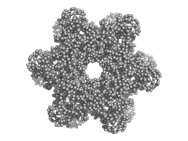
Protein DPCD trimer, 71 kDa Homo sapiens protein
RuvB-like 1 trimer, 155 kDa Homo sapiens protein
RuvB-like 2 trimer, 156 kDa Homo sapiens protein
Buffer: 20 mM HEPES, 150 mM NaCl, 1% glycerol, 5 mM TCEP, pH: 7.5
Experiment: SAXS data collected at SWING, SOLEIL on 2019 Mar 21
Deciphering cellular and molecular determinants of human DPCD protein in complex with RUVBL1/RUVBL2 AAA-ATPases. J Mol Biol :167760 (2022)
...Santos Morais R, Santo PE, Ley M, Schelcher C, Abel Y, Plassart L, Deslignière E, Chagot ME, Quinternet M, Paiva ACF, Hessmann S, Morellet N, M F Sousa P, Vandermoere F, Bertrand E, Charpentier B, Ban...
RgGuinier 5.4 nm
Dmax 15.9 nm
VolumePorod 856 nm3

SASDBH7 – Dps1 truncated, DNA binding protein under starvation conditions (SEC-SAXS)

N-terminal truncated DNA protection during starvation protein 1 experimental SAS data
GASBOR model
Sample: N-terminal truncated DNA protection during starvation protein 1 dodecamer, 216 kDa Deinococcus radiodurans R1 protein
Buffer: 20 mM Tris-HCl, 150 mM NaCl, pH: 7.5
Experiment: SAXS data collected at BM29, ESRF on 2013 Nov 22
SAXS Structural Studies of Dps from Deinococcus radiodurans Highlights the Conformation of the Mobile N-Terminal Extensions. J Mol Biol 429(5):667-687 (2017)
Santos SP, Cuypers MG, Round A, Finet S, Narayanan T, Mitchell EP, Romão CV
RgGuinier 3.9 nm
Dmax 10.0 nm
VolumePorod 291 nm3

SASDFT4 – Conformation of the R11-15 human dystrophin fragment (SANS)

Dystrophin (R11-15 human dystrophin fragment) experimental SAS data
CUSTOM IN-HOUSE model
Sample: Dystrophin (R11-15 human dystrophin fragment) monomer, 60 kDa Homo sapiens protein
Buffer: 20 mM Tris-d11, 150 mM NaCl, 0.1 mM EDTA-d16, in 100% v/v D2O, pH: 7.1
Experiment: SANS data collected at D22, Institut Laue-Langevin (ILL) on 2016 Nov 1
How the central domain of dystrophin acts to bridge F-actin to sarcolemmal lipids. J Struct Biol :107411 (2019)
...Santos Morais R, Chéron A, Lagarrigue M, Winder SJ, Chenuel T, Pérez J, Appavou MS, Martel A, Alviset G, Le Rumeur E, Combet S, Hubert JF, Delalande O
RgGuinier 6.2 nm
Dmax 27.4 nm
VolumePorod 146 nm3

SASDFG5 – His-RuvBl1/RuvBl2 dodecamer

RuvB-like 1RuvB-like 2 experimental SAS data
DAMMIF model
Sample: RuvB-like 1 hexamer, 311 kDa Homo sapiens protein
RuvB-like 2 hexamer, 311 kDa Homo sapiens protein
Buffer: 20 mM HEPES, 150 mM NaCl, 1% glycerol, 5 mM TCEP, pH: 7.5
Experiment: SAXS data collected at SWING, SOLEIL on 2019 Mar 21
Deciphering cellular and molecular determinants of human DPCD protein in complex with RUVBL1/RUVBL2 AAA-ATPases. J Mol Biol :167760 (2022)
...Santos Morais R, Santo PE, Ley M, Schelcher C, Abel Y, Plassart L, Deslignière E, Chagot ME, Quinternet M, Paiva ACF, Hessmann S, Morellet N, M F Sousa P, Vandermoere F, Bertrand E, Charpentier B, Ban...
RgGuinier 6.2 nm
Dmax 20.0 nm
VolumePorod 1490 nm3

SASDBF7 – Dps2, DNA binding protein under starvation conditions (SEC-SAXS)

N-terminal truncated DNA protection during starvation protein 2 experimental SAS data
EOM/RANCH model
Sample: N-terminal truncated DNA protection during starvation protein 2 dodecamer, 279 kDa Deinococcus radiodurans R1 protein
Buffer: 20 mM Tris-HCl, 150 mM NaCl, pH: 7.5
Experiment: SAXS data collected at BM29, ESRF on 2013 Nov 22
SAXS Structural Studies of Dps from Deinococcus radiodurans Highlights the Conformation of the Mobile N-Terminal Extensions. J Mol Biol 429(5):667-687 (2017)
Santos SP, Cuypers MG, Round A, Finet S, Narayanan T, Mitchell EP, Romão CV
RgGuinier 4.2 nm
Dmax 12.7 nm
VolumePorod 445 nm3

SASDFU4 – Conformation of R11-15 human dystrophin fragment in interaction with zwitterionic phospholipid bicelles (SANS)

Dystrophin (R11-15 human dystrophin fragment) experimental SAS data
DAMMIF model
Sample: Dystrophin (R11-15 human dystrophin fragment) monomer, 60 kDa Homo sapiens protein
Buffer: 20 mM Tris-d11, 150 mM NaCl, 0.1 mM EDTA-d16, in 100% v/v D2O, pH: 7.1
Experiment: SANS data collected at D22, Institut Laue-Langevin (ILL) on 2016 Nov 1
How the central domain of dystrophin acts to bridge F-actin to sarcolemmal lipids. J Struct Biol :107411 (2019)
...Santos Morais R, Chéron A, Lagarrigue M, Winder SJ, Chenuel T, Pérez J, Appavou MS, Martel A, Alviset G, Le Rumeur E, Combet S, Hubert JF, Delalande O
RgGuinier 6.2 nm
Dmax 28.1 nm
VolumePorod 144 nm3

SASDFV4 – Conformation of R11-15 human dystrophin fragment in interaction with anionic phospholipid bicelles (SANS)

Dystrophin (R11-15 human dystrophin fragment) experimental SAS data
DAMMIF model
Sample: Dystrophin (R11-15 human dystrophin fragment) monomer, 60 kDa Homo sapiens protein
Buffer: 20 mM Tris-d11, 150 mM NaCl, 0.1 mM EDTA-d16, in 100% v/v D2O, pH: 7.1
Experiment: SANS data collected at D22, Institut Laue-Langevin (ILL) on 2016 Nov 1
How the central domain of dystrophin acts to bridge F-actin to sarcolemmal lipids. J Struct Biol :107411 (2019)
...Santos Morais R, Chéron A, Lagarrigue M, Winder SJ, Chenuel T, Pérez J, Appavou MS, Martel A, Alviset G, Le Rumeur E, Combet S, Hubert JF, Delalande O
RgGuinier 8.1 nm
Dmax 29.6 nm
VolumePorod 231 nm3