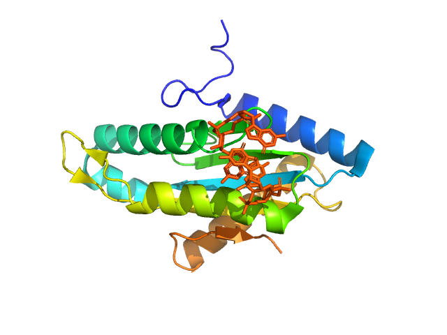
UniProt ID: A1U3W3 (None-None) Diguanylate cyclase
|
|
|
|
| Sample: |
Diguanylate cyclase monomer, 20 kDa Marinobacter hydrocarbonoclasticus protein
|
| Buffer: |
5 mM DTT 100 mM NaCl 10 mM Tris-HCl 0.02 % NaN3, pH: 7.5 |
| Experiment: |
SAXS
data collected at BL4-2, Stanford Synchrotron Radiation Lightsource (SSRL) on 2010 Feb 12
|
Small angle X-ray scattering as a complementary tool for high-throughput structural studies.
Biopolymers 95(8):517-30 (2011)
Grant TD, Luft JR, Wolfley JR, Tsuruta H, Martel A, Montelione GT, Snell EH
|
| RgGuinier |
1.9 |
nm |
| Dmax |
6.6 |
nm |
| VolumePorod |
32 |
nm3 |
|
|
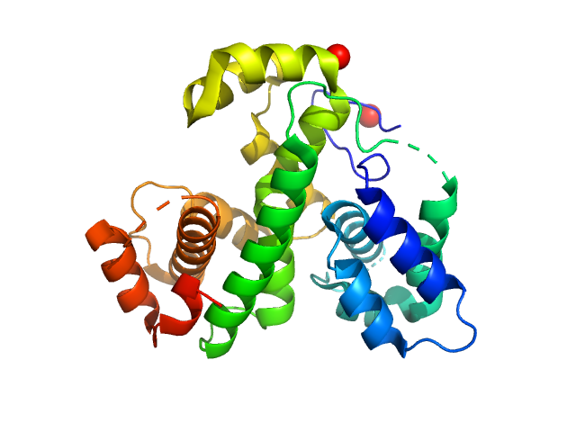
UniProt ID: Q7WZ31 (None-None) MmoQ
|
|
|
|
| Sample: |
MmoQ monomer, 32 kDa Methylococcus capsulatus protein
|
| Buffer: |
5 mM DTT 100 mM NaCl 10 mM Tris-HCl 0.02 % NaN3, pH: 7.5 |
| Experiment: |
SAXS
data collected at BL4-2, Stanford Synchrotron Radiation Lightsource (SSRL) on 2010 Feb 12
|
Small angle X-ray scattering as a complementary tool for high-throughput structural studies.
Biopolymers 95(8):517-30 (2011)
Grant TD, Luft JR, Wolfley JR, Tsuruta H, Martel A, Montelione GT, Snell EH
|
| RgGuinier |
2.3 |
nm |
| Dmax |
8.2 |
nm |
| VolumePorod |
62 |
nm3 |
|
|
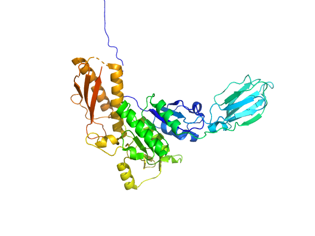
UniProt ID: Q24QE6 (None-None) Uncharacterized protein
|
|
|
|
| Sample: |
Uncharacterized protein monomer, 48 kDa Desulfitobacterium hafniense protein
|
| Buffer: |
5 mM DTT 100 mM NaCl 10 mM Tris-HCl 0.02 % NaN3, pH: 7.5 |
| Experiment: |
SAXS
data collected at BL4-2, Stanford Synchrotron Radiation Lightsource (SSRL) on 2010 Feb 12
|
Small angle X-ray scattering as a complementary tool for high-throughput structural studies.
Biopolymers 95(8):517-30 (2011)
Grant TD, Luft JR, Wolfley JR, Tsuruta H, Martel A, Montelione GT, Snell EH
|
| RgGuinier |
2.8 |
nm |
| Dmax |
9.9 |
nm |
| VolumePorod |
66 |
nm3 |
|
|
UniProt ID: P0CG48 (305-379) Polyubiquitin-C
|
|
|
|
| Sample: |
Polyubiquitin-C dimer, 17 kDa Homo sapiens protein
|
| Buffer: |
100mM NaCl, 10mM sodium acetate, pH: 6 |
| Experiment: |
SAXS
data collected at BL19U2, Shanghai Synchrotron Radiation Facility (SSRF) on 2016 Mar 24
|
Lys63-linked ubiquitin chain adopts multiple conformational states for specific target recognition.
Elife 4 (2015)
Liu Z, Gong Z, Jiang WX, Yang J, Zhu WK, Guo DC, Zhang WP, Liu ML, Tang C
|
| RgGuinier |
2.1 |
nm |
| Dmax |
6.5 |
nm |
| VolumePorod |
24 |
nm3 |
|
|
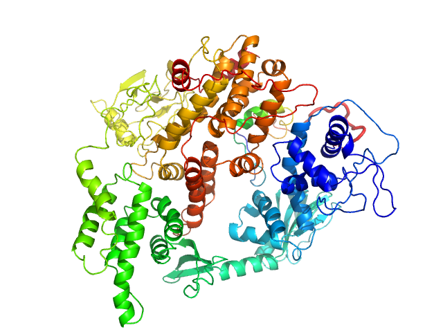
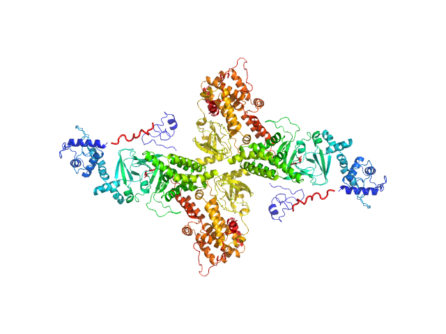
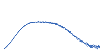
UniProt ID: O95398-3 (2-881) Rap guanine nucleotide exchange factor 3
|
|
|
|
| Sample: |
Rap guanine nucleotide exchange factor 3 monomer, 100 kDa Homo sapiens protein
|
| Buffer: |
1mM EDTA, 10mM DTT, 500mM NaCl, and 10mM Tris, pH: 9 |
| Experiment: |
SAXS
data collected at Rigaku BioSAXS-1000, Sealy Center For Structural Biology, UTMB-G on 2012 Sep 7
|
Conformational States of Exchange Protein Directly Activated by cAMP (EPAC1) Revealed by Ensemble Modeling and Integrative Structural Biology.
Cells 9(1) (2019)
White MA, Tsalkova T, Mei FC, Cheng X
|
| RgGuinier |
3.4 |
nm |
| Dmax |
11.0 |
nm |
| VolumePorod |
180 |
nm3 |
|
|
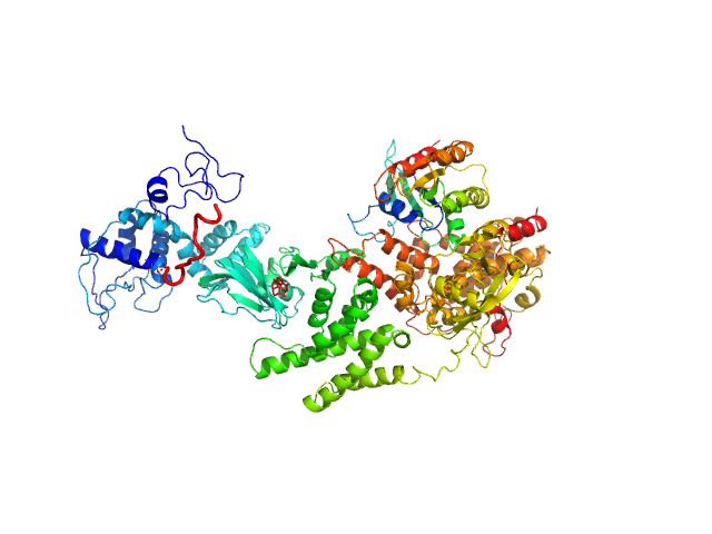
UniProt ID: O95398-3 (2-881) Rap guanine nucleotide exchange factor 3 (dimer)
|
|
|
|
| Sample: |
Rap guanine nucleotide exchange factor 3 (dimer) dimer, 200 kDa Homo sapiens protein
|
| Buffer: |
1mM EDTA, 10mM DTT, 500mM NaCl, 1mM cAMP, and 10mM Tris, pH: 9 |
| Experiment: |
SAXS
data collected at Rigaku BioSAXS-1000, Sealy Center For Structural Biology, UTMB-G on 2012 Jan 30
|
Conformational States of Exchange Protein Directly Activated by cAMP (EPAC1) Revealed by Ensemble Modeling and Integrative Structural Biology.
Cells 9(1) (2019)
White MA, Tsalkova T, Mei FC, Cheng X
|
| RgGuinier |
5.3 |
nm |
| Dmax |
15.7 |
nm |
| VolumePorod |
415 |
nm3 |
|
|
UniProt ID: O95398-3 (2-881) Rap guanine nucleotide exchange factor 3
UniProt ID: Q52L50 (1-167) RAS related protein 1b
|
|
|
|
| Sample: |
Rap guanine nucleotide exchange factor 3 monomer, 100 kDa Homo sapiens protein
RAS related protein 1b monomer, 18 kDa Mus musculus protein
|
| Buffer: |
1mM EDTA, 10mM DTT, 500mM NaCl, 1mM cAMP, and 10mM Tris, pH: 9 |
| Experiment: |
SAXS
data collected at Rigaku BioSAXS-1000, Sealy Center For Structural Biology, UTMB-G on 2013 Apr 1
|
Conformational States of Exchange Protein Directly Activated by cAMP (EPAC1) Revealed by Ensemble Modeling and Integrative Structural Biology.
Cells 9(1) (2019)
White MA, Tsalkova T, Mei FC, Cheng X
|
| RgGuinier |
4.1 |
nm |
| Dmax |
14.2 |
nm |
| VolumePorod |
207 |
nm3 |
|
|
UniProt ID: (None-None) 169 bp DNA (145 bp Widom 601, flanked by 12bp DNA)
UniProt ID: P06897 (None-None) Histone H2A type 1
UniProt ID: P02281 (None-None) Histone H2B 1.1
UniProt ID: P84233 (1-136) Histone H3.2
UniProt ID: P62799 (None-None) Histone H4
|
|
|
|
| Sample: |
169 bp DNA (145 bp Widom 601, flanked by 12bp DNA) monomer, 52 kDa DNA
Histone H2A type 1 monomer, 14 kDa Xenopus laevis protein
Histone H2B 1.1 monomer, 14 kDa Xenopus laevis protein
Histone H3.2 monomer, 15 kDa Xenopus laevis protein
Histone H4 monomer, 11 kDa Xenopus laevis protein
|
| Buffer: |
10 mM Tris, 100 mM NaCl, 2 mM MgCl2, 0.1 mM EDTA, 1 mM DTT, 60% (w/v) sucrose, ADP-BeF3 (0.5 mM ADP, 4 mM NaF, 0.6 mM BeCl2), pH: 7.8 |
| Experiment: |
SAXS
data collected at G1, Cornell High Energy Synchrotron Source (CHESS) on 2015 Oct 24
|
The ATPase motor of the Chd1 chromatin remodeler stimulates DNA unwrapping from the nucleosome.
Nucleic Acids Res 46(10):4978-4990 (2018)
Tokuda JM, Ren R, Levendosky RF, Tay RJ, Yan M, Pollack L, Bowman GD
|
| RgGuinier |
4.8 |
nm |
| Dmax |
14.0 |
nm |
|
|
UniProt ID: P32657 (118-1274) Chromodomain-helicase-DNA-binding protein 1
UniProt ID: (None-None) 169 bp DNA (145 bp Widom 601, flanked by 12bp DNA)
UniProt ID: P06897 (None-None) Histone H2A type 1
UniProt ID: P02281 (None-None) Histone H2B 1.1
UniProt ID: P84233 (1-136) Histone H3.2
UniProt ID: P62799 (None-None) Histone H4
|
|
|
|
| Sample: |
Chromodomain-helicase-DNA-binding protein 1 dimer, 266 kDa Saccharomyces cerevisiae protein
169 bp DNA (145 bp Widom 601, flanked by 12bp DNA) monomer, 52 kDa DNA
Histone H2A type 1 monomer, 14 kDa Xenopus laevis protein
Histone H2B 1.1 monomer, 14 kDa Xenopus laevis protein
Histone H3.2 monomer, 15 kDa Xenopus laevis protein
Histone H4 monomer, 11 kDa Xenopus laevis protein
|
| Buffer: |
10 mM Tris, 100 mM NaCl, 2 mM MgCl2, 0.1 mM EDTA, 1 mM DTT, 60% (w/v) sucrose, pH: 7.8 |
| Experiment: |
SAXS
data collected at G1, Cornell High Energy Synchrotron Source (CHESS) on 2015 Oct 24
|
The ATPase motor of the Chd1 chromatin remodeler stimulates DNA unwrapping from the nucleosome.
Nucleic Acids Res 46(10):4978-4990 (2018)
Tokuda JM, Ren R, Levendosky RF, Tay RJ, Yan M, Pollack L, Bowman GD
|
| RgGuinier |
5.2 |
nm |
| Dmax |
12.8 |
nm |
|
|
UniProt ID: P32657 (118-1274) Chromodomain-helicase-DNA-binding protein 1
UniProt ID: (None-None) 169 bp DNA (145 bp Widom 601, flanked by 12bp DNA)
UniProt ID: P06897 (None-None) Histone H2A type 1
UniProt ID: P02281 (None-None) Histone H2B 1.1
UniProt ID: P84233 (1-136) Histone H3.2
UniProt ID: P62799 (None-None) Histone H4
|
|
|
|
| Sample: |
Chromodomain-helicase-DNA-binding protein 1 dimer, 266 kDa Saccharomyces cerevisiae protein
169 bp DNA (145 bp Widom 601, flanked by 12bp DNA) monomer, 52 kDa DNA
Histone H2A type 1 monomer, 14 kDa Xenopus laevis protein
Histone H2B 1.1 monomer, 14 kDa Xenopus laevis protein
Histone H3.2 monomer, 15 kDa Xenopus laevis protein
Histone H4 monomer, 11 kDa Xenopus laevis protein
|
| Buffer: |
10 mM Tris, 100 mM NaCl, 2 mM MgCl2, 0.1 mM EDTA, 1 mM DTT, 60% (w/v) sucrose, ADP-BeF3 (0.5 mM ADP, 4 mM NaF, 0.6 mM BeCl2), pH: 7.8 |
| Experiment: |
SAXS
data collected at G1, Cornell High Energy Synchrotron Source (CHESS) on 2015 Oct 24
|
The ATPase motor of the Chd1 chromatin remodeler stimulates DNA unwrapping from the nucleosome.
Nucleic Acids Res 46(10):4978-4990 (2018)
Tokuda JM, Ren R, Levendosky RF, Tay RJ, Yan M, Pollack L, Bowman GD
|
| RgGuinier |
5.3 |
nm |
| Dmax |
16.5 |
nm |
|
|


 experimental SAS data")

Histone H2A type 1Histone H2B 1.1Histone H3.2Histone H4 experimental SAS data")

Histone H2A type 1Histone H2B 1.1Histone H3.2Histone H4 experimental SAS data")
 Rg histogram")
Histone H2A type 1Histone H2B 1.1Histone H3.2Histone H4 experimental SAS data")
