Search

 
Advanced search  

352 hits found for X33

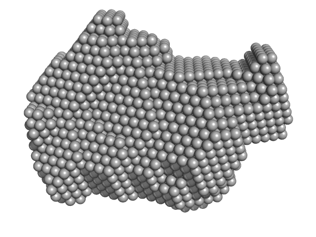
SASDN83 – Microtubule affinity regulating kinase (isoform MARK1)

Serine/threonine-protein kinase MARK1 experimental SAS data
CUSTOM IN-HOUSE model
Sample: Serine/threonine-protein kinase MARK1 monomer, 37 kDa Homo sapiens protein
Buffer: 0.1 M Bis-Tris, 0.2 M ammonium citrate, 1mM DTT, pH: 6.5
Experiment: SAXS data collected at ...X33, DORIS III, DESY on 2006 May 16
Structural Variations in the Catalytic and Ubiquitin-associated Domains of Microtubule-associated Protein/Microtubule Affinity Regulating Kinase (MARK) 1 and MARK2 Journal of Biological Chemistry 281(37):27586-27599 (2006)
Marx A, Nugoor C, Müller J, Panneerselvam S, Timm T, Bilang M, Mylonas E, Svergun D, Mandelkow E, Mandelkow E
RgGuinier 2.3 nm
Dmax 8.0 nm
VolumePorod 64 nm3

SASDQ83 – Lipid_unpublished_41

Lipid experimental SAS data
Lipid Kratky plot
Sample: Lipid None, lipid
Buffer:
Experiment: SAXS data collected at ...X33, DORIS III, DESY on 2011 Dec 19
Lipid_unpublished_41
Siawosch Schewa

SASDM93 – Native tannin macromolecules (DP7, average polymerization 6.3) in water-ethanol solution (water fraction 100%)

Native tannin macromolecules (DP7, average polymerization 6.3) in water-ethanol solution (water fraction 100%) experimental SAS data
DAMMIN model
Sample: Native tannin macromolecules (DP7, average polymerization 6.3) in water-ethanol solution (water fraction 100%) heptamer, 2 kDa
Buffer: water-ethanol solution (water fraction 100%), pH: 7
Experiment: SAXS data collected at ...X33, DORIS III, DESY on 2007 Nov 24
Rigidity, conformation, and solvation of native and oxidized tannin macromolecules in water-ethanol solution. J Chem Phys 130(24):245103 (2009)
Zanchi D, Konarev PV, Tribet C, Baron A, Svergun DI, Guyot S
RgGuinier 1.0 nm
Dmax 3.1 nm

SASDN93 – Dystrophiamyotonica kinase (DMPK)

Myotonin-protein kinase experimental SAS data
PDB (PROTEIN DATA BANK) model
Sample: Myotonin-protein kinase monomer, 92 kDa Homo sapiens protein
Buffer: 50 mM TrisHCl 50mM NaCl, 2.5 mM-mercaptoethanol, pH: 8
Experiment: SAXS data collected at ...X33, DORIS III, DESY on 2005 Mar 18
Molecular insights into the self‐assembly mechanism of dystrophia myotonica kinase The FASEB Journal 20(8):1142-1151 (2006)
Garcia P, Ucurum Z, Bucher R, Svergun D, Huber T, Lustig A, Konarev P, Marino M, Mayans O
RgGuinier 3.9 nm
Dmax 13.0 nm
VolumePorod 173 nm3

SASDQ93 – Lipid_unpublished_40

Lipid experimental SAS data
Lipid Kratky plot
Sample: Lipid None, lipid
Buffer:
Experiment: SAXS data collected at ...X33, DORIS III, DESY on 2011 Dec 20
Lipid_unpublished_40
Siawosch Schewa

SASDMA3 – Oxidized tannin macromolecules (DP7, average polymerization 6.3) in water-ethanol solution (water fraction 100%)

Oxidized tannin macromolecules (DP7, average polymerization 6.3) in water-ethanol solution (water fraction 100%) experimental SAS data
DAMMIN model
Sample: Oxidized tannin macromolecules (DP7, average polymerization 6.3) in water-ethanol solution (water fraction 100%) heptamer, 2 kDa
Buffer: water-ethanol solution (water fraction 100%), pH: 7
Experiment: SAXS data collected at ...X33, DORIS III, DESY on 2007 Oct 12
Rigidity, conformation, and solvation of native and oxidized tannin macromolecules in water-ethanol solution. J Chem Phys 130(24):245103 (2009)
Zanchi D, Konarev PV, Tribet C, Baron A, Svergun DI, Guyot S
RgGuinier 1.0 nm
Dmax 3.3 nm

SASDQA3 – Lipid_unpublished_42

Lipid experimental SAS data
Lipid Kratky plot
Sample: Lipid None, lipid
Buffer:
Experiment: SAXS data collected at ...X33, DORIS III, DESY on 2011 Dec 19
Lipid_unpublished_42
Siawosch Schewa

SASDMB3 – Native tannin macromolecules (DP7, average polymerization 6.3) in water-ethanol solution (water fraction 80%)

Native tannin macromolecules (DP7, average polymerization 6.3) in water-ethanol solution (water fraction 80%) experimental SAS data
DAMMIN model
Sample: Native tannin macromolecules (DP7, average polymerization 6.3) in water-ethanol solution (water fraction 80%) heptamer, 2 kDa
Buffer: water-ethanol solution (water fraction 80%), pH: 7
Experiment: SAXS data collected at ...X33, DORIS III, DESY on 2007 Nov 24
Rigidity, conformation, and solvation of native and oxidized tannin macromolecules in water-ethanol solution. J Chem Phys 130(24):245103 (2009)
Zanchi D, Konarev PV, Tribet C, Baron A, Svergun DI, Guyot S
RgGuinier 0.9 nm
Dmax 3.3 nm

SASDQB3 – Lipid_unpublished_43

Lipid experimental SAS data
Lipid Kratky plot
Sample: Lipid None, lipid
Buffer:
Experiment: SAXS data collected at ...X33, DORIS III, DESY on 2011 Dec 19
Lipid_unpublished_43
Siawosch Schewa

SASDZB3 – Calcium binding domains 1 and 2 from sodium-calcium exchanger 1.4, apo state

Calcium Binding Domains 1 and 2 from Sodium-Calcium Exchanger 1.4 experimental SAS data
Calcium binding domains 1 and 2 from sodium-calcium exchanger 1.4, apo state Rg histogram
Sample: Calcium Binding Domains 1 and 2 from Sodium-Calcium Exchanger 1.4 monomer, 32 kDa Canis lupus familiaris protein
Buffer: 20 mM Tris-HCl, 100 mM KCl, 20 mM -mercaptoethanol, 10 mM EDTA, pH: 7.2
Experiment: SAXS data collected at ...X33, DORIS III, DESY on 2012 Apr 30
Use of biomolecular emulator for characterizing flexible proteins by small-angle x-ray scattering. Protein Sci 35(6):e70646 (2026)
Haitin Y, Manori B, Giladi M
RgGuinier 2.9 nm

SASDBC3 – RepB, the replication initiator protein of a promiscuous Streptococcal plasmid pMV158

Replication initiator protein of a promiscuous streptococcal plasmid pMV158. experimental SAS data
NONE model
Sample: Replication initiator protein of a promiscuous streptococcal plasmid pMV158. hexamer, Streptococcus sp. protein
Buffer: 10 mM TRIS 5 mM EDTA 1.0 M KCl, pH: 8.5
Experiment: SAXS data collected at ...X33, DORIS III, DESY on 2007 Jun 16
Conformational plasticity of RepB, the replication initiator protein of promiscuous streptococcal plasmid pMV158. Sci Rep 6:20915 (2016)
Boer DR, Ruiz-Masó JA, Rueda M, Petoukhov MV, Machón C, Svergun DI, Orozco M, del Solar G, Coll M
RgGuinier 3.8 nm
Dmax 10.6 nm
VolumePorod 282 nm3

SASDMC3 – Oxidized tannin macromolecules (DP7, average polymerization 6.3) in water-ethanol solution (water fraction 0%)

Oxidized tannin macromolecules (DP7, average polymerization 6.3) in water-ethanol solution (water fraction 0%) experimental SAS data
DAMMIN model
Sample: Oxidized tannin macromolecules (DP7, average polymerization 6.3) in water-ethanol solution (water fraction 0%) heptamer, 2 kDa
Buffer: water-ethanol solution (water fraction 0%), pH: 7
Experiment: SAXS data collected at ...X33, DORIS III, DESY on 2007 Nov 24
Rigidity, conformation, and solvation of native and oxidized tannin macromolecules in water-ethanol solution. J Chem Phys 130(24):245103 (2009)
Zanchi D, Konarev PV, Tribet C, Baron A, Svergun DI, Guyot S
RgGuinier 1.3 nm
Dmax 4.5 nm

SASDQC3 – Lipid_unpublished_44

Lipid experimental SAS data
Lipid Kratky plot
Sample: Lipid None, lipid
Buffer:
Experiment: SAXS data collected at ...X33, DORIS III, DESY on 2011 Dec 19
Lipid_unpublished_44
Siawosch Schewa

SASDZC3 – Calcium binding domains 1 and 2 from sodium-calcium exchanger 1.4, calcium-bound state

Calcium Binding Domains 1 and 2 from Sodium-Calcium Exchanger 1.4 experimental SAS data
Calcium binding domains 1 and 2 from sodium-calcium exchanger 1.4, calcium-bound state Rg histogram
Sample: Calcium Binding Domains 1 and 2 from Sodium-Calcium Exchanger 1.4 monomer, 32 kDa Canis lupus familiaris protein
Buffer: 20 mM Tris-HCl, 100 mM KCl, 20 mM -mercaptoethanol, 10 mM CaCl2, pH: 7.2
Experiment: SAXS data collected at ...X33, DORIS III, DESY on 2012 Apr 30
Use of biomolecular emulator for characterizing flexible proteins by small-angle x-ray scattering. Protein Sci 35(6):e70646 (2026)
Haitin Y, Manori B, Giladi M
RgGuinier 3.2 nm

SASDMD3 – Oxidized tannin macromolecules (DP7, average polymerization 6.3) in water-ethanol solution (water fraction 20%)

Oxidized tannin macromolecules (DP7, average polymerization 6.3) in water-ethanol solution (water fraction 20%) experimental SAS data
DAMMIN model
Sample: Oxidized tannin macromolecules (DP7, average polymerization 6.3) in water-ethanol solution (water fraction 20%) heptamer, 2 kDa
Buffer: water-ethanol solution (water fraction 20%), pH: 7
Experiment: SAXS data collected at ...X33, DORIS III, DESY on 2007 Nov 24
Rigidity, conformation, and solvation of native and oxidized tannin macromolecules in water-ethanol solution. J Chem Phys 130(24):245103 (2009)
Zanchi D, Konarev PV, Tribet C, Baron A, Svergun DI, Guyot S
RgGuinier 1.1 nm
Dmax 3.5 nm

SASDQD3 – Lipid_unpublished_45

Lipid experimental SAS data
Lipid Kratky plot
Sample: Lipid None, lipid
Buffer:
Experiment: SAXS data collected at ...X33, DORIS III, DESY on 2011 Dec 3
Lipid_unpublished_45
Siawosch Schewa

SASDZD3 – Double C2-like domain-containing protein beta (DOC2B) C2AB domains from , apo state

Double C2-like domain-containing protein beta experimental SAS data
Double C2-like domain-containing protein beta (DOC2B) C2AB domains from , apo state Rg histogram
Sample: Double C2-like domain-containing protein beta monomer, 33 kDa Rattus norvegicus protein
Buffer: 20 mM Tris–HCl, 200 mM NaCl, 20 mM β-mercaptoethanol, 10 mM EDTA, pH: 6.8
Experiment: SAXS data collected at ...X33, DORIS III, DESY on 2012 Apr 30
Use of biomolecular emulator for characterizing flexible proteins by small-angle x-ray scattering. Protein Sci 35(6):e70646 (2026)
Haitin Y, Manori B, Giladi M
RgGuinier 2.6 nm

SASDME3 – Oxidized tannin macromolecules (DP7, average polymerization 6.3) in water-ethanol solution (water fraction 60%)

Oxidized tannin macromolecules (DP7, average polymerization 6.3) in water-ethanol solution (water fraction 60%) experimental SAS data
DAMMIN model
Sample: Oxidized tannin macromolecules (DP7, average polymerization 6.3) in water-ethanol solution (water fraction 60%) heptamer, 2 kDa
Buffer: water-ethanol solution (water fraction 60%), pH: 7
Experiment: SAXS data collected at ...X33, DORIS III, DESY on 2007 Nov 24
Rigidity, conformation, and solvation of native and oxidized tannin macromolecules in water-ethanol solution. J Chem Phys 130(24):245103 (2009)
Zanchi D, Konarev PV, Tribet C, Baron A, Svergun DI, Guyot S
RgGuinier 1.0 nm
Dmax 2.9 nm

SASDQE3 – Lipid_unpublished_46

Lipid experimental SAS data
Lipid Kratky plot
Sample: Lipid None, lipid
Buffer:
Experiment: SAXS data collected at ...X33, DORIS III, DESY on 2009 Dec 19
Lipid_unpublished_46
Siawosch Schewa

SASDLF3 – Staphylococcal immunoglobulin-binding protein bound to complement component C3

Protein A experimental SAS data
DAMMIN model
Sample: Protein A monomer, 28 kDa Staphylococcus aureus protein
Buffer: 10 mM HEPES, pH 7.2, 3 mM EDTA, pH: 7.2
Experiment: SAXS data collected at ...X33, DORIS III, DESY on 2005 May 12
A structural basis for Staphylococcal complement subversion: X-ray structure of the complement-binding domain of Staphylococcus aureus protein Sbi in complex with ligand C3d Molecular Immunology 48(4):452-462 (2011)
Clark E, Crennell S, Upadhyay A, Zozulya A, Mackay J, Svergun D, Bagby S, van den Elsen J
RgGuinier 4.6 nm
Dmax 16.0 nm
VolumePorod 81 nm3

SASDMF3 – Oxidized tannin macromolecules (DP7, average polymerization 6.3) in water-ethanol solution (water fraction 80%)

Oxidized tannin macromolecules (DP7, average polymerization 6.3) in water-ethanol solution (water fraction 80%) experimental SAS data
DAMMIN model
Sample: Oxidized tannin macromolecules (DP7, average polymerization 6.3) in water-ethanol solution (water fraction 80%) heptamer, 2 kDa
Buffer: water-ethanol solution (water fraction 80%), pH: 7
Experiment: SAXS data collected at ...X33, DORIS III, DESY on 2007 Oct 12
Rigidity, conformation, and solvation of native and oxidized tannin macromolecules in water-ethanol solution. J Chem Phys 130(24):245103 (2009)
Zanchi D, Konarev PV, Tribet C, Baron A, Svergun DI, Guyot S
RgGuinier 1.2 nm
Dmax 3.6 nm

SASDQF3 – Lipid_unpublished_48

Lipid experimental SAS data
Lipid Kratky plot
Sample: Lipid None, lipid
Buffer:
Experiment: SAXS data collected at ...X33, DORIS III, DESY on 2011 Mar 12
Lipid_unpublished_48
Siawosch Schewa

SASDLG3 – Thermoplasma E2 catalytic core

Regulatory protein E2 experimental SAS data
PDB (PROTEIN DATA BANK) model
Sample: Regulatory protein E2 , 1037 kDa Human papillomavirus type … protein
Buffer: 50 mM Tris ⁄ HCl, pH 8.8, 100 mM NaCl, pH: 8.8
Experiment: SAXS data collected at ...X33, DORIS III, DESY on 2009 Jul 13
The catalytic core of an archaeal 2-oxoacid dehydrogenase multienzyme complex is a 42-mer protein assembly FEBS Journal 279(5):713-723 (2012)
Marrott N, Marshall J, Svergun D, Crennell S, Hough D, Danson M, van den Elsen J
RgGuinier 8.8 nm
Dmax 22.0 nm
VolumePorod 2473 nm3

SASDQG3 – Lipid_unpublished_47

Lipid experimental SAS data
Lipid Kratky plot
Sample: Lipid None, lipid
Buffer:
Experiment: SAXS data collected at ...X33, DORIS III, DESY on 2011 Dec 19
Lipid_unpublished_47
Siawosch Schewa

SASDLH3 – Truncated Thermoplasma E2 catalytic core

Regulatory protein E2 experimental SAS data
CUSTOM IN-HOUSE model
Sample: Regulatory protein E2 trimer, 73 kDa Human papillomavirus type … protein
Buffer: 50 mM Tris ⁄ HCl, pH 8.8, 100 mM NaCl, pH: 8.8
Experiment: SAXS data collected at ...X33, DORIS III, DESY on 2011 May 27
Why are the 2-oxoacid dehydrogenase complexes so large? Generation of an active trimeric complex Biochemical Journal 463(3):405-412 (2014)
Marrott N, Marshall J, Svergun D, Crennell S, Hough D, van den Elsen J, Danson M
RgGuinier 3.1 nm
Dmax 11.0 nm
VolumePorod 149 nm3

SASDQH3 – Lipid_unpublished_49

Lipid experimental SAS data
Lipid Kratky plot
Sample: Lipid None, lipid
Buffer:
Experiment: SAXS data collected at ...X33, DORIS III, DESY on 2011 Mar 12
Lipid_unpublished_49
Siawosch Schewa

SASDQJ3 – Lipid_unpublished_50

Lipid experimental SAS data
Lipid Kratky plot
Sample: Lipid None, lipid
Buffer:
Experiment: SAXS data collected at ...X33, DORIS III, DESY on 2011 Mar 11
Lipid_unpublished_50

SASDQK3 – Lipid_unpublished_51

Lipid experimental SAS data
Lipid Kratky plot
Sample: Lipid None, lipid
Buffer:
Experiment: SAXS data collected at ...X33, DORIS III, DESY on 2011 Mar 11
Lipid_unpublished_51
Siawosch Schewa

SASDQL3 – Lipid_unpublished_88

Lipid experimental SAS data
Lipid Kratky plot
Sample: Lipid None, lipid
Buffer:
Experiment: SAXS data collected at ...X33, DORIS III, DESY on 2011 Mar 11
Lipid_unpublished_88
Siawosch Schewa

SASDQM3 – Lipid_unpublished_53

Lipid experimental SAS data
Lipid Kratky plot
Sample: Lipid None, lipid
Buffer:
Experiment: SAXS data collected at ...X33, DORIS III, DESY on 2010 Jul 10
Lipid_unpublished_53
Siawosch Schewa

SASDQN3 – Lipid_unpublished_52

Lipid experimental SAS data
Lipid Kratky plot
Sample: Lipid None, lipid
Buffer:
Experiment: SAXS data collected at ...X33, DORIS III, DESY on 2010 Jul 22
Lipid_unpublished_52
Siawosch Schewa

SASDQP3 – Lipid_unpublished_54

Lipid experimental SAS data
Lipid Kratky plot
Sample: Lipid None, lipid
Buffer:
Experiment: SAXS data collected at ...X33, DORIS III, DESY on 2010 Jun 24
Lipid_unpublished_54
Siawosch Schewa

SASDAQ3 – MutS tetramer

DNA mismatch repair protein MutS experimental SAS data
DAMMIF model
Sample: DNA mismatch repair protein MutS tetramer, 381 kDa Escherichia coli protein
Buffer: 50 mM HEPES, 50 mM KCl, pH: 7.5
Experiment: SAXS data collected at ...X33, DORIS III, DESY on 2011 May 12
Using stable MutS dimers and tetramers to quantitatively analyze DNA mismatch recognition and sliding clamp formation. Nucleic Acids Res 41(17):8166-81 (2013)
Groothuizen FS, Fish A, Petoukhov MV, Reumer A, Manelyte L, Winterwerp HH, Marinus MG, Lebbink JH, Svergun DI, Friedhoff P, Sixma TK
RgGuinier 7.8 nm
Dmax 28.0 nm
VolumePorod 700 nm3

SASDQQ3 – Lipid_unpublished_56

Lipid experimental SAS data
Lipid Kratky plot
Sample: Lipid None, lipid
Buffer:
Experiment: SAXS data collected at ...X33, DORIS III, DESY on 2011 Jul 10
Lipid_unpublished_56
Siawosch Schewa

SASDAR3 – PsrP functional binding region

Functional binding region (187-385) of the pneumococcal serine-rich repeat protein experimental SAS data
PsrP functional binding region Rg histogram
Sample: Functional binding region (187-385) of the pneumococcal serine-rich repeat protein monomer, 22 kDa Streptococcus pneumoniae protein
Buffer: 20 mM sodium citrate 250 mM NaCl 2.5 % Glycerol, pH: 5.5
Experiment: SAXS data collected at ...X33, DORIS III, DESY on 2011 Jul 2
The basic keratin 10-binding domain of the virulence-associated pneumococcal serine-rich protein PsrP adopts a novel MSCRAMM fold. Open Biol 4:130090 (2014)
Schulte T, Löfling J, Mikaelsson C, Kikhney A, Hentrich K, Diamante A, Ebel C, Normark S, Svergun D, Henriques-Normark B, Achour A
RgGuinier 2.3 nm
Dmax 7.7 nm
VolumePorod 37 nm3

SASDQR3 – Lipid_unpublished_55

Lipid experimental SAS data
Lipid Kratky plot
Sample: Lipid None, lipid
Buffer:
Experiment: SAXS data collected at ...X33, DORIS III, DESY on 2010 Jun 24
Lipid_unpublished_55
Siawosch Schewa

SASDAS3 – Fila16-17

immunoglobulin- like filamin two-domain fragment 16-17 experimental SAS data
GASBOR model
Sample: immunoglobulin- like filamin two-domain fragment 16-17 monomer, 19 kDa Escherichia coli protein
Buffer: 100 mM NaCl 10 mM dithiothreitol 20 mM Tris, pH: 8
Experiment: SAXS data collected at ...X33, DORIS III, DESY on 2007 Jun 18
Atomic structures of two novel immunoglobulin-like domain pairs in the actin cross-linking protein filamin. J Biol Chem 284(37):25450-8 (2009)
Heikkinen OK, Ruskamo S, Konarev PV, Svergun DI, Iivanainen T, Heikkinen SM, Permi P, Koskela H, Kilpeläinen I, Ylänne J
RgGuinier 1.9 nm
Dmax 6.0 nm
VolumePorod 33 nm3

SASDQS3 – Lipid_unpublished_57

Lipid experimental SAS data
Lipid Kratky plot
Sample: Lipid None, lipid
Buffer:
Experiment: SAXS data collected at ...X33, DORIS III, DESY on 2011 Jul 10
Lipid_unpublished_57
Siawosch Schewa

SASDAT3 – Fila18-19

immunoglobulin- like filamin two-domain fragment 18-19 experimental SAS data
GASBOR model
Sample: immunoglobulin- like filamin two-domain fragment 18-19 monomer, 20 kDa Escherichia coli protein
Buffer: 100 mM NaCl 10 mM dithiothreitol 20 mM Tris, pH: 8
Experiment: SAXS data collected at ...X33, DORIS III, DESY on 2007 Jun 18
Atomic structures of two novel immunoglobulin-like domain pairs in the actin cross-linking protein filamin. J Biol Chem 284(37):25450-8 (2009)
Heikkinen OK, Ruskamo S, Konarev PV, Svergun DI, Iivanainen T, Heikkinen SM, Permi P, Koskela H, Kilpeläinen I, Ylänne J
RgGuinier 2.1 nm
Dmax 6.5 nm
VolumePorod 34 nm3

SASDQT3 – Lipid_unpublished_59

Lipid experimental SAS data
Lipid Kratky plot
Sample: Lipid None, lipid
Buffer:
Experiment: SAXS data collected at ...X33, DORIS III, DESY on 2011 Jul 10
Lipid_unpublished_59
Siawosch Schewa

SASDAU3 – Fila22-23

immunoglobulin- like filamin two-domain fragment 22-23 experimental SAS data
GASBOR model
Sample: immunoglobulin- like filamin two-domain fragment 22-23 monomer, 19 kDa Escherichia coli protein
Buffer: 100 mM NaCl 10 mM dithiothreitol 20 mM Tris, pH: 8
Experiment: SAXS data collected at ...X33, DORIS III, DESY on 2007 Jun 18
Atomic structures of two novel immunoglobulin-like domain pairs in the actin cross-linking protein filamin. J Biol Chem 284(37):25450-8 (2009)
Heikkinen OK, Ruskamo S, Konarev PV, Svergun DI, Iivanainen T, Heikkinen SM, Permi P, Koskela H, Kilpeläinen I, Ylänne J
RgGuinier 2.8 nm
Dmax 9.0 nm
VolumePorod 32 nm3

SASDQU3 – Lipid_unpublished_58

Lipid experimental SAS data
Lipid Kratky plot
Sample: Lipid None, lipid
Buffer:
Experiment: SAXS data collected at ...X33, DORIS III, DESY on 2011 Jul 10
Lipid_unpublished_58
Siawosch Schewa

SASDAV3 – Geminin:Cdt1 2:1 heterotrimer

GemininDNA replication factor Cdt1 experimental SAS data
CRYSOL model
Sample: Geminin dimer, 47 kDa Homo sapiens protein
DNA replication factor Cdt1 monomer, 60 kDa Homo sapiens protein
Buffer: 25 mM Tris75 200 mM NaCl, pH: 7.5
Experiment: SAXS data collected at ...X33, DORIS III, DESY on 2006 Aug 13
Quaternary structure of the human Cdt1-Geminin complex regulates DNA replication licensing. Proc Natl Acad Sci U S A 106(47):19807-12 (2009)
De Marco V, Gillespie PJ, Li A, Karantzelis N, Christodoulou E, Klompmaker R, van Gerwen S, Fish A, Petoukhov MV, Iliou MS, Lygerou Z, Medema RH, Blow JJ, Svergun DI, Taraviras S, Perrakis A
RgGuinier 2.9 nm
Dmax 10.0 nm
VolumePorod 70 nm3

SASDQV3 – Lipid_unpublished_60

Lipid experimental SAS data
Lipid Kratky plot
Sample: Lipid None, lipid
Buffer:
Experiment: SAXS data collected at ...X33, DORIS III, DESY on 2009 Apr 30
Lipid_unpublished_60
Siawosch Schewa

SASDAW3 – Geminin:Cdt1 4:2 heterohexamer

GemininDNA replication factor Cdt1 experimental SAS data
CRYSOL model
Sample: Geminin dimer, 47 kDa Homo sapiens protein
DNA replication factor Cdt1 monomer, 60 kDa Homo sapiens protein
Buffer: 25 mM Tris75 200 mM NaCl, pH: 7.5
Experiment: SAXS data collected at ...X33, DORIS III, DESY on 2006 Oct 5
Quaternary structure of the human Cdt1-Geminin complex regulates DNA replication licensing. Proc Natl Acad Sci U S A 106(47):19807-12 (2009)
De Marco V, Gillespie PJ, Li A, Karantzelis N, Christodoulou E, Klompmaker R, van Gerwen S, Fish A, Petoukhov MV, Iliou MS, Lygerou Z, Medema RH, Blow JJ, Svergun DI, Taraviras S, Perrakis A
RgGuinier 3.8 nm
Dmax 14.0 nm
VolumePorod 120 nm3

SASDQW3 – Lipid_unpublished_61

Lipid experimental SAS data
Lipid Kratky plot
Sample: Lipid None, lipid
Buffer:
Experiment: SAXS data collected at ...X33, DORIS III, DESY on 2009 Apr 30
Lipid_unpublished_61
Siawosch Schewa

SASDAX3 – MutS tetramer

DNA mismatch repair protein MutS experimental SAS data
DNA mismatch repair protein MutS Kratky plot
Sample: DNA mismatch repair protein MutS tetramer, 381 kDa Escherichia coli protein
Buffer: 50 mM HEPES, 50 mM KCl, pH: 7.5
Experiment: SAXS data collected at ...X33, DORIS III, DESY on 2011 May 12
Using stable MutS dimers and tetramers to quantitatively analyze DNA mismatch recognition and sliding clamp formation. Nucleic Acids Res 41(17):8166-81 (2013)
Groothuizen FS, Fish A, Petoukhov MV, Reumer A, Manelyte L, Winterwerp HH, Marinus MG, Lebbink JH, Svergun DI, Friedhoff P, Sixma TK
RgGuinier 8.5 nm
Dmax 29.0 nm
VolumePorod 750 nm3

SASDQX3 – Lipid_unpublished_63

Lipid experimental SAS data
Lipid Kratky plot
Sample: Lipid None, lipid
Buffer:
Experiment: SAXS data collected at ...X33, DORIS III, DESY on 2009 Apr 30
Lipid_unpublished_63
Siawosch Schewa

SASDAY3 – MutS tetramer

DNA mismatch repair protein MutS experimental SAS data
DNA mismatch repair protein MutS Kratky plot
Sample: DNA mismatch repair protein MutS tetramer, 381 kDa Escherichia coli protein
Buffer: 50 mM HEPES, 50 mM KCl, pH: 7.5
Experiment: SAXS data collected at ...X33, DORIS III, DESY on 2011 May 12
Using stable MutS dimers and tetramers to quantitatively analyze DNA mismatch recognition and sliding clamp formation. Nucleic Acids Res 41(17):8166-81 (2013)
Groothuizen FS, Fish A, Petoukhov MV, Reumer A, Manelyte L, Winterwerp HH, Marinus MG, Lebbink JH, Svergun DI, Friedhoff P, Sixma TK
RgGuinier 8.3 nm
Dmax 29.0 nm
VolumePorod 720 nm3

SASDQY3 – Lipid_unpublished_62

Lipid experimental SAS data
Lipid Kratky plot
Sample: Lipid None, lipid
Buffer:
Experiment: SAXS data collected at ...X33, DORIS III, DESY on 2009 Apr 30
Lipid_unpublished_62
Siawosch Schewa

SASDAZ3 – MutS tetramer

DNA mismatch repair protein MutS experimental SAS data
DNA mismatch repair protein MutS Kratky plot
Sample: DNA mismatch repair protein MutS tetramer, 381 kDa Escherichia coli protein
Buffer: 50 mM HEPES, 50 mM KCl, pH: 7.5
Experiment: SAXS data collected at ...X33, DORIS III, DESY on 2011 May 12
Using stable MutS dimers and tetramers to quantitatively analyze DNA mismatch recognition and sliding clamp formation. Nucleic Acids Res 41(17):8166-81 (2013)
Groothuizen FS, Fish A, Petoukhov MV, Reumer A, Manelyte L, Winterwerp HH, Marinus MG, Lebbink JH, Svergun DI, Friedhoff P, Sixma TK
RgGuinier 8.0 nm

SASDQZ3 – Lipid_unpublished_64

Lipid experimental SAS data
Lipid Kratky plot
Sample: Lipid None, lipid
Buffer:
Experiment: SAXS data collected at ...X33, DORIS III, DESY on 2009 Apr 30
Lipid_unpublished_64
Siawosch Schewa

SASDA24 – MutS tetramer

DNA mismatch repair protein MutS experimental SAS data
DNA mismatch repair protein MutS Kratky plot
Sample: DNA mismatch repair protein MutS tetramer, 381 kDa Escherichia coli protein
Buffer: 50 mM HEPES, 50 mM KCl, pH: 7.5
Experiment: SAXS data collected at ...X33, DORIS III, DESY on 2011 May 12
Using stable MutS dimers and tetramers to quantitatively analyze DNA mismatch recognition and sliding clamp formation. Nucleic Acids Res 41(17):8166-81 (2013)
Groothuizen FS, Fish A, Petoukhov MV, Reumer A, Manelyte L, Winterwerp HH, Marinus MG, Lebbink JH, Svergun DI, Friedhoff P, Sixma TK
RgGuinier 7.8 nm
Dmax 27.0 nm

SASDQ24 – Lipid_unpublished_66

Lipid experimental SAS data
Lipid Kratky plot
Sample: Lipid None, lipid
Buffer:
Experiment: SAXS data collected at ...X33, DORIS III, DESY on 2009 Dec 19
Lipid_unpublished_66
Siawosch Schewa

SASDQ34 – Lipid_unpublished_65

Lipid experimental SAS data
Lipid Kratky plot
Sample: Lipid None, lipid
Buffer:
Experiment: SAXS data collected at ...X33, DORIS III, DESY on 2009 Dec 19
Lipid_unpublished_65
Siawosch Schewa

SASDQ44 – Lipid_unpublished_67

Lipid experimental SAS data
Lipid Kratky plot
Sample: Lipid None, lipid
Buffer:
Experiment: SAXS data collected at ...X33, DORIS III, DESY on 2010 Jul 22
Lipid_unpublished_67
Siawosch Schewa

SASDA54 – RNA Aptamer

SRB2m experimental SAS data
DAMMIN model
Sample: SRB2m dimer, 33 kDa RNA
Buffer: Hepes, pH: 7.4
Experiment: SAXS data collected at ...X33, DORIS III, DESY on 2007 Oct 24
Characterization of a fluorophore binding RNA aptamer by fluorescence correlation spectroscopy and small angle X-ray scattering. Anal Biochem 389(1):52-62 (2009)
Werner A, Konarev PV, Svergun DI, Hahn U
RgGuinier 2.8 nm
Dmax 10.0 nm
VolumePorod 38 nm3

SASDQ54 – Lipid_unpublished_69

Lipid experimental SAS data
Lipid Kratky plot
Sample: Lipid None, lipid
Buffer:
Experiment: SAXS data collected at ...X33, DORIS III, DESY on 2010 Jul 22
Lipid_unpublished_69
Siawosch Schewa

SASDQ64 – Lipid_unpublished_68

Lipid experimental SAS data
Lipid Kratky plot
Sample: Lipid None, lipid
Buffer:
Experiment: SAXS data collected at ...X33, DORIS III, DESY on 2010 Jul 22
Lipid_unpublished_68
Siawosch Schewa

SASDA74 – RNA Aptamer

SRB2m experimental SAS data
DAMMIN model
Sample: SRB2m dimer, 33 kDa RNA
Buffer: Hepes, pH: 7.4
Experiment: SAXS data collected at ...X33, DORIS III, DESY on 2007 Oct 24
Characterization of a fluorophore binding RNA aptamer by fluorescence correlation spectroscopy and small angle X-ray scattering. Anal Biochem 389(1):52-62 (2009)
Werner A, Konarev PV, Svergun DI, Hahn U
RgGuinier 2.4 nm
Dmax 8.5 nm
VolumePorod 31 nm3

SASDQ74 – Lipid_unpublished_70

Lipid experimental SAS data
Lipid Kratky plot
Sample: Lipid None, lipid
Buffer:
Experiment: SAXS data collected at ...X33, DORIS III, DESY on 2010 Jul 24
Lipid_unpublished_70
Siawosch Schewa

SASDA84 – RNA Aptamer

SRB2m experimental SAS data
DAMMIN model
Sample: SRB2m dimer, 33 kDa RNA
Buffer: Hepes, pH: 7.4
Experiment: SAXS data collected at ...X33, DORIS III, DESY on 2007 Oct 24
Characterization of a fluorophore binding RNA aptamer by fluorescence correlation spectroscopy and small angle X-ray scattering. Anal Biochem 389(1):52-62 (2009)
Werner A, Konarev PV, Svergun DI, Hahn U
RgGuinier 2.3 nm
Dmax 8.0 nm
VolumePorod 24 nm3

SASDQ84 – Lipid_unpublished_71

Lipid experimental SAS data
Lipid Kratky plot
Sample: Lipid None, lipid
Buffer:
Experiment: SAXS data collected at ...X33, DORIS III, DESY on 2010 Jul 25
Lipid_unpublished_71
Siawosch Schewa

SASDA94 – Der p21

Der p21, allegren experimental SAS data
DAMMIN model
Sample: Der p21, allegren dimer, 26 kDa Escherichia coli protein
Buffer: 10 mM Hepes , pH: 7
Experiment: SAXS data collected at ...X33, DORIS III, DESY on 2006 Jul 24
Characterization of Der p 21, a new important allergen derived from the gut of house dust mites. Allergy 63(6):758-67 (2008)
Weghofer M, Dall'Antonia Y, Grote M, Stöcklinger A, Kneidinger M, Balic N, Krauth MT, Fernández-Caldas E, Thomas WR, van Hage M, Vieths S, Spitzauer S, Horak F, Svergun DI, Konarev PV, Valent P, Thalh...
RgGuinier 2.7 nm
Dmax 8.5 nm
VolumePorod 50 nm3

SASDP94 – Lipid_unpublished_80

Lipid experimental SAS data
Lipid Kratky plot
Sample: Lipid None, lipid
Buffer:
Experiment: SAXS data collected at ...X33, DORIS III, DESY on 2011 Dec 19
Lipid_unpublished_80
Siawosch Schewa

SASDQ94 – Lipid_unpublished_72

Lipid experimental SAS data
Lipid Kratky plot
Sample: Lipid None, lipid
Buffer:
Experiment: SAXS data collected at ...X33, DORIS III, DESY on 2010 Jul 25
Lipid_unpublished_72
Siawosch Schewa

SASDAA4 – Full length GbpA

Full length GbpA experimental SAS data
GASBOR model
Sample: Full length GbpA monomer, 54 kDa Vibrio cholerae protein
Buffer: 25 mM Tris/HCl 150 mM NaCl, pH: 7.5
Experiment: SAXS data collected at ...X33, DORIS III, DESY on 2007 Oct 16
The Vibrio cholerae colonization factor GbpA possesses a modular structure that governs binding to different host surfaces. PLoS Pathog 8(1):e1002373 (2012)
Wong E, Vaaje-Kolstad G, Ghosh A, Hurtado-Guerrero R, Konarev PV, Ibrahim AF, Svergun DI, Eijsink VG, Chatterjee NS, van Aalten DM
RgGuinier 3.9 nm
Dmax 14.5 nm
VolumePorod 100 nm3

SASDQA4 – Lipid_unpublished_73

Lipid experimental SAS data
Lipid Kratky plot
Sample: Lipid None, lipid
Buffer:
Experiment: SAXS data collected at ...X33, DORIS III, DESY on 2010 Jul 25
Lipid_unpublished_73
Siawosch Schewa

SASDAB4 – Truncated GbpA

Truncated GbpA experimental SAS data
GASBOR model
Sample: Truncated GbpA monomer, 44 kDa Vibrio cholerae protein
Buffer: 25 mM Tris/HCl 150 mM NaCl, pH: 7.5
Experiment: SAXS data collected at ...X33, DORIS III, DESY on 2007 Oct 16
The Vibrio cholerae colonization factor GbpA possesses a modular structure that governs binding to different host surfaces. PLoS Pathog 8(1):e1002373 (2012)
Wong E, Vaaje-Kolstad G, Ghosh A, Hurtado-Guerrero R, Konarev PV, Ibrahim AF, Svergun DI, Eijsink VG, Chatterjee NS, van Aalten DM
RgGuinier 3.6 nm
Dmax 12.5 nm
VolumePorod 80 nm3

SASDQB4 – Lipid_unpublished_75

Lipid experimental SAS data
Lipid Kratky plot
Sample: Lipid None, lipid
Buffer:
Experiment: SAXS data collected at ...X33, DORIS III, DESY on 2011 Dec 18
Lipid_unpublished_75
Siawosch Schewa

SASDAC4 – FilaminC 23-24

Filamin C 23-24 experimental SAS data
DAMMIN model
Sample: Filamin C 23-24 dimer, 40 kDa Escherichia coli protein
Buffer: 20 mM Tris-HCl 50 mM NaCl, pH: 8
Experiment: SAXS data collected at ...X33, DORIS III, DESY on 2005 Jun 17
Crystal structure of human filamin C domain 23 and small angle scattering model for filamin C 23-24 dimer. J Mol Biol 368(4):1011-23 (2007)
Sjekloća L, Pudas R, Sjöblom B, Konarev P, Carugo O, Rybin V, Kiema TR, Svergun D, Ylänne J, Djinović Carugo K
RgGuinier 3.5 nm
Dmax 13.0 nm
VolumePorod 75 nm3

SASDQC4 – Lipid_unpublished_74

Lipid experimental SAS data
Lipid Kratky plot
Sample: Lipid None, lipid
Buffer:
Experiment: SAXS data collected at ...X33, DORIS III, DESY on 2010 Jul 25
Lipid_unpublished_74
Siawosch Schewa

SASDAD4 – Full length GtBP3

full length CtBP3 experimental SAS data
DAMMIN model
Sample: full length CtBP3 tetramer, 170 kDa protein
Buffer: 25 mM Tris/HCl 250 mM NaCl, pH: 8
Experiment: SAXS data collected at ...X33, DORIS III, DESY on 2004 Nov 18
The C-terminal domain of the transcriptional corepressor CtBP is intrinsically unstructured. Protein Sci 15(5):1042-50 (2006)
Nardini M, Svergun D, Konarev PV, Spanò S, Fasano M, Bracco C, Pesce A, Donadini A, Cericola C, Secundo F, Luini A, Corda D, Bolognesi M
RgGuinier 5.1 nm
Dmax 19.0 nm
VolumePorod 330 nm3

SASDQD4 – Lipid_unpublished_76

Lipid experimental SAS data
Lipid Kratky plot
Sample: Lipid None, lipid
Buffer:
Experiment: SAXS data collected at ...X33, DORIS III, DESY on 2011 Dec 19
Lipid_unpublished_76
Siawosch Schewa

SASDAE4 – C-terminal CtBP3

C-term part CtBP3 experimental SAS data
DAMMIN model
Sample: C-term part CtBP3 dimer, 52 kDa protein
Buffer: 25 mM Tris/HCl 250 mM NaCl, pH: 8
Experiment: SAXS data collected at ...X33, DORIS III, DESY on 2004 Nov 18
The C-terminal domain of the transcriptional corepressor CtBP is intrinsically unstructured. Protein Sci 15(5):1042-50 (2006)
Nardini M, Svergun D, Konarev PV, Spanò S, Fasano M, Bracco C, Pesce A, Donadini A, Cericola C, Secundo F, Luini A, Corda D, Bolognesi M
RgGuinier 5.5 nm
Dmax 20.0 nm
VolumePorod 126 nm3

SASDQE4 – Lipid_unpublished_77

Lipid experimental SAS data
Lipid Kratky plot
Sample: Lipid None, lipid
Buffer:
Experiment: SAXS data collected at ...X33, DORIS III, DESY on 2011 Dec 19
Lipid_unpublished_77
Siawosch Schewa

SASDAF4 – DmMfe2

Peroxisomal multifunctional enzyme type 2  experimental SAS data
Peroxisomal multifunctional enzyme type 2  Kratky plot
Sample: Peroxisomal multifunctional enzyme type 2 dimer, 128 kDa Drosophila melanogaster protein
Buffer: 20 mM Sodium Phosphate 200 mM NaCl 5% (v/v) Glycerol 1mM Na2EDTA 1 mM NaN3, pH: 7.5
Experiment: SAXS data collected at ...X33, DORIS III, DESY on 2010 Jun 12
Quaternary structure of human, Drosophila melanogaster and Caenorhabditis elegans MFE-2 in solution from synchrotron small-angle X-ray scattering. FEBS Lett 587(4):305-10 (2013)
Mehtälä ML, Haataja TJ, Blanchet CE, Hiltunen JK, Svergun DI, Glumoff T
RgGuinier 3.6 nm
Dmax 12.0 nm

SASDBF4 – Wild-type LytA (N-His6) with choline

Lytic Amidase with choline experimental SAS data
SASREF model
Sample: Lytic Amidase with choline dimer, 75 kDa Streptococcus pneumoniae protein
Buffer: 20 mM Tris 150 mM NaCl 5 mM choline chloride 1 µM ZnCl2, pH: 7.5
Experiment: SAXS data collected at ...X33, DORIS III, DESY on 2011 Jul 2
Structural and functional insights into peptidoglycan access for the lytic amidase LytA of Streptococcus pneumoniae. MBio 5(1):e01120-13 (2014)
Mellroth P, Sandalova T, Kikhney A, Vilaplana F, Hesek D, Lee M, Mobashery S, Normark S, Svergun D, Henriques-Normark B, Achour A
RgGuinier 4.9 nm
Dmax 15.0 nm
VolumePorod 115 nm3

SASDAG4 – HsMfe2

Peroxisomal multifunctional enzyme type 2 experimental SAS data
Peroxisomal multifunctional enzyme type 2 Kratky plot
Sample: Peroxisomal multifunctional enzyme type 2 dimer, 159 kDa Homo sapiens protein
Buffer: 20 mM Sodium Phosphate 200 mM NaCl 5% (v/v) Glycerol 1mM Na2EDTA 1 mM NaN3, pH: 7.5
Experiment: SAXS data collected at ...X33, DORIS III, DESY on 2011 Mar 22
Quaternary structure of human, Drosophila melanogaster and Caenorhabditis elegans MFE-2 in solution from synchrotron small-angle X-ray scattering. FEBS Lett 587(4):305-10 (2013)
Mehtälä ML, Haataja TJ, Blanchet CE, Hiltunen JK, Svergun DI, Glumoff T
RgGuinier 4.6 nm
Dmax 15.0 nm

SASDBG4 – Prophage LytA with choline

Prophage Lytic Amidase with choline experimental SAS data
SASREF model
Sample: Prophage Lytic Amidase with choline dimer, 73 kDa Streptococcus pneumoniae protein
Buffer: 20 mM Tris 150 mM NaCl 5 mM choline chloride 1 µM ZnCl2, pH: 7.5
Experiment: SAXS data collected at ...X33, DORIS III, DESY on 2011 Jul 2
Structural and functional insights into peptidoglycan access for the lytic amidase LytA of Streptococcus pneumoniae. MBio 5(1):e01120-13 (2014)
Mellroth P, Sandalova T, Kikhney A, Vilaplana F, Hesek D, Lee M, Mobashery S, Normark S, Svergun D, Henriques-Normark B, Achour A
RgGuinier 5.9 nm
Dmax 17.0 nm
VolumePorod 130 nm3

SASDAH4 – DH-PH

DH-PH module of PDZRhoGEF experimental SAS data
DAMMIN model
Sample: DH-PH module of PDZRhoGEF monomer, 41 kDa Homo sapiens protein
Buffer: 50 mM Tris-HCL 150 mM NaCl 1.0 mM DTT, pH: 7.5
Experiment: SAXS data collected at ...X33, DORIS III, DESY on 2008 Oct 30
The solution structure and dynamics of the DH-PH module of PDZRhoGEF in isolation and in complex with nucleotide-free RhoA. Protein Sci 18(10):2067-79 (2009)
Cierpicki T, Bielnicki J, Zheng M, Gruszczyk J, Kasterka M, Petoukhov M, Zhang A, Fernandez EJ, Svergun DI, Derewenda U, Bushweller JH, Derewenda ZS
RgGuinier 2.9 nm
Dmax 9.0 nm
VolumePorod 60 nm3

SASDBH4 – Wild-type LytA (N-His6) without choline

Lytic Amidase without choline experimental SAS data
SASREF model
Sample: Lytic Amidase without choline monomer, 37 kDa Streptococcus pneumoniae protein
Buffer: 20 mM Tris 150 mM NaCl 1 µM ZnCl2, pH: 7.5
Experiment: SAXS data collected at ...X33, DORIS III, DESY on 2011 Jul 3
Structural and functional insights into peptidoglycan access for the lytic amidase LytA of Streptococcus pneumoniae. MBio 5(1):e01120-13 (2014)
Mellroth P, Sandalova T, Kikhney A, Vilaplana F, Hesek D, Lee M, Mobashery S, Normark S, Svergun D, Henriques-Normark B, Achour A
RgGuinier 3.4 nm
Dmax 12.5 nm
VolumePorod 56 nm3

SASDAJ4 – CRM1

Exportin-1 experimental SAS data
DAMMIF model
Sample: Exportin-1 monomer, 123 kDa Mus musculus protein
Buffer: 50 mM Tris-HCL 150 mM NaCl 1.0 mM DTT, pH: 7.5
Experiment: SAXS data collected at ...X33, DORIS III, DESY on 2009 Feb 3
Structural determinants and mechanism of mammalian CRM1 allostery. Structure 21(8):1350-60 (2013)
Dölker N, Blanchet CE, Voß B, Haselbach D, Kappel C, Monecke T, Svergun DI, Stark H, Ficner R, Zachariae U, Grubmüller H, Dickmanns A
RgGuinier 3.8 nm
Dmax 11.0 nm
VolumePorod 190 nm3

SASDBJ4 – Wild-type LytA choline-binding domain

Lytic Amidase choline-binding domain experimental SAS data
SASREF model
Sample: Lytic Amidase choline-binding domain dimer, 17 kDa Streptococcus pneumoniae protein
Buffer: 20 mM Tris 150 mM NaCl 5 mM choline chloride 1 µM ZnCl2, pH: 7.5
Experiment: SAXS data collected at ...X33, DORIS III, DESY on 2011 Dec 11
Structural and functional insights into peptidoglycan access for the lytic amidase LytA of Streptococcus pneumoniae. MBio 5(1):e01120-13 (2014)
Mellroth P, Sandalova T, Kikhney A, Vilaplana F, Hesek D, Lee M, Mobashery S, Normark S, Svergun D, Henriques-Normark B, Achour A
RgGuinier 3.3 nm
Dmax 10.0 nm
VolumePorod 49 nm3

SASDAK4 – CRM1 RanGTP

Exportin-1GTP-binding nuclear protein Ran experimental SAS data
DAMMIF model
Sample: Exportin-1 monomer, 123 kDa Mus musculus protein
GTP-binding nuclear protein Ran monomer, 24 kDa Homo sapiens protein
Buffer: 50 mM Tris-HCL 150 mM NaCl 1.0 mM DTT, pH: 7.5
Experiment: SAXS data collected at ...X33, DORIS III, DESY on 2009 Feb 3
Structural determinants and mechanism of mammalian CRM1 allostery. Structure 21(8):1350-60 (2013)
Dölker N, Blanchet CE, Voß B, Haselbach D, Kappel C, Monecke T, Svergun DI, Stark H, Ficner R, Zachariae U, Grubmüller H, Dickmanns A
RgGuinier 3.6 nm
Dmax 10.0 nm

SASDAL4 – CRM1 RanGTP Snu1

Exportin-1GTP-binding nuclear protein RanSnurportin-1 experimental SAS data
DAMMIF model
Sample: Exportin-1 monomer, 123 kDa Mus musculus protein
GTP-binding nuclear protein Ran monomer, 24 kDa Homo sapiens protein
Snurportin-1 monomer, 41 kDa Homo sapiens protein
Buffer: 50 mM Tris-HCL 150 mM NaCl 1.0 mM DTT, pH: 7.5
Experiment: SAXS data collected at ...X33, DORIS III, DESY on 2009 Feb 3
Structural determinants and mechanism of mammalian CRM1 allostery. Structure 21(8):1350-60 (2013)
Dölker N, Blanchet CE, Voß B, Haselbach D, Kappel C, Monecke T, Svergun DI, Stark H, Ficner R, Zachariae U, Grubmüller H, Dickmanns A
RgGuinier 4.1 nm
Dmax 14.0 nm

SASDAM4 – CRM1 Snu1

Exportin-1Snurportin-1 experimental SAS data
DAMMIF model
Sample: Exportin-1 monomer, 123 kDa Mus musculus protein
Snurportin-1 monomer, 41 kDa Homo sapiens protein
Buffer: 50 mM Tris-HCL 150 mM NaCl 1.0 mM DTT, pH: 7.5
Experiment: SAXS data collected at ...X33, DORIS III, DESY on 2011 Dec 10
Structural determinants and mechanism of mammalian CRM1 allostery. Structure 21(8):1350-60 (2013)
Dölker N, Blanchet CE, Voß B, Haselbach D, Kappel C, Monecke T, Svergun DI, Stark H, Ficner R, Zachariae U, Grubmüller H, Dickmanns A
RgGuinier 4.3 nm
Dmax 15.0 nm

SASDLM4 – Prebiotic-producing GH10 xylanase (RmXyn10A, full length)

Prebiotic-producing GH10 xylanase (RmXyn10A, full length) experimental SAS data
BUNCH model
Sample: Prebiotic-producing GH10 xylanase (RmXyn10A, full length) monomer, 108 kDa Rhodothermus marinus protein
Buffer: 50 mM TRIS, 0.5 M NaCl, pH: 7.4
Experiment: SAXS data collected at ...X33, DORIS III, DESY on 2004 Mar 12
Structural insights of RmXyn10A - A prebiotic-producing GH10 xylanase with a non-conserved aglycone binding region. Biochim Biophys Acta Proteins Proteom 1866(2):292-306 (2018)
Aronsson A, Güler F, Petoukhov MV, Crennell SJ, Svergun DI, Linares-Pastén JA, Nordberg Karlsson E
RgGuinier 3.9 nm
Dmax 12.5 nm
VolumePorod 170 nm3

SASDAN4 – Calmodulin:peptide complex

CalmodulinC-terminal region of human myelin basic protein experimental SAS data
SASREF model
Sample: Calmodulin monomer, 17 kDa Homo sapiens protein
C-terminal region of human myelin basic protein monomer, 2 kDa Homo sapiens protein
Buffer: 25 mM Tris75 200 mM NaCl, pH: 7.5
Experiment: SAXS data collected at ...X33, DORIS III, DESY on 2006 Nov 28
Interaction between the C-terminal region of human myelin basic protein and calmodulin: analysis of complex formation and solution structure. BMC Struct Biol 8:10 (2008)
Majava V, Petoukhov MV, Hayashi N, Pirilä P, Svergun DI, Kursula P
RgGuinier 2.1 nm
Dmax 7.0 nm
VolumePorod 36 nm3

SASDBP4 – Neisseria meningitidis iron-regulated outer membrane lipoprotein FrpD

Iron-regulated outer membrane lipoprotein FrpD experimental SAS data
DAMMIN model
Sample: Iron-regulated outer membrane lipoprotein FrpD monomer, 27 kDa Neisseria meningitidis protein
Buffer: 10 mM Tris-HCl 150 mM NaCl 0.01% NaN3, pH: 7.4
Experiment: SAXS data collected at ...X33, DORIS III, DESY on 2011 Oct 19
Structural basis of the interaction between the putative adhesion-involved and iron-regulated FrpD and FrpC proteins of Neisseria meningitidis. Sci Rep 7:40408 (2017)
Sviridova E, Rezacova P, Bondar A, Veverka V, Novak P, Schenk G, Svergun DI, Kuta Smatanova I, Bumba L
RgGuinier 2.2 nm
Dmax 6.5 nm
VolumePorod 41 nm3

SASDAQ4 – 6:3 complex of GAD:CaM

CalmodulinGlutamate decarboxylase experimental SAS data
BUNCH model
Sample: Calmodulin monomer, 17 kDa Homo sapiens protein
Glutamate decarboxylase hexamer, 340 kDa Petunia x hybrida protein
Buffer: 50 mM HEPES, 50 mM KCl, pH: 7.5
Experiment: SAXS data collected at ...X33, DORIS III, DESY on 2004 Nov 9
A common structural basis for pH- and calmodulin-mediated regulation in plant glutamate decarboxylase. J Mol Biol 392(2):334-51 (2009)
Gut H, Dominici P, Pilati S, Astegno A, Petoukhov MV, Svergun DI, Grütter MG, Capitani G
RgGuinier 5.9 nm
Dmax 23.0 nm
VolumePorod 630 nm3

SASDBQ4 – Neisseria meningitidis iron-regulated FrpD-FrpC lipoprotein/protein complex

Iron-regulated outer membrane lipoprotein FrpDIron-regulated protein FrpC experimental SAS data
DAMMIN model
Sample: Iron-regulated outer membrane lipoprotein FrpD monomer, 27 kDa Neisseria meningitidis protein
Iron-regulated protein FrpC monomer, 46 kDa Neisseria meningitidis protein
Buffer: 50 mM Tris-HCl 150 mM NaCl 0.01% NaN3, pH: 7.4
Experiment: SAXS data collected at ...X33, DORIS III, DESY on 2011 Oct 19
Structural basis of the interaction between the putative adhesion-involved and iron-regulated FrpD and FrpC proteins of Neisseria meningitidis. Sci Rep 7:40408 (2017)
Sviridova E, Rezacova P, Bondar A, Veverka V, Novak P, Schenk G, Svergun DI, Kuta Smatanova I, Bumba L
RgGuinier 3.7 nm
Dmax 13.5 nm
VolumePorod 123 nm3

SASDAR4 – PTB full

Polypyrimidine tract-binding protein 1 experimental SAS data
BUNCH model
Sample: Polypyrimidine tract-binding protein 1 monomer, 57 kDa Homo sapiens protein
Buffer: 25 mM Tris 100 mM NaCl 5.0 mM DTT, pH: 7.2
Experiment: SAXS data collected at ...X33, DORIS III, DESY on 2004 Feb 11
Conformation of polypyrimidine tract binding protein in solution. Structure 14(6):1021-7 (2006)
Petoukhov MV, Monie TP, Allain FH, Matthews S, Curry S, Svergun DI
RgGuinier 4.0 nm
Dmax 16.5 nm

SASDMS4 – Listeria monocytogenes invasion protein internalin B

Internalin B experimental SAS data
PDB (PROTEIN DATA BANK) model
Sample: Internalin B monomer, 36 kDa Listeria monocytogenes serotype … protein
Buffer: 25mM Na-phosphate buffer, 150 mM NaCl, 1mM DTT, pH: 7.5
Experiment: SAXS data collected at ...X33, DORIS III, DESY on 2003 Nov 11
X-ray and Neutron Small-Angle Scattering Analysis of the Complex Formed by the Met Receptor and the Listeria monocytogenes Invasion Protein InlB Journal of Molecular Biology 377(2):489-500 (2008)
Niemann H, Petoukhov M, Härtlein M, Moulin M, Gherardi E, Timmins P, Heinz D, Svergun D
RgGuinier 2.8 nm
Dmax 9.6 nm
VolumePorod 54 nm3

SASDAT4 – uPAR wild-type

Urokinase plasminogen activator surface receptor experimental SAS data
DAMMIF model
Sample: Urokinase plasminogen activator surface receptor monomer, 37 kDa Homo sapiens protein
Buffer: 25 mM Sodium Phosphate 5 % Glycerol 50 mM NaSO4, pH: 7.2
Experiment: SAXS data collected at ...X33, DORIS III, DESY on 2010 Jun 10
A flexible multidomain structure drives the function of the urokinase-type plasminogen activator receptor (uPAR). J Biol Chem 287(41):34304-15 (2012)
Mertens HD, Kjaergaard M, Mysling S, Gårdsvoll H, Jørgensen TJ, Svergun DI, Ploug M
RgGuinier 2.8 nm
Dmax 9.7 nm
VolumePorod 69 nm3

SASDMT4 – Complex of Met receptor with InlB

Hepatocyte growth factor receptorInternalin B experimental SAS data
SASREF CV model
Sample: Hepatocyte growth factor receptor monomer, 100 kDa Mus musculus protein
Internalin B monomer, 36 kDa Listeria monocytogenes serotype … protein
Buffer: 25mM Na-phosphate buffer, 150 mM NaCl, 1mM DTT, pH: 7.5
Experiment: SAXS data collected at ...X33, DORIS III, DESY on 2004 Mar 1
X-ray and Neutron Small-Angle Scattering Analysis of the Complex Formed by the Met Receptor and the Listeria monocytogenes Invasion Protein InlB Journal of Molecular Biology 377(2):489-500 (2008)
Niemann H, Petoukhov M, Härtlein M, Moulin M, Gherardi E, Timmins P, Heinz D, Svergun D
RgGuinier 4.6 nm
Dmax 16.0 nm
VolumePorod 270 nm3

SASDAU4 – uPAR H47C/N259C

Urokinase plasminogen activator surface receptor experimental SAS data
DAMMIF model
Sample: Urokinase plasminogen activator surface receptor monomer, 37 kDa Homo sapiens protein
Buffer: 25 mM Sodium Phosphate 5 % Glycerol 50 mM NaSO4, pH: 7.2
Experiment: SAXS data collected at ...X33, DORIS III, DESY on 2010 Jun 10
A flexible multidomain structure drives the function of the urokinase-type plasminogen activator receptor (uPAR). J Biol Chem 287(41):34304-15 (2012)
Mertens HD, Kjaergaard M, Mysling S, Gårdsvoll H, Jørgensen TJ, Svergun DI, Ploug M
RgGuinier 2.2 nm
Dmax 7.0 nm
VolumePorod 57 nm3

SASDMU4 – Retinoic acid receptor RXR-alpha ligand binding domain (LBD)

Retinoic acid receptor RXR-alpha experimental SAS data
CUSTOM IN-HOUSE model
Sample: Retinoic acid receptor RXR-alpha dimer, 51 kDa Homo sapiens protein
Buffer: 20 mM Tris, 100 mM NaCl, 100 mM KCl, 5% glycerol, 2 mM Chaps, and 5 mM DTT, pH: 7.5
Experiment: SAXS data collected at ...X33, DORIS III, DESY on 2010 Oct 7
Solution Behavior of the Intrinsically Disordered N-Terminal Domain of Retinoid X Receptor α in the Context of the Full-Length Protein Biochemistry 55(12):1741-1748 (2016)
Belorusova A, Osz J, Petoukhov M, Peluso-Iltis C, Kieffer B, Svergun D, Rochel N
RgGuinier 2.5 nm
Dmax 8.3 nm
VolumePorod 60 nm3

SASDAV4 – uPAR wild-type AE105 complex

Urokinase plasminogen activator surface receptorsynthetic peptide AE105 experimental SAS data
Urokinase plasminogen activator surface receptor synthetic peptide AE105 Kratky plot
Sample: Urokinase plasminogen activator surface receptor monomer, 37 kDa Homo sapiens protein
synthetic peptide AE105 monomer, 1 kDa protein
Buffer: 25 mM Sodium Phosphate 5 % Glycerol 50 mM NaSO4, pH: 7.2
Experiment: SAXS data collected at ...X33, DORIS III, DESY on 2010 Jun 10
A flexible multidomain structure drives the function of the urokinase-type plasminogen activator receptor (uPAR). J Biol Chem 287(41):34304-15 (2012)
Mertens HD, Kjaergaard M, Mysling S, Gårdsvoll H, Jørgensen TJ, Svergun DI, Ploug M
RgGuinier 2.5 nm
Dmax 8.5 nm
VolumePorod 61 nm3

SASDLV4 – Ceruloplasmin ternary complex with lactoferrin and myeloperoxidase

CeruloplasminLactotransferrinMyeloperoxidase experimental SAS data
OTHER [STATIC IMAGE] model
Sample: Ceruloplasmin dimer, 244 kDa Homo sapiens protein
Lactotransferrin dimer, 156 kDa Homo sapiens protein
Myeloperoxidase dimer, 132 kDa Homo sapiens protein
Buffer: 0.1 M sodium-acetate buffer, pH: 5.6
Experiment: SAXS data collected at ...X33, DORIS III, DESY on 2007 Nov 24
Ceruloplasmin: Macromolecular Assemblies with Iron-Containing Acute Phase Proteins PLoS ONE 8(7):e67145 (2013)
Samygina V, Sokolov A, Bourenkov G, Petoukhov M, Pulina M, Zakharova E, Vasilyev V, Bartunik H, Svergun D, Tuma R
RgGuinier 6.9 nm
Dmax 22.6 nm
VolumePorod 726 nm3