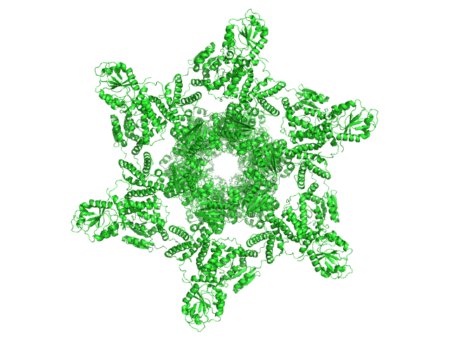
Search

 
Advanced search  

217 hits found for sans

SASDS22 – Sulfite reductase flavoprotein crosslinked octamer

Sulfite reductase [NADPH] flavoprotein alpha-component experimental SAS data
SASREF model
Sample: Sulfite reductase [NADPH] flavoprotein alpha-component octamer, 530 kDa Escherichia coli (strain … protein
Buffer: 50 mM potassium phosphate, 100 mM NaCl, 1 mM EDTA, pH: 7.8
Experiment: SANS data collected at ...SANS, Spallation Neutron Source on 2022 Jun 24
Domain crossover in the reductase subunit of NADPH-dependent assimilatory sulfite reductase. J Struct Biol 215(4):108028 (2023)
Walia N, Murray DT, Garg Y, He H, Weiss KL, Nagy G, Elizabeth Stroupe M
RgGuinier 8.4 nm
Dmax 23.0 nm
VolumePorod 767 nm3

SASDS32 – Reconstituted sulfite reductase crosslinked dodecamer with partially deuterated hemoprotein in 86% D2O

Sulfite reductase [NADPH] hemoprotein beta-componentSulfite reductase [NADPH] flavoprotein alpha-component (E121C, C162T, C552S, N556C) experimental SAS data
Sample: Sulfite reductase [NADPH] hemoprotein beta-component tetramer, 256 kDa Escherichia coli (strain … protein
Sulfite reductase [NADPH] flavoprotein alpha-component (E121C, C162T, C552S, N556C) octamer, 566 kDa Escherichia coli (strain … protein
Buffer: 50 mM potassium phosphate, 100 mM NaCl, 1 mM EDTA, pH: 7.8
Experiment: SANS data collected at ...SANS, Spallation Neutron Source on 2023 Apr 24
Domain crossover in the reductase subunit of NADPH-dependent assimilatory sulfite reductase. J Struct Biol 215(4):108028 (2023)
Walia N, Murray DT, Garg Y, He H, Weiss KL, Nagy G, Elizabeth Stroupe M
RgGuinier 7.5 nm
Dmax 22.6 nm
VolumePorod 823 nm3

SASDS42 – Reconstituted sulfite reductase crosslinked dodecamer with partially deuterated hemoprotein in 41% D2O

Sulfite reductase [NADPH] hemoprotein beta-componentSulfite reductase [NADPH] flavoprotein alpha-component (E121C, C162T, C552S, N556C) experimental SAS data
Sample: Sulfite reductase [NADPH] hemoprotein beta-component tetramer, 256 kDa Escherichia coli (strain … protein
Sulfite reductase [NADPH] flavoprotein alpha-component (E121C, C162T, C552S, N556C) octamer, 566 kDa Escherichia coli (strain … protein
Buffer: 50 mM potassium phosphate, 100 mM NaCl, 1 mM EDTA, pH: 7.8
Experiment: SANS data collected at ...SANS, Spallation Neutron Source on 2022 Aug 24
Domain crossover in the reductase subunit of NADPH-dependent assimilatory sulfite reductase. J Struct Biol 215(4):108028 (2023)
Walia N, Murray DT, Garg Y, He H, Weiss KL, Nagy G, Elizabeth Stroupe M
RgGuinier 11.1 nm
Dmax 32.4 nm
VolumePorod 138 nm3

SASDN52SANS data from cytochrome c' from Alcaligenes xylosoxidans at pD = 1.7

Cytochrome c' experimental SAS data
Cytochrome c' Kratky plot
Sample: Cytochrome c' monomer, 14 kDa Achromobacter xylosoxidans protein
Buffer: Phosphate Buffer pD 1.7, pH: 1.7
Experiment: SANS data collected at KWS1, FRM2 on 2017 Aug 12
Open-Bundle Structure as the Unfolding Intermediate of Cytochrome c′ Revealed by Small Angle Neutron Scattering Biomolecules 12(1):95 (2022)
Yamaguchi T, Akao K, Koutsioubas A, Frielinghaus H, Kohzuma T
RgGuinier 2.3 nm
Dmax 8.6 nm
VolumePorod 13 nm3

SASDS52 – Sulfite reductase crosslinked dodecamer

Sulfite reductase [NADPH] hemoprotein beta-componentSulfite reductase [NADPH] flavoprotein alpha-component (E121C, C162T, C552S, N556C) experimental SAS data
Sample: Sulfite reductase [NADPH] hemoprotein beta-component tetramer, 256 kDa Escherichia coli (strain … protein
Sulfite reductase [NADPH] flavoprotein alpha-component (E121C, C162T, C552S, N556C) octamer, 566 kDa Escherichia coli (strain … protein
Buffer: 50 mM potassium phosphate, 100 mM NaCl, 1 mM EDTA, pH: 7.8
Experiment: SANS data collected at ...SANS, Spallation Neutron Source on 2022 Jul 15
Domain crossover in the reductase subunit of NADPH-dependent assimilatory sulfite reductase. J Struct Biol 215(4):108028 (2023)
Walia N, Murray DT, Garg Y, He H, Weiss KL, Nagy G, Elizabeth Stroupe M
RgGuinier 10.3 nm
Dmax 31.0 nm
VolumePorod 1170 nm3

SASDN62SANS data from cytochrome c' from Alcaligenes xylosoxidans at pD = 6.4

Cytochrome c' experimental SAS data
Cytochrome c' Kratky plot
Sample: Cytochrome c' dimer, 27 kDa Alcaligenes protein
Buffer: Phosphate Buffer pD 6.4, pH: 6.4
Experiment: SANS data collected at KWS1, FRM2 on 2017 Aug 12
Open-Bundle Structure as the Unfolding Intermediate of Cytochrome c′ Revealed by Small Angle Neutron Scattering Biomolecules 12(1):95 (2022)
Yamaguchi T, Akao K, Koutsioubas A, Frielinghaus H, Kohzuma T
RgGuinier 1.8 nm
Dmax 5.5 nm
VolumePorod 11 nm3

SASDS62 – Sulfite reductase flavoprotein truncated linker (ΔAAPSQS) octamer

Sulfite reductase [NADPH] flavoprotein alpha-component (Δ212-217) experimental SAS data
Sulfite reductase [NADPH] flavoprotein alpha-component (Δ212-217) Kratky plot
Sample: Sulfite reductase [NADPH] flavoprotein alpha-component (Δ212-217) octamer, 526 kDa Escherichia coli (strain … protein
Buffer: 50 mM potassium phosphate, 100 mM NaCl, 1 mM EDTA, pH: 7.8
Experiment: SANS data collected at ...SANS, Spallation Neutron Source on 2022 Jun 25
Domain crossover in the reductase subunit of NADPH-dependent assimilatory sulfite reductase. J Struct Biol 215(4):108028 (2023)
Walia N, Murray DT, Garg Y, He H, Weiss KL, Nagy G, Elizabeth Stroupe M
RgGuinier 13.7 nm
Dmax 25.5 nm
VolumePorod 961 nm3

SASDN72SANS data from cytochrome c' from Alcaligenes xylosoxidans at pD = 9.6

Cytochrome c' experimental SAS data
Cytochrome c' Kratky plot
Sample: Cytochrome c' dimer, 27 kDa Alcaligenes protein
Buffer: Phosphate Buffer pD 9.6, pH: 9.6
Experiment: SANS data collected at KWS1, FRM2 on 2017 Aug 12
Open-Bundle Structure as the Unfolding Intermediate of Cytochrome c′ Revealed by Small Angle Neutron Scattering Biomolecules 12(1):95 (2022)
Yamaguchi T, Akao K, Koutsioubas A, Frielinghaus H, Kohzuma T
RgGuinier 1.9 nm
Dmax 5.3 nm
VolumePorod 10 nm3

SASDS72 – Sulfite reductase truncated linker (ΔAAPSQS) dodecamer

Sulfite reductase [NADPH] hemoprotein beta-componentSulfite reductase [NADPH] flavoprotein alpha-component (Δ212-217) experimental SAS data
Sulfite reductase [NADPH] hemoprotein beta-component Sulfite reductase [NADPH] flavoprotein alpha-component (Δ212-217) Kratky plot
Sample: Sulfite reductase [NADPH] hemoprotein beta-component tetramer, 256 kDa Escherichia coli (strain … protein
Sulfite reductase [NADPH] flavoprotein alpha-component (Δ212-217) octamer, 528 kDa Escherichia coli (strain … protein
Buffer: 50 mM potassium phosphate, 100 mM NaCl, 1 mM EDTA, pH: 7.8
Experiment: SANS data collected at ...SANS, Spallation Neutron Source on 2022 Jun 26
Domain crossover in the reductase subunit of NADPH-dependent assimilatory sulfite reductase. J Struct Biol 215(4):108028 (2023)
Walia N, Murray DT, Garg Y, He H, Weiss KL, Nagy G, Elizabeth Stroupe M
RgGuinier 10.8 nm
Dmax 30.3 nm
VolumePorod 1260 nm3

SASDN82SANS data from cytochrome c' from Alcaligenes xylosoxidans at pD = 13

Cytochrome c' experimental SAS data
Cytochrome c' Kratky plot
Sample: Cytochrome c' monomer, 14 kDa Achromobacter xylosoxidans protein
Buffer: Phosphate Buffer pD 13, pH: 13
Experiment: SANS data collected at KWS1, FRM2 on 2017 Aug 12
Open-Bundle Structure as the Unfolding Intermediate of Cytochrome c′ Revealed by Small Angle Neutron Scattering Biomolecules 12(1):95 (2022)
Yamaguchi T, Akao K, Koutsioubas A, Frielinghaus H, Kohzuma T
RgGuinier 4.8 nm
Dmax 9.0 nm
VolumePorod 20 nm3

SASDS82 – Reconstituted sulfite reductase crosslinked dodecamer with partially deuterated hemoprotein in 0% D2O

Sulfite reductase [NADPH] flavoprotein alpha-component (E121C, C162T, C552S, N556C)Sulfite reductase [NADPH] hemoprotein beta-component experimental SAS data
Sulfite reductase [NADPH] flavoprotein alpha-component (E121C, C162T, C552S, N556C) Sulfite reductase [NADPH] hemoprotein beta-component Kratky plot
Sample: Sulfite reductase [NADPH] flavoprotein alpha-component (E121C, C162T, C552S, N556C) octamer, 566 kDa Escherichia coli (strain … protein
Sulfite reductase [NADPH] hemoprotein beta-component tetramer, 256 kDa Escherichia coli (strain … protein
Buffer: 50 mM potassium phosphate, 100 mM NaCl, 1 mM EDTA, pH: 7.8
Experiment: SANS data collected at ...SANS, Spallation Neutron Source on 2022 Jul 2
Domain crossover in the reductase subunit of NADPH-dependent assimilatory sulfite reductase. J Struct Biol 215(4):108028 (2023)
Walia N, Murray DT, Garg Y, He H, Weiss KL, Nagy G, Elizabeth Stroupe M
RgGuinier 9.5 nm
Dmax 28.3 nm
VolumePorod 1010 nm3

SASDS92 – Reconstituted sulfite reductase crosslinked dodecamer with partially deuterated hemoprotein in 100% D2O

Sulfite reductase [NADPH] hemoprotein beta-componentSulfite reductase [NADPH] flavoprotein alpha-component (E121C, C162T, C552S, N556C) experimental SAS data
Sulfite reductase [NADPH] hemoprotein beta-component Sulfite reductase [NADPH] flavoprotein alpha-component (E121C, C162T, C552S, N556C) Kratky plot
Sample: Sulfite reductase [NADPH] hemoprotein beta-component tetramer, 256 kDa Escherichia coli (strain … protein
Sulfite reductase [NADPH] flavoprotein alpha-component (E121C, C162T, C552S, N556C) octamer, 566 kDa Escherichia coli (strain … protein
Buffer: 50 mM potassium phosphate, 100 mM NaCl, 1 mM EDTA, pH: 7.8
Experiment: SANS data collected at ...SANS, Spallation Neutron Source on 2022 Jun 27
Domain crossover in the reductase subunit of NADPH-dependent assimilatory sulfite reductase. J Struct Biol 215(4):108028 (2023)
Walia N, Murray DT, Garg Y, He H, Weiss KL, Nagy G, Elizabeth Stroupe M
RgGuinier 8.2 nm
Dmax 25.1 nm
VolumePorod 839 nm3

SASDSA2 – Sulfite reductase flavoprotein reduced with 10 molar equivalents sodium dithionite

Sulfite reductase [NADPH] flavoprotein alpha-component experimental SAS data
Sulfite reductase [NADPH] flavoprotein alpha-component Kratky plot
Sample: Sulfite reductase [NADPH] flavoprotein alpha-component octamer, 530 kDa Escherichia coli (strain … protein
Buffer: 50 mM potassium phosphate, 100 mM NaCl, 1 mM EDTA, pH: 7.8
Experiment: SANS data collected at ...SANS, Spallation Neutron Source on 2022 Jul 15
Domain crossover in the reductase subunit of NADPH-dependent assimilatory sulfite reductase. J Struct Biol 215(4):108028 (2023)
Walia N, Murray DT, Garg Y, He H, Weiss KL, Nagy G, Elizabeth Stroupe M
RgGuinier 7.9 nm
Dmax 27.6 nm
VolumePorod 1060 nm3

SASDSB2 – Sulfite reductase flavoprotein crosslinked reduced with 10 molar equivalents sodium dithionite

Sulfite reductase [NADPH] flavoprotein alpha-component (E121C, C162T, C552S, N556C) experimental SAS data
Sulfite reductase [NADPH] flavoprotein alpha-component (E121C, C162T, C552S, N556C) Kratky plot
Sample: Sulfite reductase [NADPH] flavoprotein alpha-component (E121C, C162T, C552S, N556C) octamer, 566 kDa Escherichia coli (strain … protein
Buffer: 50 mM potassium phosphate, 100 mM NaCl, 1 mM EDTA, pH: 7.8
Experiment: SANS data collected at ...SANS, Spallation Neutron Source on 2022 Jul 15
Domain crossover in the reductase subunit of NADPH-dependent assimilatory sulfite reductase. J Struct Biol 215(4):108028 (2023)
Walia N, Murray DT, Garg Y, He H, Weiss KL, Nagy G, Elizabeth Stroupe M
RgGuinier 8.0 nm
Dmax 25.3 nm
VolumePorod 827 nm3

SASDSC2 – Sulfite reductase truncated linker (ΔAAPSQS) flavoprotein reduced with 10 molar equivalents sodium dithionite

Sulfite reductase [NADPH] flavoprotein alpha-component (Δ212-217) experimental SAS data
Sulfite reductase [NADPH] flavoprotein alpha-component (Δ212-217) Kratky plot
Sample: Sulfite reductase [NADPH] flavoprotein alpha-component (Δ212-217) octamer, 526 kDa Escherichia coli (strain … protein
Buffer: 50 mM potassium phosphate, 100 mM NaCl, 1 mM EDTA, pH: 7.8
Experiment: SANS data collected at ...SANS, Spallation Neutron Source on 2022 Jul 15
Domain crossover in the reductase subunit of NADPH-dependent assimilatory sulfite reductase. J Struct Biol 215(4):108028 (2023)
Walia N, Murray DT, Garg Y, He H, Weiss KL, Nagy G, Elizabeth Stroupe M
RgGuinier 7.6 nm
Dmax 25.0 nm
VolumePorod 952 nm3

SASDNK2 – ...SANS in 100% D2O

75% deuterated Circadian clock protein KaiBCircadian clock protein KaiA75% deuterated Circadian clock protein kinase KaiC (S431D mutant) experimental SAS data
OTHER model
Sample: 75% deuterated Circadian clock protein KaiB hexamer, 71 kDa Synechococcus elongatus (strain … protein
Circadian clock protein KaiA dodecamer, 393 kDa Synechococcus elongatus (strain … protein
75% deuterated Circadian clock protein kinase KaiC (S431D mutant) hexamer, 357 kDa Synechococcus elongatus (strain … protein
Buffer: 50 mM sodium phosphate buffer, 150 mm NaCl, 5 mM MgCl2, 0.5 mM EDTA, 1 mM ATP, 1 mM DTT, 50 mM arginine, 50 mM glutamine, in 100% D2O, pH: 7.8
Experiment: SANS data collected at D22, Institut Laue-Langevin (ILL) on 2018 Sep 19
Overall structure of fully assembled cyanobacterial KaiABC circadian clock complex by an integrated experimental-computational approach. Commun Biol 5(1):184 (2022)
Yunoki Y, Matsumoto A, Morishima K, Martel A, Porcar L, Sato N, Yogo R, Tominaga T, Inoue R, Yagi-Utsumi M, Okuda A, Shimizu M, Urade R, Terauchi K, Kono H, Yagi H, Kato K, Sugiyama M
RgGuinier 7.8 nm
Dmax 25.6 nm
VolumePorod 1620 nm3

SASDMU2 – Hydrolytically Degradable Polymer Micelles for Drug Delivery (structure of hydrophobic substituent - 5α-cholestan-3-one) without Dox

Hydrolytically Degradable Polymer Micelles for Drug Delivery (structure of hydrophobic substituent - 5α-cholestan-3-one) without Dox experimental SAS data
DAMMIF model
Sample: Hydrolytically Degradable Polymer Micelles for Drug Delivery (structure of hydrophobic substituent - 5α-cholestan-3-one) without Dox monomer, 125 kDa
Buffer: phosphate buffer saline (PBS) (pH 5.0), pH: 5
Experiment: SAXS data collected at EMBL X33, DORIS III, DESY on 2012 Aug 19
...SANS kinetic study. Biomacromolecules 14(11):4061-70 (2013)
Filippov SK, Franklin JM, Konarev PV, Chytil P, Etrych T, Bogomolova A, Dyakonova M, Papadakis CM, Radulescu A, Ulbrich K, Stepanek P, Svergun DI
RgGuinier 6.1 nm
Dmax 20.5 nm

SASDGV2 – ...SANS data in 42% v/v D2O)

Fibrillarin-like rRNA/tRNA 2'-O-methyltransferase50S ribosomal protein L7AeNOP5/NOP56 related proteinpyrococcus furiosus sR26 stabilized constructPyrococcus furiosus sR26 substrate D' experimental SAS data
Fibrillarin-like rRNA/tRNA 2'-O-methyltransferase 50S ribosomal protein L7Ae NOP5/NOP56 related protein pyrococcus furiosus sR26 stabilized construct Pyrococcus furiosus sR26 substrate D' Kratky plot
Sample: Fibrillarin-like rRNA/tRNA 2'-O-methyltransferase dimer, 52 kDa Pyrococcus furiosus protein
50S ribosomal protein L7Ae dimer, 27 kDa Pyrococcus furiosus protein
NOP5/NOP56 related protein dimer, 94 kDa Pyrococcus furiosus protein
pyrococcus furiosus sR26 stabilized construct monomer, 24 kDa Pyrococcus furiosus RNA
Pyrococcus furiosus sR26 substrate D' monomer, 4 kDa Pyrococcus furiosus RNA
Buffer: 50 mM phosphate 500 mM NaCl 42%D2O, pH: 6.6
Experiment: SANS data collected at KWS1, FRM2 on 2015 May 24
The guide sRNA sequence determines the activity level of box C/D RNPs. Elife 9 (2020)
Graziadei A, Gabel F, Kirkpatrick J, Carlomagno T
RgGuinier 4.9 nm
Dmax 16.0 nm
VolumePorod 87 nm3

SASDMV2 – Hydrolytically Degradable Polymer Micelles for Drug Delivery (structure of hydrophobic substituent - 5α-cholestan-3-one) with Dox

Hydrolytically Degradable Polymer Micelles for Drug Delivery (structure of hydrophobic substituent - 5α-cholestan-3-one) with Dox (10%) experimental SAS data
DAMMIF model
Sample: Hydrolytically Degradable Polymer Micelles for Drug Delivery (structure of hydrophobic substituent - 5α-cholestan-3-one) with Dox (10%) monomer, 220 kDa
Buffer: phosphate buffer saline (PBS) (pH 5.0), pH: 5
Experiment: SAXS data collected at EMBL X33, DORIS III, DESY on 2012 Aug 19
...SANS kinetic study. Biomacromolecules 14(11):4061-70 (2013)
Filippov SK, Franklin JM, Konarev PV, Chytil P, Etrych T, Bogomolova A, Dyakonova M, Papadakis CM, Radulescu A, Ulbrich K, Stepanek P, Svergun DI
RgGuinier 7.5 nm
Dmax 25.5 nm

SASDGW2 – ...SANS data in 42% v/v D2O)

Fibrillarin-like rRNA/tRNA 2'-O-methyltransferase50S ribosomal protein L7AeNOP5/NOP56 related proteinpyrococcus furiosus sR26 stabilized constructPyrococcus furiosus sR26 substrate D' experimental SAS data
Fibrillarin-like rRNA/tRNA 2'-O-methyltransferase 50S ribosomal protein L7Ae NOP5/NOP56 related protein pyrococcus furiosus sR26 stabilized construct Pyrococcus furiosus sR26 substrate D' Kratky plot
Sample: Fibrillarin-like rRNA/tRNA 2'-O-methyltransferase dimer, 52 kDa Pyrococcus furiosus protein
50S ribosomal protein L7Ae dimer, 27 kDa Pyrococcus furiosus protein
NOP5/NOP56 related protein dimer, 94 kDa Pyrococcus furiosus protein
pyrococcus furiosus sR26 stabilized construct monomer, 24 kDa Pyrococcus furiosus RNA
Pyrococcus furiosus sR26 substrate D' monomer, 4 kDa Pyrococcus furiosus RNA
Buffer: 50 mM phosphate 500 mM NaCl 42%D2O, pH: 6.6
Experiment: SANS data collected at D22, Institut Laue-Langevin (ILL) on 2015 Sep 21
The guide sRNA sequence determines the activity level of box C/D RNPs. Elife 9 (2020)
Graziadei A, Gabel F, Kirkpatrick J, Carlomagno T
RgGuinier 4.1 nm
Dmax 14.0 nm
VolumePorod 210 nm3

SASDMW2 – Hydrolytically Degradable Polymer Micelles for Drug Delivery (structure of hydrophobic substituent - Opb-Chol) without Dox

Hydrolytically Degradable Polymer Micelles for Drug Delivery (structure of hydrophobic substituent - Opb-Chol) without Dox experimental SAS data
DAMMIF model
Sample: Hydrolytically Degradable Polymer Micelles for Drug Delivery (structure of hydrophobic substituent - Opb-Chol) without Dox monomer, 125 kDa
Buffer: phosphate buffer saline (PBS) (pH 5.0), pH: 5
Experiment: SAXS data collected at EMBL X33, DORIS III, DESY on 2012 Aug 19
...SANS kinetic study. Biomacromolecules 14(11):4061-70 (2013)
Filippov SK, Franklin JM, Konarev PV, Chytil P, Etrych T, Bogomolova A, Dyakonova M, Papadakis CM, Radulescu A, Ulbrich K, Stepanek P, Svergun DI
RgGuinier 6.4 nm
Dmax 22.5 nm

SASDGX2 – ...SANS data in 42% v/v D2O)

Fibrillarin-like rRNA/tRNA 2'-O-methyltransferase50S ribosomal protein L7AeNOP5/NOP56 related proteinpyrococcus furiosus sR26 stabilized constructPyrococcus furiosus sR26 substrate D' experimental SAS data
Fibrillarin-like rRNA/tRNA 2'-O-methyltransferase 50S ribosomal protein L7Ae NOP5/NOP56 related protein pyrococcus furiosus sR26 stabilized construct Pyrococcus furiosus sR26 substrate D' Kratky plot
Sample: Fibrillarin-like rRNA/tRNA 2'-O-methyltransferase dimer, 52 kDa Pyrococcus furiosus protein
50S ribosomal protein L7Ae dimer, 27 kDa Pyrococcus furiosus protein
NOP5/NOP56 related protein dimer, 94 kDa Pyrococcus furiosus protein
pyrococcus furiosus sR26 stabilized construct monomer, 24 kDa Pyrococcus furiosus RNA
Pyrococcus furiosus sR26 substrate D' monomer, 4 kDa Pyrococcus furiosus RNA
Buffer: 50 mM phosphate 500 mM NaCl 42%D2O, pH: 6.6
Experiment: SANS data collected at D22, Institut Laue-Langevin (ILL) on 2015 Nov 23
The guide sRNA sequence determines the activity level of box C/D RNPs. Elife 9 (2020)
Graziadei A, Gabel F, Kirkpatrick J, Carlomagno T
RgGuinier 4.0 nm
Dmax 13.3 nm
VolumePorod 42 nm3

SASDMX2 – Hydrolytically Degradable Polymer Micelles for Drug Delivery (structure of hydrophobic substituent - Opb-Chol) with Dox (10%)

Hydrolytically Degradable Polymer Micelles for Drug Delivery (structure of hydrophobic substituent - Opb-Chol) with Dox (10%) experimental SAS data
DAMMIF model
Sample: Hydrolytically Degradable Polymer Micelles for Drug Delivery (structure of hydrophobic substituent - Opb-Chol) with Dox (10%) monomer, 225 kDa
Buffer: phosphate buffer saline (PBS) (pH 5.0), pH: 5
Experiment: SAXS data collected at EMBL X33, DORIS III, DESY on 2012 Aug 19
...SANS kinetic study. Biomacromolecules 14(11):4061-70 (2013)
Filippov SK, Franklin JM, Konarev PV, Chytil P, Etrych T, Bogomolova A, Dyakonova M, Papadakis CM, Radulescu A, Ulbrich K, Stepanek P, Svergun DI
RgGuinier 10.0 nm
Dmax 21.9 nm

SASDGY2 – ...SANS data in 42% v/v D2O)

Fibrillarin-like rRNA/tRNA 2'-O-methyltransferase50S ribosomal protein L7AeNOP5/NOP56 related proteinpyrococcus furiosus sR26 stabilized constructPyrococcus furiosus sR26 substrate D' experimental SAS data
Fibrillarin-like rRNA/tRNA 2'-O-methyltransferase 50S ribosomal protein L7Ae NOP5/NOP56 related protein pyrococcus furiosus sR26 stabilized construct Pyrococcus furiosus sR26 substrate D' Kratky plot
Sample: Fibrillarin-like rRNA/tRNA 2'-O-methyltransferase dimer, 52 kDa Pyrococcus furiosus protein
50S ribosomal protein L7Ae dimer, 27 kDa Pyrococcus furiosus protein
NOP5/NOP56 related protein dimer, 94 kDa Pyrococcus furiosus protein
pyrococcus furiosus sR26 stabilized construct monomer, 24 kDa Pyrococcus furiosus RNA
Pyrococcus furiosus sR26 substrate D' monomer, 4 kDa Pyrococcus furiosus RNA
Buffer: 50 mM phosphate 500 mM NaCl 42%D2O, pH: 6.6
Experiment: SANS data collected at D22, Institut Laue-Langevin (ILL) on 2019 Jul 21
The guide sRNA sequence determines the activity level of box C/D RNPs. Elife 9 (2020)
Graziadei A, Gabel F, Kirkpatrick J, Carlomagno T
RgGuinier 2.8 nm
Dmax 9.9 nm
VolumePorod 52 nm3

SASDMY2 – Hydrolytically Degradable Polymer Micelles for Drug Delivery (structure of hydrophobic substituent - Lev-Chol) without Dox

Hydrolytically Degradable Polymer Micelles for Drug Delivery (structure of hydrophobic substituent - Lev-Chol) without Dox experimental SAS data
DAMMIF model
Sample: Hydrolytically Degradable Polymer Micelles for Drug Delivery (structure of hydrophobic substituent - Lev-Chol) without Dox monomer, 125 kDa
Buffer: phosphate buffer saline (PBS) (pH 5.0), pH: 5
Experiment: SAXS data collected at EMBL X33, DORIS III, DESY on 2012 Aug 19
...SANS kinetic study. Biomacromolecules 14(11):4061-70 (2013)
Filippov SK, Franklin JM, Konarev PV, Chytil P, Etrych T, Bogomolova A, Dyakonova M, Papadakis CM, Radulescu A, Ulbrich K, Stepanek P, Svergun DI
RgGuinier 6.5 nm
Dmax 22.9 nm

SASDGZ2 – ...SANS data in 42% v/v D2O)

Fibrillarin-like rRNA/tRNA 2'-O-methyltransferase50S ribosomal protein L7AeNOP5/NOP56 related proteinpyrococcus furiosus sR26 stabilized constructPyrococcus furiosus sR26 substrate D' experimental SAS data
Fibrillarin-like rRNA/tRNA 2'-O-methyltransferase 50S ribosomal protein L7Ae NOP5/NOP56 related protein pyrococcus furiosus sR26 stabilized construct Pyrococcus furiosus sR26 substrate D' Kratky plot
Sample: Fibrillarin-like rRNA/tRNA 2'-O-methyltransferase dimer, 52 kDa Pyrococcus furiosus protein
50S ribosomal protein L7Ae dimer, 27 kDa Pyrococcus furiosus protein
NOP5/NOP56 related protein dimer, 94 kDa Pyrococcus furiosus protein
pyrococcus furiosus sR26 stabilized construct monomer, 24 kDa Pyrococcus furiosus RNA
Pyrococcus furiosus sR26 substrate D' monomer, 4 kDa Pyrococcus furiosus RNA
Buffer: 50 mM phosphate 500 mM NaCl 42%D2O, pH: 6.6
Experiment: SANS data collected at KWS1, FRM2 on 2015 May 23
The guide sRNA sequence determines the activity level of box C/D RNPs. Elife 9 (2020)
Graziadei A, Gabel F, Kirkpatrick J, Carlomagno T
RgGuinier 4.8 nm
Dmax 16.6 nm
VolumePorod 82 nm3

SASDMZ2 – Hydrolytically Degradable Polymer Micelles for Drug Delivery (structure of hydrophobic substituent - Lev-Chol) with Dox (10%)

Hydrolytically Degradable Polymer Micelles for Drug Delivery (structure of hydrophobic substituent - Lev-Chol) with Dox (10%) experimental SAS data
DAMMIF model
Sample: Hydrolytically Degradable Polymer Micelles for Drug Delivery (structure of hydrophobic substituent - Lev-Chol) with Dox (10%) monomer, 125 kDa
Buffer: phosphate buffer saline (PBS) (pH 5.0), pH: 5
Experiment: SAXS data collected at EMBL X33, DORIS III, DESY on 2012 Aug 19
...SANS kinetic study. Biomacromolecules 14(11):4061-70 (2013)
Filippov SK, Franklin JM, Konarev PV, Chytil P, Etrych T, Bogomolova A, Dyakonova M, Papadakis CM, Radulescu A, Ulbrich K, Stepanek P, Svergun DI
RgGuinier 6.9 nm
Dmax 23.8 nm

SASDG23 – ...SANS data in 42% v/v D2O)

Fibrillarin-like rRNA/tRNA 2'-O-methyltransferase50S ribosomal protein L7AeNOP5/NOP56 related proteinpyrococcus furiosus sR26 stabilized constructPyrococcus furiosus sR26 substrate D' experimental SAS data
Fibrillarin-like rRNA/tRNA 2'-O-methyltransferase 50S ribosomal protein L7Ae NOP5/NOP56 related protein pyrococcus furiosus sR26 stabilized construct Pyrococcus furiosus sR26 substrate D' Kratky plot
Sample: Fibrillarin-like rRNA/tRNA 2'-O-methyltransferase dimer, 52 kDa Pyrococcus furiosus protein
50S ribosomal protein L7Ae dimer, 27 kDa Pyrococcus furiosus protein
NOP5/NOP56 related protein dimer, 94 kDa Pyrococcus furiosus protein
pyrococcus furiosus sR26 stabilized construct monomer, 24 kDa Pyrococcus furiosus RNA
Pyrococcus furiosus sR26 substrate D' monomer, 4 kDa Pyrococcus furiosus RNA
Buffer: 50 mM phosphate 500 mM NaCl 42%D2O, pH: 6.6
Experiment: SANS data collected at D22, Institut Laue-Langevin (ILL) on 2018 Apr 8
The guide sRNA sequence determines the activity level of box C/D RNPs. Elife 9 (2020)
Graziadei A, Gabel F, Kirkpatrick J, Carlomagno T
RgGuinier 4.0 nm
Dmax 13.8 nm
VolumePorod 214 nm3

SASDM23 – Hydrolytically Degradable Polymer Micelles for Drug Delivery (structure of hydrophobic substituent - cholest-4-en-3-one) without Dox

Hydrolytically Degradable Polymer Micelles for Drug Delivery (structure of hydrophobic substituent - cholest-4-en-3-one) without Dox experimental SAS data
DAMMIF model
Sample: Hydrolytically Degradable Polymer Micelles for Drug Delivery (structure of hydrophobic substituent - cholest-4-en-3-one) without Dox monomer, 125 kDa
Buffer: phosphate buffer saline (PBS) (pH 5.0), pH: 5
Experiment: SAXS data collected at EMBL X33, DORIS III, DESY on 2012 Aug 16
...SANS kinetic study. Biomacromolecules 14(11):4061-70 (2013)
Filippov SK, Franklin JM, Konarev PV, Chytil P, Etrych T, Bogomolova A, Dyakonova M, Papadakis CM, Radulescu A, Ulbrich K, Stepanek P, Svergun DI
RgGuinier 5.9 nm
Dmax 19.7 nm

SASDG33 – ...SANS data in 42% v/v D2O)

Fibrillarin-like rRNA/tRNA 2'-O-methyltransferase50S ribosomal protein L7AeNOP5/NOP56 related proteinpyrococcus furiosus sR26 stabilized constructPyrococcus furiosus sR26 substrate D experimental SAS data
Fibrillarin-like rRNA/tRNA 2'-O-methyltransferase 50S ribosomal protein L7Ae NOP5/NOP56 related protein pyrococcus furiosus sR26 stabilized construct Pyrococcus furiosus sR26 substrate D Kratky plot
Sample: Fibrillarin-like rRNA/tRNA 2'-O-methyltransferase dimer, 52 kDa Pyrococcus furiosus protein
50S ribosomal protein L7Ae dimer, 27 kDa Pyrococcus furiosus protein
NOP5/NOP56 related protein dimer, 94 kDa Pyrococcus furiosus protein
pyrococcus furiosus sR26 stabilized construct monomer, 24 kDa Pyrococcus furiosus RNA
Pyrococcus furiosus sR26 substrate D monomer, 4 kDa Pyrococcus furiosus RNA
Buffer: 50 mM phosphate 500 mM NaCl 42%D2O, pH: 6.6
Experiment: SANS data collected at D22, Institut Laue-Langevin (ILL) on 2018 Apr 8
The guide sRNA sequence determines the activity level of box C/D RNPs. Elife 9 (2020)
Graziadei A, Gabel F, Kirkpatrick J, Carlomagno T
RgGuinier 4.0 nm
Dmax 13.9 nm
VolumePorod 231 nm3

SASDL33 – ...SANS

Proton-gated ion channel experimental SAS data
GROMACS model
Sample: Proton-gated ion channel pentamer, 183 kDa Gloeobacter violaceus (strain … protein
Buffer: D2O, 20 mM Tris, 150 mM NaCl, 0.5 mM matched-out deuterated DDM,, pH: 7.5
Experiment: SANS data collected at D22, Institut Laue-Langevin (ILL) on 2020 Aug 22
Probing solution structure of the pentameric ligand-gated ion channel GLIC by small-angle neutron scattering Proceedings of the National Academy of Sciences 118(37):e2108006118 (2021)
Lycksell M, Rovšnik U, Bergh C, Johansen N, Martel A, Porcar L, Arleth L, Howard R, Lindahl E
RgGuinier 3.8 nm
Dmax 13.5 nm
VolumePorod 274 nm3

SASDM33 – Hydrolytically Degradable Polymer Micelles for Drug Delivery (structure of hydrophobic substituent - cholest-4-en-3-one) with Dox (10%)

Hydrolytically Degradable Polymer Micelles for Drug Delivery (structure of hydrophobic substituent - cholest-4-en-3-one) with Dox (10%) experimental SAS data
DAMMIF model
Sample: Hydrolytically Degradable Polymer Micelles for Drug Delivery (structure of hydrophobic substituent - cholest-4-en-3-one) with Dox (10%) monomer, 125 kDa
Buffer: phosphate buffer saline (PBS) (pH 5.0), pH: 5
Experiment: SAXS data collected at EMBL X33, DORIS III, DESY on 2012 Aug 19
...SANS kinetic study. Biomacromolecules 14(11):4061-70 (2013)
Filippov SK, Franklin JM, Konarev PV, Chytil P, Etrych T, Bogomolova A, Dyakonova M, Papadakis CM, Radulescu A, Ulbrich K, Stepanek P, Svergun DI
RgGuinier 5.5 nm
Dmax 18.3 nm

SASDG43 – ...SANS data in 42% v/v D2O)

Fibrillarin-like rRNA/tRNA 2'-O-methyltransferase50S ribosomal protein L7AeNOP5/NOP56 related proteinpyrococcus furiosus sR26 stabilized constructPyrococcus furiosus sR26 substrate D experimental SAS data
Fibrillarin-like rRNA/tRNA 2'-O-methyltransferase 50S ribosomal protein L7Ae NOP5/NOP56 related protein pyrococcus furiosus sR26 stabilized construct Pyrococcus furiosus sR26 substrate D Kratky plot
Sample: Fibrillarin-like rRNA/tRNA 2'-O-methyltransferase dimer, 52 kDa Pyrococcus furiosus protein
50S ribosomal protein L7Ae dimer, 27 kDa Pyrococcus furiosus protein
NOP5/NOP56 related protein dimer, 94 kDa Pyrococcus furiosus protein
pyrococcus furiosus sR26 stabilized construct monomer, 24 kDa Pyrococcus furiosus RNA
Pyrococcus furiosus sR26 substrate D monomer, 4 kDa Pyrococcus furiosus RNA
Buffer: 50 mM phosphate 500 mM NaCl 42%D2O, pH: 6.6
Experiment: SANS data collected at KWS1, FRM2 on 2015 May 24
The guide sRNA sequence determines the activity level of box C/D RNPs. Elife 9 (2020)
Graziadei A, Gabel F, Kirkpatrick J, Carlomagno T
RgGuinier 4.9 nm
Dmax 16.6 nm
VolumePorod 65 nm3

SASDL43 – ...SANS

Proton-gated ion channel experimental SAS data
GROMACS model
Sample: Proton-gated ion channel pentamer, 183 kDa Gloeobacter violaceus (strain … protein
Buffer: D2O, 20 mM citrate, 150 mM NaCl, 0.5 mM match-out deuterated DDM, pH: 3
Experiment: SANS data collected at D22, Institut Laue-Langevin (ILL) on 2020 Aug 22
Probing solution structure of the pentameric ligand-gated ion channel GLIC by small-angle neutron scattering Proceedings of the National Academy of Sciences 118(37):e2108006118 (2021)
Lycksell M, Rovšnik U, Bergh C, Johansen N, Martel A, Porcar L, Arleth L, Howard R, Lindahl E
RgGuinier 3.8 nm
Dmax 12.7 nm
VolumePorod 279 nm3

SASDG53 – ...SANS data in 42% v/v D2O)

Fibrillarin-like rRNA/tRNA 2'-O-methyltransferase50S ribosomal protein L7AeNOP5/NOP56 related proteinpyrococcus furiosus sR26 stabilized constructPyrococcus furiosus sR26 substrate D experimental SAS data
Fibrillarin-like rRNA/tRNA 2'-O-methyltransferase 50S ribosomal protein L7Ae NOP5/NOP56 related protein pyrococcus furiosus sR26 stabilized construct Pyrococcus furiosus sR26 substrate D Kratky plot
Sample: Fibrillarin-like rRNA/tRNA 2'-O-methyltransferase dimer, 52 kDa Pyrococcus furiosus protein
50S ribosomal protein L7Ae dimer, 27 kDa Pyrococcus furiosus protein
NOP5/NOP56 related protein dimer, 94 kDa Pyrococcus furiosus protein
pyrococcus furiosus sR26 stabilized construct monomer, 24 kDa Pyrococcus furiosus RNA
Pyrococcus furiosus sR26 substrate D monomer, 4 kDa Pyrococcus furiosus RNA
Buffer: 50 mM phosphate 500 mM NaCl 42%D2O, pH: 6.6
Experiment: SANS data collected at D22, Institut Laue-Langevin (ILL) on 2015 Sep 21
The guide sRNA sequence determines the activity level of box C/D RNPs. Elife 9 (2020)
Graziadei A, Gabel F, Kirkpatrick J, Carlomagno T
RgGuinier 4.1 nm
Dmax 14.0 nm
VolumePorod 222 nm3

SASDL53 – ...SANS

Proton-gated ion channel experimental SAS data
Proton-gated ion channel Kratky plot
Sample: Proton-gated ion channel pentamer, 183 kDa Gloeobacter violaceus (strain … protein
Buffer: D2O, 20 mM Tris, 150 mM NaCl, 0.5 mM matched-out deuterated DDM,, pH: 7.5
Experiment: SANS data collected at D22, Institut Laue-Langevin (ILL) on 2019 Jun 20
Probing solution structure of the pentameric ligand-gated ion channel GLIC by small-angle neutron scattering Proceedings of the National Academy of Sciences 118(37):e2108006118 (2021)
Lycksell M, Rovšnik U, Bergh C, Johansen N, Martel A, Porcar L, Arleth L, Howard R, Lindahl E
RgGuinier 3.8 nm
Dmax 12.0 nm
VolumePorod 235 nm3

SASDG63 – ...SANS data in 42% v/v D2O)

Fibrillarin-like rRNA/tRNA 2'-O-methyltransferase50S ribosomal protein L7AeNOP5/NOP56 related proteinpyrococcus furiosus sR26 stabilized constructPyrococcus furiosus sR26 substrate D experimental SAS data
Fibrillarin-like rRNA/tRNA 2'-O-methyltransferase 50S ribosomal protein L7Ae NOP5/NOP56 related protein pyrococcus furiosus sR26 stabilized construct Pyrococcus furiosus sR26 substrate D Kratky plot
Sample: Fibrillarin-like rRNA/tRNA 2'-O-methyltransferase dimer, 52 kDa Pyrococcus furiosus protein
50S ribosomal protein L7Ae dimer, 27 kDa Pyrococcus furiosus protein
NOP5/NOP56 related protein dimer, 94 kDa Pyrococcus furiosus protein
pyrococcus furiosus sR26 stabilized construct monomer, 24 kDa Pyrococcus furiosus RNA
Pyrococcus furiosus sR26 substrate D monomer, 4 kDa Pyrococcus furiosus RNA
Buffer: 50 mM phosphate 500 mM NaCl 42%D2O, pH: 6.6
Experiment: SANS data collected at D22, Institut Laue-Langevin (ILL) on 2015 Nov 23
The guide sRNA sequence determines the activity level of box C/D RNPs. Elife 9 (2020)
Graziadei A, Gabel F, Kirkpatrick J, Carlomagno T
RgGuinier 3.9 nm
Dmax 12.5 nm
VolumePorod 49 nm3

SASDJ63 – Streptococcus pneumoniae NADPH oxidase - Tag-free SpNOX

FAD-binding FR-type domain-containing protein experimental SAS data
OTHER model
Sample: FAD-binding FR-type domain-containing protein monomer, 47 kDa Streptococcus pneumoniae protein
Buffer: 50 mM Tris-HCl pH 7, 300 mM NaCl, 5 mM LMNG, 10 µM FAD, 21.4% D₂O, pH: 7
Experiment: SANS data collected at D22, Institut Laue-Langevin (ILL) on 2018 Jun 26
...SANS Using LMNG Stealth Carrier. Biophys J 119(3):605-618 (2020)
Vermot A, Petit-Härtlein I, Breyton C, Le Roy A, Thépaut M, Vivès C, Moulin M, Härtlein M, Grudinin S, Smith SME, Ebel C, Martel A, Fieschi F
RgGuinier 3.0 nm
Dmax 10.0 nm
VolumePorod 89 nm3

SASDL63 – ...SANS

Proton-gated ion channel experimental SAS data
Proton-gated ion channel Kratky plot
Sample: Proton-gated ion channel pentamer, 183 kDa Gloeobacter violaceus (strain … protein
Buffer: D2O, 20 mM Tris, 150 mM NaCl, 0.5 mM matched-out deuterated DDM,, pH: 7.5
Experiment: SANS data collected at D22, Institut Laue-Langevin (ILL) on 2019 Jun 21
Probing solution structure of the pentameric ligand-gated ion channel GLIC by small-angle neutron scattering Proceedings of the National Academy of Sciences 118(37):e2108006118 (2021)
Lycksell M, Rovšnik U, Bergh C, Johansen N, Martel A, Porcar L, Arleth L, Howard R, Lindahl E
RgGuinier 4.0 nm
Dmax 17.7 nm
VolumePorod 225 nm3

SASDG73 – ...SANS data in 42% v/v D2O)

Fibrillarin-like rRNA/tRNA 2'-O-methyltransferase50S ribosomal protein L7AeNOP5/NOP56 related proteinpyrococcus furiosus sR26 stabilized constructPyrococcus furiosus sR26 substrate D experimental SAS data
Fibrillarin-like rRNA/tRNA 2'-O-methyltransferase 50S ribosomal protein L7Ae NOP5/NOP56 related protein pyrococcus furiosus sR26 stabilized construct Pyrococcus furiosus sR26 substrate D Kratky plot
Sample: Fibrillarin-like rRNA/tRNA 2'-O-methyltransferase dimer, 52 kDa Pyrococcus furiosus protein
50S ribosomal protein L7Ae dimer, 27 kDa Pyrococcus furiosus protein
NOP5/NOP56 related protein dimer, 94 kDa Pyrococcus furiosus protein
pyrococcus furiosus sR26 stabilized construct monomer, 24 kDa Pyrococcus furiosus RNA
Pyrococcus furiosus sR26 substrate D monomer, 4 kDa Pyrococcus furiosus RNA
Buffer: 50 mM phosphate 500 mM NaCl 42%D2O, pH: 6.6
Experiment: SANS data collected at D22, Institut Laue-Langevin (ILL) on 2019 Jul 21
The guide sRNA sequence determines the activity level of box C/D RNPs. Elife 9 (2020)
Graziadei A, Gabel F, Kirkpatrick J, Carlomagno T
RgGuinier 2.7 nm
Dmax 9.5 nm
VolumePorod 25 nm3

SASDG83 – ...SANS data in 42% v/v D2O)

Fibrillarin-like rRNA/tRNA 2'-O-methyltransferase50S ribosomal protein L7AeNOP5/NOP56 related proteinpyrococcus furiosus sR26 stabilized constructPyrococcus furiosus sR26 substrate D experimental SAS data
Fibrillarin-like rRNA/tRNA 2'-O-methyltransferase 50S ribosomal protein L7Ae NOP5/NOP56 related protein pyrococcus furiosus sR26 stabilized construct Pyrococcus furiosus sR26 substrate D Kratky plot
Sample: Fibrillarin-like rRNA/tRNA 2'-O-methyltransferase dimer, 52 kDa Pyrococcus furiosus protein
50S ribosomal protein L7Ae dimer, 27 kDa Pyrococcus furiosus protein
NOP5/NOP56 related protein dimer, 94 kDa Pyrococcus furiosus protein
pyrococcus furiosus sR26 stabilized construct monomer, 24 kDa Pyrococcus furiosus RNA
Pyrococcus furiosus sR26 substrate D monomer, 4 kDa Pyrococcus furiosus RNA
Buffer: 50 mM phosphate 500 mM NaCl 42%D2O, pH: 6.6
Experiment: SANS data collected at KWS1, FRM2 on 2015 May 23
The guide sRNA sequence determines the activity level of box C/D RNPs. Elife 9 (2020)
Graziadei A, Gabel F, Kirkpatrick J, Carlomagno T
RgGuinier 5.1 nm
Dmax 15.5 nm
VolumePorod 125 nm3

SASDRE3 – Tissue Transglutaminase + Ca: Time-resolved 0ms

Protein-glutamine gamma-glutamyltransferase 2 experimental SAS data
Protein-glutamine gamma-glutamyltransferase 2 Kratky plot
Sample: Protein-glutamine gamma-glutamyltransferase 2 , 77 kDa Homo sapiens protein
Buffer: 20 mM HEPES, 100 mM NaCl, 10% glycerol, 1 mM DTT, pH: 7.5
Experiment: SAXS data collected at ID7A1 BioSAXS / HP-Bio Beamline, Cornell High Energy Synchrotron Source (CHESS) on 2021 Nov 19
Chaotic advection mixer for capturing transient states of diverse biological macromolecular systems with time-resolved small-angle X-ray scattering IUCrJ 10(3):363-375 (2023)
Zielinski K, Katz A, Calvey G, Pabit S, Milano S, Aplin C, San Emeterio J, Cerione R, Pollack L
RgGuinier 4.1 nm
Dmax 16.0 nm
VolumePorod 150 nm3

SASDRF3 – Tissue Transglutaminase + Ca: Time-resolved 32 ms

Protein-glutamine gamma-glutamyltransferase 2 experimental SAS data
Protein-glutamine gamma-glutamyltransferase 2 Kratky plot
Sample: Protein-glutamine gamma-glutamyltransferase 2 , 77 kDa Homo sapiens protein
Buffer: 20 mM HEPES, 100 mM NaCl, 10% glycerol, 1 mM DTT, pH: 7.5
Experiment: SAXS data collected at ID7A1 BioSAXS / HP-Bio Beamline, Cornell High Energy Synchrotron Source (CHESS) on 2021 Nov 19
Chaotic advection mixer for capturing transient states of diverse biological macromolecular systems with time-resolved small-angle X-ray scattering IUCrJ 10(3):363-375 (2023)
Zielinski K, Katz A, Calvey G, Pabit S, Milano S, Aplin C, San Emeterio J, Cerione R, Pollack L
RgGuinier 4.1 nm
Dmax 17.0 nm
VolumePorod 170 nm3

SASDRG3 – Tissue Transglutaminase + Ca: Time-resolved 63 ms

Protein-glutamine gamma-glutamyltransferase 2 experimental SAS data
Protein-glutamine gamma-glutamyltransferase 2 Kratky plot
Sample: Protein-glutamine gamma-glutamyltransferase 2 , 77 kDa Homo sapiens protein
Buffer: 20 mM HEPES, 100 mM NaCl, 10% glycerol, 1 mM DTT, pH: 7.5
Experiment: SAXS data collected at ID7A1 BioSAXS / HP-Bio Beamline, Cornell High Energy Synchrotron Source (CHESS) on 2021 Nov 19
Chaotic advection mixer for capturing transient states of diverse biological macromolecular systems with time-resolved small-angle X-ray scattering IUCrJ 10(3):363-375 (2023)
Zielinski K, Katz A, Calvey G, Pabit S, Milano S, Aplin C, San Emeterio J, Cerione R, Pollack L
RgGuinier 4.1 nm
Dmax 18.0 nm
VolumePorod 155 nm3

SASDRH3 – Tissue Transglutaminase + Ca: Time-resolved 100 ms

Protein-glutamine gamma-glutamyltransferase 2 experimental SAS data
Protein-glutamine gamma-glutamyltransferase 2 Kratky plot
Sample: Protein-glutamine gamma-glutamyltransferase 2 , 77 kDa Homo sapiens protein
Buffer: 20 mM HEPES, 100 mM NaCl, 10% glycerol, 1 mM DTT, pH: 7.5
Experiment: SAXS data collected at ID7A1 BioSAXS / HP-Bio Beamline, Cornell High Energy Synchrotron Source (CHESS) on 2021 Nov 19
Chaotic advection mixer for capturing transient states of diverse biological macromolecular systems with time-resolved small-angle X-ray scattering IUCrJ 10(3):363-375 (2023)
Zielinski K, Katz A, Calvey G, Pabit S, Milano S, Aplin C, San Emeterio J, Cerione R, Pollack L
RgGuinier 4.1 nm
Dmax 20.0 nm
VolumePorod 190 nm3

SASDRJ3 – Tissue Transglutaminase + Ca: Time-resolved 316 ms

Protein-glutamine gamma-glutamyltransferase 2 experimental SAS data
Protein-glutamine gamma-glutamyltransferase 2 Kratky plot
Sample: Protein-glutamine gamma-glutamyltransferase 2 , 77 kDa Homo sapiens protein
Buffer: 20 mM HEPES, 100 mM NaCl, 10% glycerol, 1 mM DTT, pH: 7.5
Experiment: SAXS data collected at ID7A1 BioSAXS / HP-Bio Beamline, Cornell High Energy Synchrotron Source (CHESS) on 2021 Nov 19
Chaotic advection mixer for capturing transient states of diverse biological macromolecular systems with time-resolved small-angle X-ray scattering IUCrJ 10(3):363-375 (2023)
Zielinski K, Katz A, Calvey G, Pabit S, Milano S, Aplin C, San Emeterio J, Cerione R, Pollack L
RgGuinier 4.5 nm
Dmax 24.0 nm
VolumePorod 200 nm3

SASDRK3 – Tissue Transglutaminase + Ca: Time-resolved 631 ms

Protein-glutamine gamma-glutamyltransferase 2 experimental SAS data
Protein-glutamine gamma-glutamyltransferase 2 Kratky plot
Sample: Protein-glutamine gamma-glutamyltransferase 2 , 77 kDa Homo sapiens protein
Buffer: 20 mM HEPES, 100 mM NaCl, 10% glycerol, 1 mM DTT, pH: 7.5
Experiment: SAXS data collected at ID7A1 BioSAXS / HP-Bio Beamline, Cornell High Energy Synchrotron Source (CHESS) on 2021 Nov 19
Chaotic advection mixer for capturing transient states of diverse biological macromolecular systems with time-resolved small-angle X-ray scattering IUCrJ 10(3):363-375 (2023)
Zielinski K, Katz A, Calvey G, Pabit S, Milano S, Aplin C, San Emeterio J, Cerione R, Pollack L
RgGuinier 4.6 nm
Dmax 26.0 nm
VolumePorod 215 nm3

SASDFL3 – All 1H histone acetyltransferase Rtt109 complex with histones H3 and H4 and histone chaperones Asf1 and Vps75 (acquired in 100% v/v D2O)

Vacuolar protein sorting-associated protein 75 (1-225 aa)Histone acetyltransferase RTT109Histone chaperone ASF1Histone H3.2 (35-135 aa)Histone H4 experimental SAS data
HADDOCK model
Sample: Vacuolar protein sorting-associated protein 75 (1-225 aa) dimer, 53 kDa Saccharomyces cerevisiae protein
Histone acetyltransferase RTT109 monomer, 50 kDa Saccharomyces cerevisiae protein
Histone chaperone ASF1 monomer, 19 kDa protein
Histone H3.2 (35-135 aa) monomer, 12 kDa Xenopus laevis protein
Histone H4 monomer, 11 kDa Xenopus laevis protein
Buffer: 50 mM citrate, 150 mM NaCl, 5 mM BME, 100% D2O, pH: 6.5
Experiment: SANS data collected at KWS1, FRM2 on 2017 Mar 3
Histone chaperone exploits intrinsic disorder to switch acetylation specificity. Nat Commun 10(1):3435 (2019)
Danilenko N, Lercher L, Kirkpatrick J, Gabel F, Codutti L, Carlomagno T
RgGuinier 3.5 nm
Dmax 11.8 nm

SASDRL3 – Tissue Transglutaminase + Ca: Time-resolved 1500 ms

Protein-glutamine gamma-glutamyltransferase 2 experimental SAS data
Protein-glutamine gamma-glutamyltransferase 2 Kratky plot
Sample: Protein-glutamine gamma-glutamyltransferase 2 , 77 kDa Homo sapiens protein
Buffer: 20 mM HEPES, 100 mM NaCl, 10% glycerol, 1 mM DTT, pH: 7.5
Experiment: SAXS data collected at ID7A1 BioSAXS / HP-Bio Beamline, Cornell High Energy Synchrotron Source (CHESS) on 2021 Nov 19
Chaotic advection mixer for capturing transient states of diverse biological macromolecular systems with time-resolved small-angle X-ray scattering IUCrJ 10(3):363-375 (2023)
Zielinski K, Katz A, Calvey G, Pabit S, Milano S, Aplin C, San Emeterio J, Cerione R, Pollack L
RgGuinier 4.6 nm
Dmax 28.0 nm
VolumePorod 230 nm3

SASDFM3 – Complex with 1H histone chaperone Asf1 and histones H3 and H4, 2H histone acetyltransferase Rtt109 and histone chaperone Vps75 (1H Asf1-H3:H4, 2H Rtt109-Vps75) acquired in 100% v/v D2O

Vacuolar protein sorting-associated protein 75 (1-225 aa)Histone acetyltransferase RTT109Histone chaperone ASF1Histone H3.2 (35-135 aa)Histone H4 experimental SAS data
HADDOCK model
Sample: Vacuolar protein sorting-associated protein 75 (1-225 aa) dimer, 53 kDa Saccharomyces cerevisiae protein
Histone acetyltransferase RTT109 monomer, 50 kDa Saccharomyces cerevisiae protein
Histone chaperone ASF1 monomer, 19 kDa protein
Histone H3.2 (35-135 aa) monomer, 12 kDa Xenopus laevis protein
Histone H4 monomer, 11 kDa Xenopus laevis protein
Buffer: 50 mM citrate, 150 mM NaCl, 5 mM BME, 100% D2O, pH: 6.5
Experiment: SANS data collected at KWS1, FRM2 on 2017 Mar 4
Histone chaperone exploits intrinsic disorder to switch acetylation specificity. Nat Commun 10(1):3435 (2019)
Danilenko N, Lercher L, Kirkpatrick J, Gabel F, Codutti L, Carlomagno T
RgGuinier -2.8 nm

SASDRM3 – Tissue Transglutaminase + Ca: 5min Equilibrium

Protein-glutamine gamma-glutamyltransferase 2 experimental SAS data
Protein-glutamine gamma-glutamyltransferase 2 Kratky plot
Sample: Protein-glutamine gamma-glutamyltransferase 2 , 77 kDa Homo sapiens protein
Buffer: 20 mM HEPES, 100 mM NaCl, 10% glycerol, 1 mM DTT, pH: 7.5
Experiment: SAXS data collected at ID7A1 BioSAXS / HP-Bio Beamline, Cornell High Energy Synchrotron Source (CHESS) on 2021 Sep 29
Chaotic advection mixer for capturing transient states of diverse biological macromolecular systems with time-resolved small-angle X-ray scattering IUCrJ 10(3):363-375 (2023)
Zielinski K, Katz A, Calvey G, Pabit S, Milano S, Aplin C, San Emeterio J, Cerione R, Pollack L
RgGuinier 5.0 nm
Dmax 27.0 nm
VolumePorod 241 nm3

SASDFN3 – Complex with 1H histone chaperones Asf1 and Vps75 and histones H3 and H4, 70%-2H histone acetyltransferase Rtt109 (1H Asf1-H3:H4-Vps75, 2H(70%) Rtt109) acquired in 100% v/v D2O

Vacuolar protein sorting-associated protein 75 (1-225 aa)Histone acetyltransferase RTT109Histone chaperone ASF1Histone H3.2 (35-135 aa)Histone H4 experimental SAS data
HADDOCK model
Sample: Vacuolar protein sorting-associated protein 75 (1-225 aa) dimer, 53 kDa Saccharomyces cerevisiae protein
Histone acetyltransferase RTT109 monomer, 50 kDa Saccharomyces cerevisiae protein
Histone chaperone ASF1 monomer, 19 kDa protein
Histone H3.2 (35-135 aa) monomer, 12 kDa Xenopus laevis protein
Histone H4 monomer, 11 kDa Xenopus laevis protein
Buffer: 50 mM citrate, 150 mM NaCl, 5 mM BME, 100% D2O, pH: 6.5
Experiment: SANS data collected at KWS1, FRM2 on 2017 Mar 4
Histone chaperone exploits intrinsic disorder to switch acetylation specificity. Nat Commun 10(1):3435 (2019)
Danilenko N, Lercher L, Kirkpatrick J, Gabel F, Codutti L, Carlomagno T
RgGuinier 3.3 nm
Dmax 10.5 nm

SASDRN3 – Tissue Transglutaminase + Ca: 10min Equilibrium

Protein-glutamine gamma-glutamyltransferase 2 experimental SAS data
Protein-glutamine gamma-glutamyltransferase 2 Kratky plot
Sample: Protein-glutamine gamma-glutamyltransferase 2 , 77 kDa Homo sapiens protein
Buffer: 20 mM HEPES, 100 mM NaCl, 10% glycerol, 1 mM DTT, pH: 7.5
Experiment: SAXS data collected at ID7A1 BioSAXS / HP-Bio Beamline, Cornell High Energy Synchrotron Source (CHESS) on 2021 Nov 19
Chaotic advection mixer for capturing transient states of diverse biological macromolecular systems with time-resolved small-angle X-ray scattering IUCrJ 10(3):363-375 (2023)
Zielinski K, Katz A, Calvey G, Pabit S, Milano S, Aplin C, San Emeterio J, Cerione R, Pollack L
RgGuinier 5.5 nm
Dmax 26.0 nm
VolumePorod 300 nm3

SASDRP3 – Tissue Transglutaminase + Ca: 30min Equilibrium

Protein-glutamine gamma-glutamyltransferase 2 experimental SAS data
Protein-glutamine gamma-glutamyltransferase 2 Kratky plot
Sample: Protein-glutamine gamma-glutamyltransferase 2 , 77 kDa Homo sapiens protein
Buffer: 20 mM HEPES, 100 mM NaCl, 10% glycerol, 1 mM DTT, pH: 7.5
Experiment: SAXS data collected at ID7A1 BioSAXS / HP-Bio Beamline, Cornell High Energy Synchrotron Source (CHESS) on 2021 Sep 29
Chaotic advection mixer for capturing transient states of diverse biological macromolecular systems with time-resolved small-angle X-ray scattering IUCrJ 10(3):363-375 (2023)
Zielinski K, Katz A, Calvey G, Pabit S, Milano S, Aplin C, San Emeterio J, Cerione R, Pollack L
RgGuinier 6.9 nm
Dmax 31.5 nm
VolumePorod 490 nm3

SASDFQ3 – Complex with 1H histone acetyltransferase Rtt109 and histones H3 and H4, 2H histone chaperones Asf1 and Vps75 (1H Rtt109-H3:H4, 2H Asf1-Vps75) acquired in 42% v/v D2O

Vacuolar protein sorting-associated protein 75 (1-225 aa)Histone acetyltransferase RTT109Histone chaperone ASF1Histone H3.2 (35-135 aa)Histone H4 experimental SAS data
HADDOCK model
Sample: Vacuolar protein sorting-associated protein 75 (1-225 aa) dimer, 53 kDa Saccharomyces cerevisiae protein
Histone acetyltransferase RTT109 monomer, 50 kDa Saccharomyces cerevisiae protein
Histone chaperone ASF1 monomer, 19 kDa protein
Histone H3.2 (35-135 aa) monomer, 12 kDa Xenopus laevis protein
Histone H4 monomer, 11 kDa Xenopus laevis protein
Buffer: 50 mM citrate, 150 mM NaCl, 5 mM BME, 42% D2O, pH: 6.5
Experiment: SANS data collected at KWS1, FRM2 on 2017 Mar 5
Histone chaperone exploits intrinsic disorder to switch acetylation specificity. Nat Commun 10(1):3435 (2019)
Danilenko N, Lercher L, Kirkpatrick J, Gabel F, Codutti L, Carlomagno T
RgGuinier 3.5 nm
Dmax 11.0 nm

SASDRQ3 – Trypsin + Aprotinin: Time-Resolved 10 ms

Serine protease 1Pancreatic trypsin inhibitor experimental SAS data
Serine protease 1 Pancreatic trypsin inhibitor Kratky plot
Sample: Serine protease 1 monomer, 26 kDa Bos taurus protein
Pancreatic trypsin inhibitor monomer, 11 kDa Bos taurus protein
Buffer: 20 mM TRIS, 40 mM KCl, 20 mM CaCl2, pH: 7
Experiment: SAXS data collected at G1, Cornell High Energy Synchrotron Source (CHESS) on 2018 May 9
Chaotic advection mixer for capturing transient states of diverse biological macromolecular systems with time-resolved small-angle X-ray scattering IUCrJ 10(3):363-375 (2023)
Zielinski K, Katz A, Calvey G, Pabit S, Milano S, Aplin C, San Emeterio J, Cerione R, Pollack L
RgGuinier 1.9 nm
Dmax 5.3 nm
VolumePorod 23 nm3

SASDFR3 – Complex with 1H histone chaperone Vps75 and histones H3 and H4, 2H histone acetyltransferase Rtt109 and histone chaperone Asf1 (1H Vps75-H3:H4, 2H Rtt109-Asf1) acquired in 42% v/v D2O

Vacuolar protein sorting-associated protein 75 (1-225 aa)Histone acetyltransferase RTT109Histone chaperone ASF1Histone H3.2 (35-135 aa)Histone H4 experimental SAS data
HADDOCK model
Sample: Vacuolar protein sorting-associated protein 75 (1-225 aa) dimer, 53 kDa Saccharomyces cerevisiae protein
Histone acetyltransferase RTT109 monomer, 50 kDa Saccharomyces cerevisiae protein
Histone chaperone ASF1 monomer, 19 kDa protein
Histone H3.2 (35-135 aa) monomer, 12 kDa Xenopus laevis protein
Histone H4 monomer, 11 kDa Xenopus laevis protein
Buffer: 50 mM citrate, 150 mM NaCl, 5 mM BME, 42% D2O, pH: 6.5
Experiment: SANS data collected at KWS1, FRM2 on 2017 Mar 5
Histone chaperone exploits intrinsic disorder to switch acetylation specificity. Nat Commun 10(1):3435 (2019)
Danilenko N, Lercher L, Kirkpatrick J, Gabel F, Codutti L, Carlomagno T
RgGuinier 3.1 nm
Dmax 10.5 nm

SASDRR3 – Trypsin + Aprotinin: Time-Resolved 30 ms

Serine protease 1Pancreatic trypsin inhibitor experimental SAS data
Serine protease 1 Pancreatic trypsin inhibitor Kratky plot
Sample: Serine protease 1 monomer, 26 kDa Bos taurus protein
Pancreatic trypsin inhibitor monomer, 11 kDa Bos taurus protein
Buffer: 20 mM TRIS, 40 mM KCl, 20 mM CaCl2, pH: 7
Experiment: SAXS data collected at G1, Cornell High Energy Synchrotron Source (CHESS) on 2018 May 9
Chaotic advection mixer for capturing transient states of diverse biological macromolecular systems with time-resolved small-angle X-ray scattering IUCrJ 10(3):363-375 (2023)
Zielinski K, Katz A, Calvey G, Pabit S, Milano S, Aplin C, San Emeterio J, Cerione R, Pollack L
RgGuinier 1.9 nm
Dmax 5.3 nm
VolumePorod 28 nm3

SASDRS3 – Trypsin + Aprotinin: Time-Resolved 100 ms

Serine protease 1Pancreatic trypsin inhibitor experimental SAS data
Serine protease 1 Pancreatic trypsin inhibitor Kratky plot
Sample: Serine protease 1 monomer, 26 kDa Bos taurus protein
Pancreatic trypsin inhibitor monomer, 11 kDa Bos taurus protein
Buffer: 20 mM TRIS, 40 mM KCl, 20 mM CaCl2, pH: 7
Experiment: SAXS data collected at G1, Cornell High Energy Synchrotron Source (CHESS) on 2018 May 9
Chaotic advection mixer for capturing transient states of diverse biological macromolecular systems with time-resolved small-angle X-ray scattering IUCrJ 10(3):363-375 (2023)
Zielinski K, Katz A, Calvey G, Pabit S, Milano S, Aplin C, San Emeterio J, Cerione R, Pollack L
RgGuinier 1.8 nm
Dmax 5.3 nm
VolumePorod 30 nm3

SASDRT3 – Trypsin + Aprotinin: Time-Resolved 400 ms

Serine protease 1Pancreatic trypsin inhibitor experimental SAS data
Serine protease 1 Pancreatic trypsin inhibitor Kratky plot
Sample: Serine protease 1 monomer, 26 kDa Bos taurus protein
Pancreatic trypsin inhibitor monomer, 11 kDa Bos taurus protein
Buffer: 20 mM TRIS, 40 mM KCl, 20 mM CaCl2, pH: 7
Experiment: SAXS data collected at G1, Cornell High Energy Synchrotron Source (CHESS) on 2018 May 9
Chaotic advection mixer for capturing transient states of diverse biological macromolecular systems with time-resolved small-angle X-ray scattering IUCrJ 10(3):363-375 (2023)
Zielinski K, Katz A, Calvey G, Pabit S, Milano S, Aplin C, San Emeterio J, Cerione R, Pollack L
RgGuinier 1.9 nm
Dmax 5.3 nm
VolumePorod 27 nm3

SASDRU3 – Trypsin + Aprotinin: Time-Resolved 630 ms

Serine protease 1Pancreatic trypsin inhibitor experimental SAS data
Serine protease 1 Pancreatic trypsin inhibitor Kratky plot
Sample: Serine protease 1 monomer, 26 kDa Bos taurus protein
Pancreatic trypsin inhibitor monomer, 11 kDa Bos taurus protein
Buffer: 20 mM TRIS, 40 mM KCl, 20 mM CaCl2, pH: 7
Experiment: SAXS data collected at G1, Cornell High Energy Synchrotron Source (CHESS) on 2018 May 9
Chaotic advection mixer for capturing transient states of diverse biological macromolecular systems with time-resolved small-angle X-ray scattering IUCrJ 10(3):363-375 (2023)
Zielinski K, Katz A, Calvey G, Pabit S, Milano S, Aplin C, San Emeterio J, Cerione R, Pollack L
RgGuinier 1.9 nm
Dmax 5.3 nm
VolumePorod 25 nm3

SASDRV3 – Trypsin + Aprotinin: Time-Resolved 1000 ms

Serine protease 1Pancreatic trypsin inhibitor experimental SAS data
Serine protease 1 Pancreatic trypsin inhibitor Kratky plot
Sample: Serine protease 1 monomer, 26 kDa Bos taurus protein
Pancreatic trypsin inhibitor monomer, 11 kDa Bos taurus protein
Buffer: 20 mM TRIS, 40 mM KCl, 20 mM CaCl2, pH: 7
Experiment: SAXS data collected at G1, Cornell High Energy Synchrotron Source (CHESS) on 2018 May 9
Chaotic advection mixer for capturing transient states of diverse biological macromolecular systems with time-resolved small-angle X-ray scattering IUCrJ 10(3):363-375 (2023)
Zielinski K, Katz A, Calvey G, Pabit S, Milano S, Aplin C, San Emeterio J, Cerione R, Pollack L
RgGuinier 1.8 nm
Dmax 5.3 nm
VolumePorod 28 nm3

SASDRW3 – Trypsin + Aprotinin: Time-Resolved 2000 ms

Serine protease 1Pancreatic trypsin inhibitor experimental SAS data
Serine protease 1 Pancreatic trypsin inhibitor Kratky plot
Sample: Serine protease 1 monomer, 26 kDa Bos taurus protein
Pancreatic trypsin inhibitor monomer, 11 kDa Bos taurus protein
Buffer: 20 mM TRIS, 40 mM KCl, 20 mM CaCl2, pH: 7
Experiment: SAXS data collected at G1, Cornell High Energy Synchrotron Source (CHESS) on 2018 May 9
Chaotic advection mixer for capturing transient states of diverse biological macromolecular systems with time-resolved small-angle X-ray scattering IUCrJ 10(3):363-375 (2023)
Zielinski K, Katz A, Calvey G, Pabit S, Milano S, Aplin C, San Emeterio J, Cerione R, Pollack L
RgGuinier 2.0 nm
Dmax 5.3 nm
VolumePorod 29 nm3

SASDRX3 – Trypsin + Aprotinin: Complex equilibrium

Serine protease 1Pancreatic trypsin inhibitor experimental SAS data
Serine protease 1 Pancreatic trypsin inhibitor Kratky plot
Sample: Serine protease 1 monomer, 26 kDa Bos taurus protein
Pancreatic trypsin inhibitor monomer, 11 kDa Bos taurus protein
Buffer: 20 mM TRIS, 40 mM KCl, 20 mM CaCl2, pH: 7
Experiment: SAXS data collected at G1, Cornell High Energy Synchrotron Source (CHESS) on 2018 May 9
Chaotic advection mixer for capturing transient states of diverse biological macromolecular systems with time-resolved small-angle X-ray scattering IUCrJ 10(3):363-375 (2023)
Zielinski K, Katz A, Calvey G, Pabit S, Milano S, Aplin C, San Emeterio J, Cerione R, Pollack L
RgGuinier 1.9 nm
Dmax 5.3 nm
VolumePorod 31 nm3

SASDRY3 – Kenics GAC rRNA + Mg: Time-Resolved 10 ms

58 nucleotide RNA L11-binding domain from E. coli 23S rRNA experimental SAS data
58 nucleotide RNA L11-binding domain from E. coli 23S rRNA Kratky plot
Sample: 58 nucleotide RNA L11-binding domain from E. coli 23S rRNA monomer, 19 kDa Escherichia coli RNA
Buffer: 10 mM Na-MOPSO, 100 mM KCl, pH: 6.5
Experiment: SAXS data collected at G1, Cornell High Energy Synchrotron Source (CHESS) on 2018 May 9
Chaotic advection mixer for capturing transient states of diverse biological macromolecular systems with time-resolved small-angle X-ray scattering IUCrJ 10(3):363-375 (2023)
Zielinski K, Katz A, Calvey G, Pabit S, Milano S, Aplin C, San Emeterio J, Cerione R, Pollack L
RgGuinier 2.2 nm
Dmax 10.0 nm
VolumePorod 28 nm3

SASDRZ3 – Kenics GAC rRNA + Mg: Time-Resolved 32 ms

58 nucleotide RNA L11-binding domain from E. coli 23S rRNA experimental SAS data
58 nucleotide RNA L11-binding domain from E. coli 23S rRNA Kratky plot
Sample: 58 nucleotide RNA L11-binding domain from E. coli 23S rRNA monomer, 19 kDa Escherichia coli RNA
Buffer: 10 mM Na-MOPSO, 100 mM KCl, pH: 6.5
Experiment: SAXS data collected at G1, Cornell High Energy Synchrotron Source (CHESS) on 2018 May 9
Chaotic advection mixer for capturing transient states of diverse biological macromolecular systems with time-resolved small-angle X-ray scattering IUCrJ 10(3):363-375 (2023)
Zielinski K, Katz A, Calvey G, Pabit S, Milano S, Aplin C, San Emeterio J, Cerione R, Pollack L
RgGuinier 2.2 nm
Dmax 10.0 nm
VolumePorod 27 nm3

SASDR24 – Kenics GAC rRNA + Mg: Time-Resolved 63 ms

58 nucleotide RNA L11-binding domain from E. coli 23S rRNA experimental SAS data
58 nucleotide RNA L11-binding domain from E. coli 23S rRNA Kratky plot
Sample: 58 nucleotide RNA L11-binding domain from E. coli 23S rRNA monomer, 19 kDa Escherichia coli RNA
Buffer: 10 mM Na-MOPSO, 100 mM KCl, pH: 6.5
Experiment: SAXS data collected at G1, Cornell High Energy Synchrotron Source (CHESS) on 2018 May 9
Chaotic advection mixer for capturing transient states of diverse biological macromolecular systems with time-resolved small-angle X-ray scattering IUCrJ 10(3):363-375 (2023)
Zielinski K, Katz A, Calvey G, Pabit S, Milano S, Aplin C, San Emeterio J, Cerione R, Pollack L
RgGuinier 2.2 nm
Dmax 9.5 nm
VolumePorod 27 nm3

SASDP34 – ...SANS data at 0% v/v D2O, plus additional SANS with contrast variation and SAXS datasets)

Ubiquitin-conjugating enzyme E2 D1 (S22R, C85K, D87S)Ubiquitin experimental SAS data
MONSA model
Sample: Ubiquitin-conjugating enzyme E2 D1 (S22R, C85K, D87S) monomer, 17 kDa Homo sapiens protein
Ubiquitin monomer, 9 kDa Homo sapiens protein
Buffer: 50 mM Tris, 150 mM NaCl, 1 mM TCEP, pH: 8
Experiment: SANS data collected at Quokka - Small Angle Neutron Scattering, Australian Centre for Neutron Scattering (ANSTO) on 2019 May 24
Production and characterisation of modularly deuterated UBE2D1–Ub conjugate by small angle neutron and X-ray scattering European Biophysics Journal (2022)
Pietras Z, Duff A, Morad V, Wood K, Jeffries C, Sunnerhagen M
RgGuinier 2.1 nm
Dmax 7.4 nm

SASDR34 – Kenics GAC rRNA + Mg: Time-Resolved 100 ms

58 nucleotide RNA L11-binding domain from E. coli 23S rRNA experimental SAS data
58 nucleotide RNA L11-binding domain from E. coli 23S rRNA Kratky plot
Sample: 58 nucleotide RNA L11-binding domain from E. coli 23S rRNA monomer, 19 kDa Escherichia coli RNA
Buffer: 10 mM Na-MOPSO, 100 mM KCl, pH: 6.5
Experiment: SAXS data collected at G1, Cornell High Energy Synchrotron Source (CHESS) on 2018 May 9
Chaotic advection mixer for capturing transient states of diverse biological macromolecular systems with time-resolved small-angle X-ray scattering IUCrJ 10(3):363-375 (2023)
Zielinski K, Katz A, Calvey G, Pabit S, Milano S, Aplin C, San Emeterio J, Cerione R, Pollack L
RgGuinier 2.2 nm
Dmax 10.0 nm
VolumePorod 28 nm3

SASDR44 – Kenics GAC rRNA + Mg: Time-Resolved 316 ms

58 nucleotide RNA L11-binding domain from E. coli 23S rRNA experimental SAS data
58 nucleotide RNA L11-binding domain from E. coli 23S rRNA Kratky plot
Sample: 58 nucleotide RNA L11-binding domain from E. coli 23S rRNA monomer, 19 kDa Escherichia coli RNA
Buffer: 10 mM Na-MOPSO, 100 mM KCl, pH: 6.5
Experiment: SAXS data collected at G1, Cornell High Energy Synchrotron Source (CHESS) on 2018 May 9
Chaotic advection mixer for capturing transient states of diverse biological macromolecular systems with time-resolved small-angle X-ray scattering IUCrJ 10(3):363-375 (2023)
Zielinski K, Katz A, Calvey G, Pabit S, Milano S, Aplin C, San Emeterio J, Cerione R, Pollack L
RgGuinier 2.2 nm
Dmax 12.0 nm
VolumePorod 27 nm3

SASDR54 – Kenics GAC rRNA + Mg: Time-Resolved 631 ms

58 nucleotide RNA L11-binding domain from E. coli 23S rRNA experimental SAS data
58 nucleotide RNA L11-binding domain from E. coli 23S rRNA Kratky plot
Sample: 58 nucleotide RNA L11-binding domain from E. coli 23S rRNA monomer, 19 kDa Escherichia coli RNA
Buffer: 10 mM Na-MOPSO, 100 mM KCl, pH: 6.5
Experiment: SAXS data collected at G1, Cornell High Energy Synchrotron Source (CHESS) on 2018 May 9
Chaotic advection mixer for capturing transient states of diverse biological macromolecular systems with time-resolved small-angle X-ray scattering IUCrJ 10(3):363-375 (2023)
Zielinski K, Katz A, Calvey G, Pabit S, Milano S, Aplin C, San Emeterio J, Cerione R, Pollack L
RgGuinier 2.3 nm
Dmax 9.8 nm
VolumePorod 26 nm3

SASDR64 – Kenics GAC rRNA + Mg: Time-Resolved 1000 ms

58 nucleotide RNA L11-binding domain from E. coli 23S rRNA experimental SAS data
58 nucleotide RNA L11-binding domain from E. coli 23S rRNA Kratky plot
Sample: 58 nucleotide RNA L11-binding domain from E. coli 23S rRNA monomer, 19 kDa Escherichia coli RNA
Buffer: 10 mM Na-MOPSO, 100 mM KCl, pH: 6.5
Experiment: SAXS data collected at G1, Cornell High Energy Synchrotron Source (CHESS) on 2018 May 9
Chaotic advection mixer for capturing transient states of diverse biological macromolecular systems with time-resolved small-angle X-ray scattering IUCrJ 10(3):363-375 (2023)
Zielinski K, Katz A, Calvey G, Pabit S, Milano S, Aplin C, San Emeterio J, Cerione R, Pollack L
RgGuinier 2.3 nm
Dmax 11.0 nm
VolumePorod 26 nm3

SASDR74 – Kenics GAC rRNA + Mg: Equilibrium initial

58 nucleotide RNA L11-binding domain from E. coli 23S rRNA experimental SAS data
58 nucleotide RNA L11-binding domain from E. coli 23S rRNA Kratky plot
Sample: 58 nucleotide RNA L11-binding domain from E. coli 23S rRNA monomer, 19 kDa Escherichia coli RNA
Buffer: 10 mM Na-MOPSO, 100 mM KCl, pH: 6.5
Experiment: SAXS data collected at G1, Cornell High Energy Synchrotron Source (CHESS) on 2018 May 9
Chaotic advection mixer for capturing transient states of diverse biological macromolecular systems with time-resolved small-angle X-ray scattering IUCrJ 10(3):363-375 (2023)
Zielinski K, Katz A, Calvey G, Pabit S, Milano S, Aplin C, San Emeterio J, Cerione R, Pollack L
RgGuinier 2.4 nm
Dmax 10.0 nm
VolumePorod 32 nm3

SASDR84 – Kenics GAC rRNA + Mg: Equilibrium final

58 nucleotide RNA L11-binding domain from E. coli 23S rRNA experimental SAS data
58 nucleotide RNA L11-binding domain from E. coli 23S rRNA Kratky plot
Sample: 58 nucleotide RNA L11-binding domain from E. coli 23S rRNA monomer, 19 kDa Escherichia coli RNA
Buffer: 10 mM Na-MOPSO, 100 mM KCl, pH: 6.5
Experiment: SAXS data collected at G1, Cornell High Energy Synchrotron Source (CHESS) on 2018 May 9
Chaotic advection mixer for capturing transient states of diverse biological macromolecular systems with time-resolved small-angle X-ray scattering IUCrJ 10(3):363-375 (2023)
Zielinski K, Katz A, Calvey G, Pabit S, Milano S, Aplin C, San Emeterio J, Cerione R, Pollack L
RgGuinier 2.2 nm
Dmax 10.0 nm
VolumePorod 26 nm3

SASDR94 – GAC rRNA + L11 Protein: Time-Resolved 30 ms

58 nucleotide RNA L11-binding domain from E. coli 23S rRNA50S ribosomal protein L11 experimental SAS data
58 nucleotide RNA L11-binding domain from E. coli 23S rRNA 50S ribosomal protein L11 Kratky plot
Sample: 58 nucleotide RNA L11-binding domain from E. coli 23S rRNA monomer, 19 kDa Escherichia coli RNA
50S ribosomal protein L11 monomer, 16 kDa Thermus thermophilus protein
Buffer: 10 mM Na-MOPSO, 100 mM KCl, pH: 6.5
Experiment: SAXS data collected at G1, Cornell High Energy Synchrotron Source (CHESS) on 2018 May 9
Chaotic advection mixer for capturing transient states of diverse biological macromolecular systems with time-resolved small-angle X-ray scattering IUCrJ 10(3):363-375 (2023)
Zielinski K, Katz A, Calvey G, Pabit S, Milano S, Aplin C, San Emeterio J, Cerione R, Pollack L
RgGuinier 2.4 nm
Dmax 12.0 nm
VolumePorod 34 nm3

SASDRA4 – GAC rRNA + L11 Protein: Time-Resolved 50 ms

58 nucleotide RNA L11-binding domain from E. coli 23S rRNA50S ribosomal protein L11 experimental SAS data
58 nucleotide RNA L11-binding domain from E. coli 23S rRNA 50S ribosomal protein L11 Kratky plot
Sample: 58 nucleotide RNA L11-binding domain from E. coli 23S rRNA monomer, 19 kDa Escherichia coli RNA
50S ribosomal protein L11 monomer, 16 kDa Thermus thermophilus protein
Buffer: 10 mM Na-MOPSO, 100 mM KCl, pH: 6.5
Experiment: SAXS data collected at G1, Cornell High Energy Synchrotron Source (CHESS) on 2018 May 9
Chaotic advection mixer for capturing transient states of diverse biological macromolecular systems with time-resolved small-angle X-ray scattering IUCrJ 10(3):363-375 (2023)
Zielinski K, Katz A, Calvey G, Pabit S, Milano S, Aplin C, San Emeterio J, Cerione R, Pollack L
RgGuinier 2.5 nm
Dmax 13.5 nm
VolumePorod 36 nm3

SASDRB4 – GAC rRNA + L11 Protein: Time-Resolved 63 ms

58 nucleotide RNA L11-binding domain from E. coli 23S rRNA50S ribosomal protein L11 experimental SAS data
58 nucleotide RNA L11-binding domain from E. coli 23S rRNA 50S ribosomal protein L11 Kratky plot
Sample: 58 nucleotide RNA L11-binding domain from E. coli 23S rRNA monomer, 19 kDa Escherichia coli RNA
50S ribosomal protein L11 monomer, 16 kDa Thermus thermophilus protein
Buffer: 10 mM Na-MOPSO, 100 mM KCl, pH: 6.5
Experiment: SAXS data collected at G1, Cornell High Energy Synchrotron Source (CHESS) on 2018 May 9
Chaotic advection mixer for capturing transient states of diverse biological macromolecular systems with time-resolved small-angle X-ray scattering IUCrJ 10(3):363-375 (2023)
Zielinski K, Katz A, Calvey G, Pabit S, Milano S, Aplin C, San Emeterio J, Cerione R, Pollack L
RgGuinier 2.4 nm
Dmax 11.5 nm
VolumePorod 33 nm3

SASDRC4 – GAC rRNA + L11 Protein: Time-Resolved 100 ms

58 nucleotide RNA L11-binding domain from E. coli 23S rRNA50S ribosomal protein L11 experimental SAS data
58 nucleotide RNA L11-binding domain from E. coli 23S rRNA 50S ribosomal protein L11 Kratky plot
Sample: 58 nucleotide RNA L11-binding domain from E. coli 23S rRNA monomer, 19 kDa Escherichia coli RNA
50S ribosomal protein L11 monomer, 16 kDa Thermus thermophilus protein
Buffer: 10 mM Na-MOPSO, 100 mM KCl, pH: 6.5
Experiment: SAXS data collected at G1, Cornell High Energy Synchrotron Source (CHESS) on 2018 May 9
Chaotic advection mixer for capturing transient states of diverse biological macromolecular systems with time-resolved small-angle X-ray scattering IUCrJ 10(3):363-375 (2023)
Zielinski K, Katz A, Calvey G, Pabit S, Milano S, Aplin C, San Emeterio J, Cerione R, Pollack L
RgGuinier 2.4 nm
Dmax 11.5 nm
VolumePorod 35 nm3

SASDJD4 – Bacteriorhodopsin solubilized in contrast-matched octyl glucoside

Bacteriorhodopsin experimental SAS data
Bacteriorhodopsin Kratky plot
Sample: Bacteriorhodopsin monomer, 27 kDa Halobacterium salinarum protein
Buffer: 25 mM NaH2PO4, 1.35 mM KOH, 40 mM partially-deuterated octyl glucoside mixture, pH: 5.6
Experiment: SANS data collected at ...SANS, NIST Center for High Resolution Neutron Scattering (CHRNS) on 2017 Jan 20
Direct localization of detergents and bacteriorhodopsin in the lipidic cubic phase by small-angle neutron scattering IUCrJ 8(1) (2021)
Cleveland IV T, Blick E, Krueger S, Leung A, Darwish T, Butler P
RgGuinier 2.7 nm
Dmax 5.4 nm
VolumePorod 68 nm3

SASDRD4 – GAC rRNA + L11 Protein: Time-Resolved 200 ms

58 nucleotide RNA L11-binding domain from E. coli 23S rRNA50S ribosomal protein L11 experimental SAS data
58 nucleotide RNA L11-binding domain from E. coli 23S rRNA 50S ribosomal protein L11 Kratky plot
Sample: 58 nucleotide RNA L11-binding domain from E. coli 23S rRNA monomer, 19 kDa Escherichia coli RNA
50S ribosomal protein L11 monomer, 16 kDa Thermus thermophilus protein
Buffer: 10 mM Na-MOPSO, 100 mM KCl, pH: 6.5
Experiment: SAXS data collected at G1, Cornell High Energy Synchrotron Source (CHESS) on 2018 May 9
Chaotic advection mixer for capturing transient states of diverse biological macromolecular systems with time-resolved small-angle X-ray scattering IUCrJ 10(3):363-375 (2023)
Zielinski K, Katz A, Calvey G, Pabit S, Milano S, Aplin C, San Emeterio J, Cerione R, Pollack L
RgGuinier 2.5 nm
Dmax 12.5 nm
VolumePorod 37 nm3

SASDRE4 – GAC rRNA + L11 Protein: Time-Resolved 316 ms

58 nucleotide RNA L11-binding domain from E. coli 23S rRNA50S ribosomal protein L11 experimental SAS data
58 nucleotide RNA L11-binding domain from E. coli 23S rRNA 50S ribosomal protein L11 Kratky plot
Sample: 58 nucleotide RNA L11-binding domain from E. coli 23S rRNA monomer, 19 kDa Escherichia coli RNA
50S ribosomal protein L11 monomer, 16 kDa Thermus thermophilus protein
Buffer: 10 mM Na-MOPSO, 100 mM KCl, pH: 6.5
Experiment: SAXS data collected at G1, Cornell High Energy Synchrotron Source (CHESS) on 2018 May 9
Chaotic advection mixer for capturing transient states of diverse biological macromolecular systems with time-resolved small-angle X-ray scattering IUCrJ 10(3):363-375 (2023)
Zielinski K, Katz A, Calvey G, Pabit S, Milano S, Aplin C, San Emeterio J, Cerione R, Pollack L
RgGuinier 2.5 nm
Dmax 10.0 nm
VolumePorod 38 nm3

SASDRF4 – GAC rRNA + L11 Protein: Time-Resolved 631 ms

58 nucleotide RNA L11-binding domain from E. coli 23S rRNA50S ribosomal protein L11 experimental SAS data
58 nucleotide RNA L11-binding domain from E. coli 23S rRNA 50S ribosomal protein L11 Kratky plot
Sample: 58 nucleotide RNA L11-binding domain from E. coli 23S rRNA monomer, 19 kDa Escherichia coli RNA
50S ribosomal protein L11 monomer, 16 kDa Thermus thermophilus protein
Buffer: 10 mM Na-MOPSO, 100 mM KCl, pH: 6.5
Experiment: SAXS data collected at G1, Cornell High Energy Synchrotron Source (CHESS) on 2018 May 9
Chaotic advection mixer for capturing transient states of diverse biological macromolecular systems with time-resolved small-angle X-ray scattering IUCrJ 10(3):363-375 (2023)
Zielinski K, Katz A, Calvey G, Pabit S, Milano S, Aplin C, San Emeterio J, Cerione R, Pollack L
RgGuinier 2.5 nm
Dmax 13.0 nm
VolumePorod 38 nm3

SASDRG4 – GAC rRNA + L11 Protein: Time-Resolved 1000 ms

58 nucleotide RNA L11-binding domain from E. coli 23S rRNA50S ribosomal protein L11 experimental SAS data
58 nucleotide RNA L11-binding domain from E. coli 23S rRNA 50S ribosomal protein L11 Kratky plot
Sample: 58 nucleotide RNA L11-binding domain from E. coli 23S rRNA monomer, 19 kDa Escherichia coli RNA
50S ribosomal protein L11 monomer, 16 kDa Thermus thermophilus protein
Buffer: 10 mM Na-MOPSO, 100 mM KCl, pH: 6.5
Experiment: SAXS data collected at G1, Cornell High Energy Synchrotron Source (CHESS) on 2018 May 9
Chaotic advection mixer for capturing transient states of diverse biological macromolecular systems with time-resolved small-angle X-ray scattering IUCrJ 10(3):363-375 (2023)
Zielinski K, Katz A, Calvey G, Pabit S, Milano S, Aplin C, San Emeterio J, Cerione R, Pollack L
RgGuinier 2.5 nm
Dmax 12.5 nm
VolumePorod 40 nm3

SASDRH4 – GAC rRNA + L11 Protein: Time-Resolved 2000 ms

58 nucleotide RNA L11-binding domain from E. coli 23S rRNA50S ribosomal protein L11 experimental SAS data
58 nucleotide RNA L11-binding domain from E. coli 23S rRNA 50S ribosomal protein L11 Kratky plot
Sample: 58 nucleotide RNA L11-binding domain from E. coli 23S rRNA monomer, 19 kDa Escherichia coli RNA
50S ribosomal protein L11 monomer, 16 kDa Thermus thermophilus protein
Buffer: 10 mM Na-MOPSO, 100 mM KCl, pH: 6.5
Experiment: SAXS data collected at G1, Cornell High Energy Synchrotron Source (CHESS) on 2018 May 9
Chaotic advection mixer for capturing transient states of diverse biological macromolecular systems with time-resolved small-angle X-ray scattering IUCrJ 10(3):363-375 (2023)
Zielinski K, Katz A, Calvey G, Pabit S, Milano S, Aplin C, San Emeterio J, Cerione R, Pollack L
RgGuinier 2.6 nm
Dmax 12.8 nm
VolumePorod 39 nm3

SASDRJ4 – GAC rRNA + L11 Protein: SEC-SAXS final

58 nucleotide RNA L11-binding domain from E. coli 23S rRNA50S ribosomal protein L11 experimental SAS data
58 nucleotide RNA L11-binding domain from E. coli 23S rRNA 50S ribosomal protein L11 Kratky plot
Sample: 58 nucleotide RNA L11-binding domain from E. coli 23S rRNA monomer, 19 kDa Escherichia coli RNA
50S ribosomal protein L11 monomer, 16 kDa Thermus thermophilus protein
Buffer: 10 mM Na-MOPSO, 100 mM KCl, pH: 6.5
Experiment: SAXS data collected at G1, Cornell High Energy Synchrotron Source (CHESS) on 2017 Nov 19
Chaotic advection mixer for capturing transient states of diverse biological macromolecular systems with time-resolved small-angle X-ray scattering IUCrJ 10(3):363-375 (2023)
Zielinski K, Katz A, Calvey G, Pabit S, Milano S, Aplin C, San Emeterio J, Cerione R, Pollack L
RgGuinier 2.6 nm
Dmax 13.0 nm
VolumePorod 48 nm3

SASDRK4 – Diffusive GAC rRNA + Mg: Time-Resolved 10 ms

58 nucleotide RNA L11-binding domain from E. coli 23S rRNA experimental SAS data
58 nucleotide RNA L11-binding domain from E. coli 23S rRNA Kratky plot
Sample: 58 nucleotide RNA L11-binding domain from E. coli 23S rRNA monomer, 19 kDa Escherichia coli RNA
Buffer: 10 mM sodium cacodylate, 100 mM KCl, pH: 6.5
Experiment: SAXS data collected at G1, Cornell High Energy Synchrotron Source (CHESS) on 2016 Dec 9
Chaotic advection mixer for capturing transient states of diverse biological macromolecular systems with time-resolved small-angle X-ray scattering IUCrJ 10(3):363-375 (2023)
Zielinski K, Katz A, Calvey G, Pabit S, Milano S, Aplin C, San Emeterio J, Cerione R, Pollack L
RgGuinier 2.2 nm
Dmax 12.0 nm
VolumePorod 29 nm3

SASDRL4 – Diffusive GAC rRNA + Mg: Time-Resolved 30 ms

58 nucleotide RNA L11-binding domain from E. coli 23S rRNA experimental SAS data
58 nucleotide RNA L11-binding domain from E. coli 23S rRNA Kratky plot
Sample: 58 nucleotide RNA L11-binding domain from E. coli 23S rRNA monomer, 19 kDa Escherichia coli RNA
Buffer: 10 mM sodium cacodylate, 100 mM KCl, pH: 6.5
Experiment: SAXS data collected at G1, Cornell High Energy Synchrotron Source (CHESS) on 2016 Dec 9
Chaotic advection mixer for capturing transient states of diverse biological macromolecular systems with time-resolved small-angle X-ray scattering IUCrJ 10(3):363-375 (2023)
Zielinski K, Katz A, Calvey G, Pabit S, Milano S, Aplin C, San Emeterio J, Cerione R, Pollack L
RgGuinier 2.2 nm
Dmax 12.0 nm
VolumePorod 29 nm3

SASDRM4 – Diffusive GAC rRNA + Mg: Time-Resolved 100 ms

58 nucleotide RNA L11-binding domain from E. coli 23S rRNA experimental SAS data
58 nucleotide RNA L11-binding domain from E. coli 23S rRNA Kratky plot
Sample: 58 nucleotide RNA L11-binding domain from E. coli 23S rRNA monomer, 19 kDa Escherichia coli RNA
Buffer: 10 mM sodium cacodylate, 100 mM KCl, pH: 6.5
Experiment: SAXS data collected at G1, Cornell High Energy Synchrotron Source (CHESS) on 2016 Dec 9
Chaotic advection mixer for capturing transient states of diverse biological macromolecular systems with time-resolved small-angle X-ray scattering IUCrJ 10(3):363-375 (2023)
Zielinski K, Katz A, Calvey G, Pabit S, Milano S, Aplin C, San Emeterio J, Cerione R, Pollack L
RgGuinier 2.2 nm
Dmax 9.0 nm
VolumePorod 29 nm3

SASDRN4 – Diffusive GAC rRNA + Mg: Time-Resolved 300 ms

58 nucleotide RNA L11-binding domain from E. coli 23S rRNA experimental SAS data
58 nucleotide RNA L11-binding domain from E. coli 23S rRNA Kratky plot
Sample: 58 nucleotide RNA L11-binding domain from E. coli 23S rRNA monomer, 19 kDa Escherichia coli RNA
Buffer: 10 mM sodium cacodylate, 100 mM KCl, pH: 6.5
Experiment: SAXS data collected at G1, Cornell High Energy Synchrotron Source (CHESS) on 2016 Dec 9
Chaotic advection mixer for capturing transient states of diverse biological macromolecular systems with time-resolved small-angle X-ray scattering IUCrJ 10(3):363-375 (2023)
Zielinski K, Katz A, Calvey G, Pabit S, Milano S, Aplin C, San Emeterio J, Cerione R, Pollack L
RgGuinier 2.2 nm
Dmax 12.0 nm
VolumePorod 29 nm3

SASDRP4 – Diffusive GAC rRNA + Mg: Time-Resolved 1000 ms

58 nucleotide RNA L11-binding domain from E. coli 23S rRNA experimental SAS data
58 nucleotide RNA L11-binding domain from E. coli 23S rRNA Kratky plot
Sample: 58 nucleotide RNA L11-binding domain from E. coli 23S rRNA monomer, 19 kDa Escherichia coli RNA
Buffer: 10 mM sodium cacodylate, 100 mM KCl, pH: 6.5
Experiment: SAXS data collected at G1, Cornell High Energy Synchrotron Source (CHESS) on 2016 Dec 9
Chaotic advection mixer for capturing transient states of diverse biological macromolecular systems with time-resolved small-angle X-ray scattering IUCrJ 10(3):363-375 (2023)
Zielinski K, Katz A, Calvey G, Pabit S, Milano S, Aplin C, San Emeterio J, Cerione R, Pollack L
RgGuinier 2.4 nm
Dmax 13.0 nm
VolumePorod 30 nm3

SASDRQ4 – Diffusive GAC rRNA + Mg: Equilibrium initial

58 nucleotide RNA L11-binding domain from E. coli 23S rRNA experimental SAS data
58 nucleotide RNA L11-binding domain from E. coli 23S rRNA Kratky plot
Sample: 58 nucleotide RNA L11-binding domain from E. coli 23S rRNA monomer, 19 kDa Escherichia coli RNA
Buffer: 10 mM sodium cacodylate, 100 mM KCl, pH: 6.5
Experiment: SAXS data collected at G1, Cornell High Energy Synchrotron Source (CHESS) on 2016 Dec 9
Chaotic advection mixer for capturing transient states of diverse biological macromolecular systems with time-resolved small-angle X-ray scattering IUCrJ 10(3):363-375 (2023)
Zielinski K, Katz A, Calvey G, Pabit S, Milano S, Aplin C, San Emeterio J, Cerione R, Pollack L
RgGuinier 2.4 nm
Dmax 10.0 nm
VolumePorod 34 nm3

SASDRR4 – Diffusive GAC rRNA + Mg: Equilibrium final

50S ribosomal protein L11 experimental SAS data
50S ribosomal protein L11 Kratky plot
Sample: 50S ribosomal protein L11 monomer, 16 kDa Thermus thermophilus protein
Buffer: 10 mM sodium cacodylate, 100 mM KCl, pH: 6.5
Experiment: SAXS data collected at G1, Cornell High Energy Synchrotron Source (CHESS) on 2016 Dec 9
Chaotic advection mixer for capturing transient states of diverse biological macromolecular systems with time-resolved small-angle X-ray scattering IUCrJ 10(3):363-375 (2023)
Zielinski K, Katz A, Calvey G, Pabit S, Milano S, Aplin C, San Emeterio J, Cerione R, Pollack L
RgGuinier 2.2 nm
Dmax 11.0 nm
VolumePorod 27 nm3

SASDES4 – ...SANS, 100% D2O)

Membrane scaffold protein 1D1 (deuterated, 75%)1-palmitoyl-2-palmitoleoyl-sn-glycero-3-phosphocholine (deuteration: 78% head, 92% acyl)Calcium-transporting ATPase 8, plasma membrane-type experimental SAS data
Membrane scaffold protein 1D1 (deuterated, 75%) 1-palmitoyl-2-palmitoleoyl-sn-glycero-3-phosphocholine (deuteration: 78% head, 92% acyl) Calcium-transporting ATPase 8, plasma membrane-type Kratky plot
Sample: Membrane scaffold protein 1D1 (deuterated, 75%) dimer, 49 kDa protein
1-palmitoyl-2-palmitoleoyl-sn-glycero-3-phosphocholine (deuteration: 78% head, 92% acyl) , 1 kDa Escherichia coli
Calcium-transporting ATPase 8, plasma membrane-type monomer, 118 kDa Arabidopsis thaliana protein
Buffer: 30 mM Tris, 150 mM NaCl, 1mM MgCl2, 1 mM CaCl2, pH: 7.5
Experiment: SANS data collected at SANS-1, Heinz Maier-Leibnitz Zentrum on 2017 Aug 28
Structural basis for activation of plasma-membrane Ca2+-ATPase by calmodulin. Commun Biol 1:206 (2018)
Nitsche J, Josts I, Heidemann J, Mertens HD, Maric S, Moulin M, Haertlein M, Busch S, Forsyth VT, Svergun DI, Uetrecht C, Tidow H
RgGuinier 4.0 nm
Dmax 13.0 nm
VolumePorod 202 nm3

SASDDT4 – Fc region of Immunoglobulin G1 (IgG1 Fc)

Immunoglobulin heavy constant gamma 1 experimental SAS data
BILBOMD model
Sample: Immunoglobulin heavy constant gamma 1 dimer, 53 kDa Homo sapiens protein
Buffer: 20mM HEPES, 50mM NaCl, pH: 7.5
Experiment: SAXS data collected at 12.3.1 (SIBYLS), Advanced Light Source (ALS) on 2016 Feb 17
Conformational Plasticity of the Immunoglobulin Fc Domain in Solution. Structure 26(7):1007-1014.e2 (2018)
Remesh SG, Armstrong AA, Mahan AD, Luo J, Hammel M
RgGuinier 2.6 nm
Dmax 10.0 nm
VolumePorod 70 nm3

SASDET4 – ...SANS, 100% D2O)

Membrane scaffold protein 1D1 (deuterated, 75%)1-palmitoyl-2-palmitoleoyl-sn-glycero-3-phosphocholine (deuteration: 78% head, 92% acyl)Calcium-transporting ATPase 8, plasma membrane-typeCalmodulin-7 (deuterated 75%) experimental SAS data
ACA8 complex with Calmodulin (75% deuterated) in stealth nanodisc (SANS, 100% D2O) Rg histogram
Sample: Membrane scaffold protein 1D1 (deuterated, 75%) dimer, 49 kDa protein
1-palmitoyl-2-palmitoleoyl-sn-glycero-3-phosphocholine (deuteration: 78% head, 92% acyl) , 1 kDa Escherichia coli
Calcium-transporting ATPase 8, plasma membrane-type monomer, 118 kDa Arabidopsis thaliana protein
Calmodulin-7 (deuterated 75%) monomer, Arabidopsis thaliana protein
Buffer: 30 mM Tris, 150 mM NaCl, 1mM MgCl2, 1 mM CaCl2, pH: 7.5
Experiment: SANS data collected at SANS-1, Heinz Maier-Leibnitz Zentrum on 2017 Aug 28
Structural basis for activation of plasma-membrane Ca2+-ATPase by calmodulin. Commun Biol 1:206 (2018)
Nitsche J, Josts I, Heidemann J, Mertens HD, Maric S, Moulin M, Haertlein M, Busch S, Forsyth VT, Svergun DI, Uetrecht C, Tidow H
RgGuinier 4.3 nm
Dmax 15.0 nm
VolumePorod 217 nm3

SASDDU4 – Fc region of Immunoglobulin G2 (IgG2 Fc)

Immunoglobulin heavy constant gamma 2 experimental SAS data
BILBOMD model
Sample: Immunoglobulin heavy constant gamma 2 dimer, 52 kDa Homo sapiens protein
Buffer: 20mM HEPES, 50mM NaCl, pH: 7.5
Experiment: SAXS data collected at 12.3.1 (SIBYLS), Advanced Light Source (ALS) on 2016 Feb 17
Conformational Plasticity of the Immunoglobulin Fc Domain in Solution. Structure 26(7):1007-1014.e2 (2018)
Remesh SG, Armstrong AA, Mahan AD, Luo J, Hammel M
RgGuinier 2.8 nm
Dmax 9.0 nm
VolumePorod 67 nm3

SASDEU4 – ...SANS, 100% D2O)

Membrane scaffold protein 1D1 (deuterated, 75%)1-palmitoyl-2-palmitoleoyl-sn-glycero-3-phosphocholine (deuteration: 78% head, 92% acyl)Calcium-transporting ATPase 8, plasma membrane-typeCalmodulin-7 experimental SAS data
Membrane scaffold protein 1D1 (deuterated, 75%) 1-palmitoyl-2-palmitoleoyl-sn-glycero-3-phosphocholine (deuteration: 78% head, 92% acyl) Calcium-transporting ATPase 8, plasma membrane-type Calmodulin-7 Kratky plot
Sample: Membrane scaffold protein 1D1 (deuterated, 75%) dimer, 49 kDa protein
1-palmitoyl-2-palmitoleoyl-sn-glycero-3-phosphocholine (deuteration: 78% head, 92% acyl) , 1 kDa Escherichia coli
Calcium-transporting ATPase 8, plasma membrane-type monomer, 118 kDa Arabidopsis thaliana protein
Calmodulin-7 monomer, Arabidopsis thaliana protein
Buffer: 30 mM Tris, 150 mM NaCl, 1mM MgCl2, 1 mM CaCl2, pH: 7.5
Experiment: SANS data collected at SANS-1, Heinz Maier-Leibnitz Zentrum on 2017 Aug 28
Structural basis for activation of plasma-membrane Ca2+-ATPase by calmodulin. Commun Biol 1:206 (2018)
Nitsche J, Josts I, Heidemann J, Mertens HD, Maric S, Moulin M, Haertlein M, Busch S, Forsyth VT, Svergun DI, Uetrecht C, Tidow H
RgGuinier 4.8 nm
Dmax 18.0 nm
VolumePorod 297 nm3

SASDPU4 – ...SANS Profile - Ribonuclease A in 100% v/v D2O buffer

Ribonuclease pancreatic experimental SAS data
PDB (PROTEIN DATA BANK) model
Sample: Ribonuclease pancreatic monomer, 14 kDa Bos taurus protein
Buffer: 50 mM Tris, 100 mM NaCl, pH: 7.5
Experiment: SANS data collected at (Consensus SAS), Multi-facility, Multiple countries on 2022 Jun 6
A round-robin approach provides a detailed assessment of biomolecular small-angle scattering data reproducibility and yields consensus curves for benchmarking Acta Crystallographica Section D Structural Biology 78(11) (2022)
Trewhella J, Vachette P, Bierma J, Blanchet C, Brookes E, Chakravarthy S, Chatzimagas L, Cleveland T, Cowieson N, Crossett B, Duff A, Franke D, Gabel F, Gillilan R, Graewert M, Grishaev A, Guss J, Ham...
RgGuinier 1.4 nm
Dmax 4.4 nm

SASDDV4 – Fc-region of Immunoglobulin G1, M135Y/S137T/T139E mutant (IgG1 Fc-YTE)

Immunoglobulin heavy constant gamma 1 M255Y/S257T/T259E experimental SAS data
BILBOMD model
Sample: Immunoglobulin heavy constant gamma 1 M255Y/S257T/T259E dimer, 53 kDa Homo sapiens protein
Buffer: 20 mM HEPES, 50mM NaCl, pH: 7.5
Experiment: SAXS data collected at 12.3.1 (SIBYLS), Advanced Light Source (ALS) on 2016 Feb 17
Conformational Plasticity of the Immunoglobulin Fc Domain in Solution. Structure 26(7):1007-1014.e2 (2018)
Remesh SG, Armstrong AA, Mahan AD, Luo J, Hammel M
RgGuinier 2.7 nm
Dmax 10.0 nm
VolumePorod 74 nm3

SASDPV4 – ...SANS Profile - Lysozyme in 100% v/v D2O buffer

Lysozyme C experimental SAS data
PDB (PROTEIN DATA BANK) model
Sample: Lysozyme C monomer, 14 kDa Gallus gallus protein
Buffer: 50 mM sodium citrate, 150 mM NaCl, pH: 4.5
Experiment: SANS data collected at (Consensus SAS), Multi-facility, Multiple countries on 2022 Jun 6
A round-robin approach provides a detailed assessment of biomolecular small-angle scattering data reproducibility and yields consensus curves for benchmarking Acta Crystallographica Section D Structural Biology 78(11) (2022)
Trewhella J, Vachette P, Bierma J, Blanchet C, Brookes E, Chakravarthy S, Chatzimagas L, Cleveland T, Cowieson N, Crossett B, Duff A, Franke D, Gabel F, Gillilan R, Graewert M, Grishaev A, Guss J, Ham...
RgGuinier 1.2 nm
Dmax 3.8 nm