Search

 
Advanced search  

70 hits found for Park

SASDL82 – Human acetylcholinesterase, apo

acetylcholinesteraseacetylcholinesterase experimental SAS data
OTHER model
Sample: acetylcholinesterase dimer, 120 kDa Homo sapiens protein
acetylcholinesterase monomer, 60 kDa Homo sapiens protein
Buffer: 50 mM Tris/HCl, 100 mM NaCl, pH: 7.4
Experiment: SAXS data collected at BL4-2, Stanford Synchrotron Radiation Lightsource (SSRL) on 2015 May 29
Covalent inhibition of hAChE by organophosphates causes homodimer dissociation through long-range allosteric effects. J Biol Chem :101007 (2021)
Blumenthal DK, Cheng X, Fajer M, Ho KY, Rohrer J, Gerlits O, Taylor P, Juneja P, Kovalevsky A, Radić Z
RgGuinier 3.9 nm
Dmax 13.0 nm
VolumePorod 162 nm3

SASDL92 – Human acetylcholinesterase, covalently bound to paraoxon

acetylcholinesteraseacetylcholinesterase experimental SAS data
PDB (PROTEIN DATA BANK) model
Sample: acetylcholinesterase dimer, 120 kDa Homo sapiens protein
acetylcholinesterase monomer, 60 kDa Homo sapiens protein
Buffer: 50 mM Tris/HCl, 100 mM NaCl, pH: 7.4
Experiment: SAXS data collected at BL4-2, Stanford Synchrotron Radiation Lightsource (SSRL) on 2015 May 29
Covalent inhibition of hAChE by organophosphates causes homodimer dissociation through long-range allosteric effects. J Biol Chem :101007 (2021)
Blumenthal DK, Cheng X, Fajer M, Ho KY, Rohrer J, Gerlits O, Taylor P, Juneja P, Kovalevsky A, Radić Z
RgGuinier 3.4 nm
Dmax 12.0 nm
VolumePorod 142 nm3

SASDHD2 – Urate Oxidase (Uricase) from Aspergillus flavus, measured by SEC-SAXS

Urate Oxidase (Uricase) from Aspergillus flavus experimental SAS data
CORAL model
Sample: Urate Oxidase (Uricase) from Aspergillus flavus tetramer, 137 kDa Aspergillus flavus protein
Buffer: 20 mM Tris. 150 mM NaCl, 1 mM EDTA, 5 mM DTT, pH: 8
Experiment: SAXS data collected at BL4-2, Stanford Synchrotron Radiation Lightsource (SSRL) on 2019 Jul 1
Urate Oxidase (Uricase) from Aspergillus flavus
Tsutomu Matsui
RgGuinier 3.3 nm
Dmax 9.3 nm
VolumePorod 225 nm3

SASDFF2 – Cell wall synthesis protein Wag31(T73E) polymer (SEC-frames 245-249)

Cell wall synthesis protein Wag31 experimental SAS data
Cell wall synthesis protein Wag31 Kratky plot
Sample: Cell wall synthesis protein Wag31 , 30 kDa Mycobacterium tuberculosis protein
Buffer: 50mM Tris pH7.5, 300mM NaCl, 10% Glycerol, 1mM EDTA (ethylene diamine tetra acetic acid), 5mM β-mercaptoethanol (BME), pH: 7.5
Experiment: SAXS data collected at BL4-2, Stanford Synchrotron Radiation Lightsource (SSRL) on 2018 Jan 29
Higher order assembling of the mycobacterial polar growth factor DivIVA/Wag31. J Struct Biol :107429 (2019)
Choukate K, Gupta A, Basu B, Virk K, Ganguli M, Chaudhuri B
RgGuinier 10.5 nm

SASDFG2 – Cell wall synthesis protein Wag31(T73E) polymer (SEC-frames 250-254)

Cell wall synthesis protein Wag31 experimental SAS data
Cell wall synthesis protein Wag31 Kratky plot
Sample: Cell wall synthesis protein Wag31 , 30 kDa Mycobacterium tuberculosis protein
Buffer: 50mM Tris pH7.5, 300mM NaCl, 10% Glycerol, 1mM EDTA (ethylene diamine tetra acetic acid), 5mM β-mercaptoethanol (BME), pH: 7.5
Experiment: SAXS data collected at BL4-2, Stanford Synchrotron Radiation Lightsource (SSRL) on 2018 Jan 29
Higher order assembling of the mycobacterial polar growth factor DivIVA/Wag31. J Struct Biol :107429 (2019)
Choukate K, Gupta A, Basu B, Virk K, Ganguli M, Chaudhuri B
RgGuinier 11.4 nm

SASDFH2 – Cell wall synthesis protein Wag31(T73E) polymer (SEC-frames 255-259)

Cell wall synthesis protein Wag31 experimental SAS data
Cell wall synthesis protein Wag31 Kratky plot
Sample: Cell wall synthesis protein Wag31 , 30 kDa Mycobacterium tuberculosis protein
Buffer: 50mM Tris pH7.5, 300mM NaCl, 10% Glycerol, 1mM EDTA (ethylene diamine tetra acetic acid), 5mM β-mercaptoethanol (BME), pH: 7.5
Experiment: SAXS data collected at BL4-2, Stanford Synchrotron Radiation Lightsource (SSRL) on 2018 Jan 29
Higher order assembling of the mycobacterial polar growth factor DivIVA/Wag31. J Struct Biol :107429 (2019)
Choukate K, Gupta A, Basu B, Virk K, Ganguli M, Chaudhuri B
RgGuinier 10.5 nm

SASDCJ2 – Solution structure of recombinant prion protein (89–230) in complex with Fab-P

Major prion proteinP-Clone Fab, Chimera experimental SAS data
PDB (PROTEIN DATA BANK) model
Sample: Major prion protein monomer, 23 kDa Mus musculus protein
P-Clone Fab, Chimera monomer, 47 kDa Homo sapiens protein
Buffer: sodium acetate buffer (20 mM sodium acetate, pH 5.1; 150 mM NaCl), pH: 5.1
Experiment: SAXS data collected at BL4-2, Stanford Synchrotron Radiation Lightsource (SSRL) on 2013 Dec 5
Prion Protein-Antibody Complexes Characterized by Chromatography-Coupled Small-Angle X-Ray Scattering. Biophys J 109(4):793-805 (2015)
Carter L, Kim SJ, Schneidman-Duhovny D, Stöhr J, Poncet-Montange G, Weiss TM, Tsuruta H, Prusiner SB, Sali A
RgGuinier 3.9 nm
Dmax 14.5 nm
VolumePorod 106 nm3

SASDFJ2 – Cell wall synthesis protein Wag31(T73E) polymer (SEC-frames 260-264)

Cell wall synthesis protein Wag31 experimental SAS data
Cell wall synthesis protein Wag31 Kratky plot
Sample: Cell wall synthesis protein Wag31 , 30 kDa Mycobacterium tuberculosis protein
Buffer: 50mM Tris pH7.5, 300mM NaCl, 10% Glycerol, 1mM EDTA (ethylene diamine tetra acetic acid), 5mM β-mercaptoethanol (BME), pH: 7.5
Experiment: SAXS data collected at BL4-2, Stanford Synchrotron Radiation Lightsource (SSRL) on 2018 Jan 29
Higher order assembling of the mycobacterial polar growth factor DivIVA/Wag31. J Struct Biol :107429 (2019)
Choukate K, Gupta A, Basu B, Virk K, Ganguli M, Chaudhuri B
RgGuinier 10.1 nm

SASDFK2 – Cell wall synthesis protein Wag31(T73E) polymer (SEC-frames 265-269)

Cell wall synthesis protein Wag31 experimental SAS data
Cell wall synthesis protein Wag31 Kratky plot
Sample: Cell wall synthesis protein Wag31 , 30 kDa Mycobacterium tuberculosis protein
Buffer: 50mM Tris pH7.5, 300mM NaCl, 10% Glycerol, 1mM EDTA (ethylene diamine tetra acetic acid), 5mM β-mercaptoethanol (BME), pH: 7.5
Experiment: SAXS data collected at BL4-2, Stanford Synchrotron Radiation Lightsource (SSRL) on 2018 Jan 29
Higher order assembling of the mycobacterial polar growth factor DivIVA/Wag31. J Struct Biol :107429 (2019)
Choukate K, Gupta A, Basu B, Virk K, Ganguli M, Chaudhuri B
RgGuinier 9.9 nm

SASDFL2 – Cell wall synthesis protein Wag31(T73E) polymer (SEC-frames 270-274)

Cell wall synthesis protein Wag31 experimental SAS data
Cell wall synthesis protein Wag31 Kratky plot
Sample: Cell wall synthesis protein Wag31 , 30 kDa Mycobacterium tuberculosis protein
Buffer: 50mM Tris pH7.5, 300mM NaCl, 10% Glycerol, 1mM EDTA (ethylene diamine tetra acetic acid), 5mM β-mercaptoethanol (BME), pH: 7.5
Experiment: SAXS data collected at BL4-2, Stanford Synchrotron Radiation Lightsource (SSRL) on 2018 Jan 29
Higher order assembling of the mycobacterial polar growth factor DivIVA/Wag31. J Struct Biol :107429 (2019)
Choukate K, Gupta A, Basu B, Virk K, Ganguli M, Chaudhuri B
RgGuinier 9.0 nm

SASDFM2 – Cell wall synthesis protein Wag31(T73E) polymer (SEC-frames 275-279)

Cell wall synthesis protein Wag31 experimental SAS data
Cell wall synthesis protein Wag31 Kratky plot
Sample: Cell wall synthesis protein Wag31 , 30 kDa Mycobacterium tuberculosis protein
Buffer: 50mM Tris pH7.5, 300mM NaCl, 10% Glycerol, 1mM EDTA (ethylene diamine tetra acetic acid), 5mM β-mercaptoethanol (BME), pH: 7.5
Experiment: SAXS data collected at BL4-2, Stanford Synchrotron Radiation Lightsource (SSRL) on 2018 Jan 29
Higher order assembling of the mycobacterial polar growth factor DivIVA/Wag31. J Struct Biol :107429 (2019)
Choukate K, Gupta A, Basu B, Virk K, Ganguli M, Chaudhuri B
RgGuinier 9.8 nm

SASDFN2 – Cell wall synthesis protein Wag31(T73E) polymer (SEC-frames 280-284)

Cell wall synthesis protein Wag31 experimental SAS data
Cell wall synthesis protein Wag31 Kratky plot
Sample: Cell wall synthesis protein Wag31 , 30 kDa Mycobacterium tuberculosis protein
Buffer: 50mM Tris pH7.5, 300mM NaCl, 10% Glycerol, 1mM EDTA (ethylene diamine tetra acetic acid), 5mM β-mercaptoethanol (BME), pH: 7.5
Experiment: SAXS data collected at BL4-2, Stanford Synchrotron Radiation Lightsource (SSRL) on 2018 Jan 29
Higher order assembling of the mycobacterial polar growth factor DivIVA/Wag31. J Struct Biol :107429 (2019)
Choukate K, Gupta A, Basu B, Virk K, Ganguli M, Chaudhuri B
RgGuinier 9.2 nm

SASDMN2 – Mouse Kirrel2 ectodomain

Kin of IRRE-like protein 2 experimental SAS data
Kin of IRRE-like protein 2 Kratky plot
Sample: Kin of IRRE-like protein 2 dimer, 106 kDa Mus musculus protein
Buffer: 10 mM HEPES pH 7.2, 150 mM NaCl, pH: 7.2
Experiment: SAXS data collected at BioCAT 18ID, Advanced Photon Source (APS), Argonne National Laboratory on 2020 Aug 3
Molecular and structural basis of olfactory sensory neuron axon coalescence by Kirrel receptors. Cell Rep 37(5):109940 (2021)
...Park Y, Quilez S, Roman CA, Dumontier E, Thornton JW, Cloutier JF, Özkan E
RgGuinier 8.9 nm
Dmax 39.0 nm

SASDFP2 – Cell wall synthesis protein Wag31(T73E) polymer (SEC-frames 285-289)

Cell wall synthesis protein Wag31 experimental SAS data
Cell wall synthesis protein Wag31 Kratky plot
Sample: Cell wall synthesis protein Wag31 , 30 kDa Mycobacterium tuberculosis protein
Buffer: 50mM Tris pH7.5, 300mM NaCl, 10% Glycerol, 1mM EDTA (ethylene diamine tetra acetic acid), 5mM β-mercaptoethanol (BME), pH: 7.5
Experiment: SAXS data collected at BL4-2, Stanford Synchrotron Radiation Lightsource (SSRL) on 2018 Jan 29
Higher order assembling of the mycobacterial polar growth factor DivIVA/Wag31. J Struct Biol :107429 (2019)
Choukate K, Gupta A, Basu B, Virk K, Ganguli M, Chaudhuri B
RgGuinier 8.1 nm

SASDMP2 – Mouse Kirrel3 ectodomain

Kin of IRRE-like protein 3 experimental SAS data
SASREF model
Sample: Kin of IRRE-like protein 3 dimer, 106 kDa Mus musculus protein
Buffer: 10 mM HEPES pH 7.2, 150 mM NaCl, pH: 7.2
Experiment: SAXS data collected at BioCAT 18ID, Advanced Photon Source (APS), Argonne National Laboratory on 2020 Aug 3
Molecular and structural basis of olfactory sensory neuron axon coalescence by Kirrel receptors. Cell Rep 37(5):109940 (2021)
...Park Y, Quilez S, Roman CA, Dumontier E, Thornton JW, Cloutier JF, Özkan E
RgGuinier 9.3 nm
Dmax 34.5 nm

SASDFQ2 – Cell wall synthesis protein Wag31(T73E) polymer (SEC-frames 290-294)

Cell wall synthesis protein Wag31 experimental SAS data
Cell wall synthesis protein Wag31 Kratky plot
Sample: Cell wall synthesis protein Wag31 , 30 kDa Mycobacterium tuberculosis protein
Buffer: 50mM Tris pH7.5, 300mM NaCl, 10% Glycerol, 1mM EDTA (ethylene diamine tetra acetic acid), 5mM β-mercaptoethanol (BME), pH: 7.5
Experiment: SAXS data collected at BL4-2, Stanford Synchrotron Radiation Lightsource (SSRL) on 2018 Jan 29
Higher order assembling of the mycobacterial polar growth factor DivIVA/Wag31. J Struct Biol :107429 (2019)
Choukate K, Gupta A, Basu B, Virk K, Ganguli M, Chaudhuri B
RgGuinier 8.1 nm

SASDMQ2 – Mouse Kirrel3 ectodomain - Q128A mutant

Kin of IRRE-like protein 3 (Q128A) experimental SAS data
Mouse Kirrel3 ectodomain - Q128A mutant Rg histogram
Sample: Kin of IRRE-like protein 3 (Q128A) monomer, 53 kDa Mus musculus protein
Buffer: 10 mM HEPES pH 7.2, 150 mM NaCl, pH: 7.2
Experiment: SAXS data collected at BioCAT 18ID, Advanced Photon Source (APS), Argonne National Laboratory on 2020 Aug 3
Molecular and structural basis of olfactory sensory neuron axon coalescence by Kirrel receptors. Cell Rep 37(5):109940 (2021)
...Park Y, Quilez S, Roman CA, Dumontier E, Thornton JW, Cloutier JF, Özkan E
RgGuinier 5.4 nm
Dmax 21.3 nm

SASDFR2 – Cell wall synthesis protein Wag31(T73A) polymer (SEC-frames 275-279)

Cell wall synthesis protein Wag31 experimental SAS data
Cell wall synthesis protein Wag31 Kratky plot
Sample: Cell wall synthesis protein Wag31 , 30 kDa Mycobacterium tuberculosis protein
Buffer: 50mM Tris pH7.5, 300mM NaCl, 10% Glycerol, 1mM EDTA (ethylene diamine tetra acetic acid), 5mM β-mercaptoethanol (BME), pH: 7.5
Experiment: SAXS data collected at BL4-2, Stanford Synchrotron Radiation Lightsource (SSRL) on 2018 Jan 29
Higher order assembling of the mycobacterial polar growth factor DivIVA/Wag31. J Struct Biol :107429 (2019)
Choukate K, Gupta A, Basu B, Virk K, Ganguli M, Chaudhuri B
RgGuinier 15.2 nm

SASDFS2 – Cell wall synthesis protein Wag31(T73A) polymer (SEC-frames 280-284)

Cell wall synthesis protein Wag31 experimental SAS data
Cell wall synthesis protein Wag31 Kratky plot
Sample: Cell wall synthesis protein Wag31 , 30 kDa Mycobacterium tuberculosis protein
Buffer: 50mM Tris pH7.5, 300mM NaCl, 10% Glycerol, 1mM EDTA (ethylene diamine tetra acetic acid), 5mM β-mercaptoethanol (BME), pH: 7.5
Experiment: SAXS data collected at BL4-2, Stanford Synchrotron Radiation Lightsource (SSRL) on 2018 Jan 29
Higher order assembling of the mycobacterial polar growth factor DivIVA/Wag31. J Struct Biol :107429 (2019)
Choukate K, Gupta A, Basu B, Virk K, Ganguli M, Chaudhuri B
RgGuinier 13.2 nm

SASDFT2 – Cell wall synthesis protein Wag31(T73A) polymer (SEC-frames 285-289)

Cell wall synthesis protein Wag31 experimental SAS data
Cell wall synthesis protein Wag31 Kratky plot
Sample: Cell wall synthesis protein Wag31 , 30 kDa Mycobacterium tuberculosis protein
Buffer: 50mM Tris pH7.5, 300mM NaCl, 10% Glycerol, 1mM EDTA (ethylene diamine tetra acetic acid), 5mM β-mercaptoethanol (BME), pH: 7.5
Experiment: SAXS data collected at BL4-2, Stanford Synchrotron Radiation Lightsource (SSRL) on 2018 Jan 29
Higher order assembling of the mycobacterial polar growth factor DivIVA/Wag31. J Struct Biol :107429 (2019)
Choukate K, Gupta A, Basu B, Virk K, Ganguli M, Chaudhuri B
RgGuinier 15.3 nm

SASDFU2 – Cell wall synthesis protein Wag31(T73A) polymer (SEC-frames 290-294)

Cell wall synthesis protein Wag31 experimental SAS data
Cell wall synthesis protein Wag31 Kratky plot
Sample: Cell wall synthesis protein Wag31 , 30 kDa Mycobacterium tuberculosis protein
Buffer: 50mM Tris pH7.5, 300mM NaCl, 10% Glycerol, 1mM EDTA (ethylene diamine tetra acetic acid), 5mM β-mercaptoethanol (BME), pH: 7.5
Experiment: SAXS data collected at BL4-2, Stanford Synchrotron Radiation Lightsource (SSRL) on 2018 Jan 29
Higher order assembling of the mycobacterial polar growth factor DivIVA/Wag31. J Struct Biol :107429 (2019)
Choukate K, Gupta A, Basu B, Virk K, Ganguli M, Chaudhuri B
RgGuinier 14.5 nm

SASDFV2 – Cell wall synthesis protein Wag31(T73A) polymer (SEC-frames 295-299)

Cell wall synthesis protein Wag31 experimental SAS data
Cell wall synthesis protein Wag31 Kratky plot
Sample: Cell wall synthesis protein Wag31 , 30 kDa Mycobacterium tuberculosis protein
Buffer: 50mM Tris pH7.5, 300mM NaCl, 10% Glycerol, 1mM EDTA (ethylene diamine tetra acetic acid), 5mM β-mercaptoethanol (BME), pH: 7.5
Experiment: SAXS data collected at BL4-2, Stanford Synchrotron Radiation Lightsource (SSRL) on 2018 Jan 29
Higher order assembling of the mycobacterial polar growth factor DivIVA/Wag31. J Struct Biol :107429 (2019)
Choukate K, Gupta A, Basu B, Virk K, Ganguli M, Chaudhuri B
RgGuinier 12.1 nm

SASDFW2 – Cell wall synthesis protein Wag31(T73A) polymer (SEC-frames 300-304)

Cell wall synthesis protein Wag31 experimental SAS data
Cell wall synthesis protein Wag31 Kratky plot
Sample: Cell wall synthesis protein Wag31 , 30 kDa Mycobacterium tuberculosis protein
Buffer: 50mM Tris pH7.5, 300mM NaCl, 10% Glycerol, 1mM EDTA (ethylene diamine tetra acetic acid), 5mM β-mercaptoethanol (BME), pH: 7.5
Experiment: SAXS data collected at BL4-2, Stanford Synchrotron Radiation Lightsource (SSRL) on 2018 Jan 29
Higher order assembling of the mycobacterial polar growth factor DivIVA/Wag31. J Struct Biol :107429 (2019)
Choukate K, Gupta A, Basu B, Virk K, Ganguli M, Chaudhuri B
RgGuinier 13.1 nm

SASDFX2 – Cell wall synthesis protein Wag31(T73A) polymer (SEC-frames 305-309)

Cell wall synthesis protein Wag31 experimental SAS data
Cell wall synthesis protein Wag31 Kratky plot
Sample: Cell wall synthesis protein Wag31 , 30 kDa Mycobacterium tuberculosis protein
Buffer: 50mM Tris pH7.5, 300mM NaCl, 10% Glycerol, 1mM EDTA (ethylene diamine tetra acetic acid), 5mM β-mercaptoethanol (BME), pH: 7.5
Experiment: SAXS data collected at BL4-2, Stanford Synchrotron Radiation Lightsource (SSRL) on 2018 Jan 29
Higher order assembling of the mycobacterial polar growth factor DivIVA/Wag31. J Struct Biol :107429 (2019)
Choukate K, Gupta A, Basu B, Virk K, Ganguli M, Chaudhuri B
RgGuinier 11.1 nm

SASDFY2 – Cell wall synthesis protein Wag31(T73A) polymer (SEC-frames 310-314)

Cell wall synthesis protein Wag31 experimental SAS data
Cell wall synthesis protein Wag31 Kratky plot
Sample: Cell wall synthesis protein Wag31 , 30 kDa Mycobacterium tuberculosis protein
Buffer: 50mM Tris pH7.5, 300mM NaCl, 10% Glycerol, 1mM EDTA (ethylene diamine tetra acetic acid), 5mM β-mercaptoethanol (BME), pH: 7.5
Experiment: SAXS data collected at BL4-2, Stanford Synchrotron Radiation Lightsource (SSRL) on 2018 Jan 29
Higher order assembling of the mycobacterial polar growth factor DivIVA/Wag31. J Struct Biol :107429 (2019)
Choukate K, Gupta A, Basu B, Virk K, Ganguli M, Chaudhuri B
RgGuinier 10.6 nm

SASDFZ2 – Cell wall synthesis protein Wag31(T73A) polymer (SEC-frames 315-319)

Cell wall synthesis protein Wag31 experimental SAS data
Cell wall synthesis protein Wag31 Kratky plot
Sample: Cell wall synthesis protein Wag31 , 30 kDa Mycobacterium tuberculosis protein
Buffer: 50mM Tris pH7.5, 300mM NaCl, 10% Glycerol, 1mM EDTA (ethylene diamine tetra acetic acid), 5mM β-mercaptoethanol (BME), pH: 7.5
Experiment: SAXS data collected at BL4-2, Stanford Synchrotron Radiation Lightsource (SSRL) on 2018 Jan 29
Higher order assembling of the mycobacterial polar growth factor DivIVA/Wag31. J Struct Biol :107429 (2019)
Choukate K, Gupta A, Basu B, Virk K, Ganguli M, Chaudhuri B
RgGuinier 12.1 nm

SASDF23 – Cell wall synthesis protein Wag31(T73A) polymer (SEC-frames 320-324)

Cell wall synthesis protein Wag31 experimental SAS data
Cell wall synthesis protein Wag31 Kratky plot
Sample: Cell wall synthesis protein Wag31 , 30 kDa Mycobacterium tuberculosis protein
Buffer: 50mM Tris pH7.5, 300mM NaCl, 10% Glycerol, 1mM EDTA (ethylene diamine tetra acetic acid), 5mM β-mercaptoethanol (BME), pH: 7.5
Experiment: SAXS data collected at BL4-2, Stanford Synchrotron Radiation Lightsource (SSRL) on 2018 Jan 29
Higher order assembling of the mycobacterial polar growth factor DivIVA/Wag31. J Struct Biol :107429 (2019)
Choukate K, Gupta A, Basu B, Virk K, Ganguli M, Chaudhuri B
RgGuinier 10.2 nm

SASDEN4 – Protein tyrosine phosphatase SHP2

Tyrosine-protein phosphatase non-receptor type 11 experimental SAS data
ALLOSMOD model
Sample: Tyrosine-protein phosphatase non-receptor type 11 monomer, 61 kDa Homo sapiens protein
Buffer: 50 mM ADA, 2 mM TCEP, pH: 6.5
Experiment: SAXS data collected at BL4-2, Stanford Synchrotron Radiation Lightsource (SSRL) on 2016 Mar 28
Mechanism of activating mutations and allosteric drug inhibition of the phosphatase SHP2. Nat Commun 9(1):4507 (2018)
Pádua RAP, Sun Y, Marko I, Pitsawong W, Stiller JB, Otten R, Kern D
RgGuinier 2.7 nm
Dmax 8.9 nm

SASDEP4 – Protein tyrosine phosphatase SHP2 with E76K activating mutation

Tyrosine-protein phosphatase non-receptor type 11 E76K experimental SAS data
ALLOSMOD model
Sample: Tyrosine-protein phosphatase non-receptor type 11 E76K monomer, 61 kDa Homo sapiens protein
Buffer: 50 mM ADA, 2 mM TCEP, pH: 6.5
Experiment: SAXS data collected at BL4-2, Stanford Synchrotron Radiation Lightsource (SSRL) on 2016 Mar 28
Mechanism of activating mutations and allosteric drug inhibition of the phosphatase SHP2. Nat Commun 9(1):4507 (2018)
Pádua RAP, Sun Y, Marko I, Pitsawong W, Stiller JB, Otten R, Kern D
RgGuinier 2.9 nm
Dmax 9.5 nm

SASDDS4 – cGMP-dependent protein kinase 1: ∆53 PKG Iα

cGMP-dependent protein kinase 1 experimental SAS data
DAMFILT model
Sample: cGMP-dependent protein kinase 1 monomer, 70 kDa Bos taurus protein
Buffer: 50 mM MES, 300 mM NaCl, 1 mM TCEP, 5 mM DTT, pH: 6.9
Experiment: SAXS data collected at BL4-2, Stanford Synchrotron Radiation Lightsource (SSRL) on 2015 Jun 6
An N-terminally truncated form of cyclic GMP-dependent protein kinase Iα (PKG Iα) is monomeric and autoinhibited and provides a model for activation. J Biol Chem 293(21):7916-7929 (2018)
Moon TM, Sheehe JL, Nukareddy P, Nausch LW, Wohlfahrt J, Matthews DE, Blumenthal DK, Dostmann WR
RgGuinier 3.0 nm
Dmax 9.7 nm
VolumePorod 105 nm3

SASDBY4 – Pentameric Nucleoplasmin-histone H2A/H2B complex

Nucleoplasmin core + A2Histone H2A (ΔAla127)Histone H2B 1.1 (Ser33Thr) experimental SAS data
DAMFILT model
Sample: Nucleoplasmin core + A2 pentamer, 81 kDa Xenopus laevis protein
Histone H2A (ΔAla127) pentamer, 69 kDa Xenopus laevis protein
Histone H2B 1.1 (Ser33Thr) pentamer, 67 kDa Xenopus laevis protein
Buffer: 20 mM Tris. 150 mM NaCl, 1 mM EDTA, 5 mM DTT, pH: 8
Experiment: SAXS data collected at BL4-2, Stanford Synchrotron Radiation Lightsource (SSRL) on 2016 Jan 7
Dynamic intramolecular regulation of the histone chaperone nucleoplasmin controls histone binding and release. Nat Commun 8(1):2215 (2017)
Warren C, Matsui T, Karp JM, Onikubo T, Cahill S, Brenowitz M, Cowburn D, Girvin M, Shechter D
RgGuinier 4.4 nm
Dmax 14.0 nm
VolumePorod 402 nm3

SASDJ35 – Glucose-6-phosphate dehydrogenase dimer

Glucose-6-phosphate 1-dehydrogenase experimental SAS data
PYMOL model
Sample: Glucose-6-phosphate 1-dehydrogenase dimer, 119 kDa Homo sapiens protein
Buffer: 20 mM Tris, 150 mM NaCl, pH: 8
Experiment: SAXS data collected at BL4-2, Stanford Synchrotron Radiation Lightsource (SSRL) on 2019 Jul 24
Long-range structural defects by pathogenic mutations in most severe glucose-6-phosphate dehydrogenase deficiency Proceedings of the National Academy of Sciences 118(4) (2021)
Horikoshi N, Hwang S, Gati C, Matsui T, Castillo-Orellana C, Raub A, Garcia A, Jabbarpour F, Batyuk A, Broweleit J, Xiang X, Chiang A, Broweleit R, Vöhringer-Martinez E, Mochly-Rosen D, Wakatsuki S
RgGuinier 3.6 nm
Dmax 12.1 nm
VolumePorod 160 nm3

SASDJ45 – Glucose-6-phosphate dehydrogenase P396L mutant

Glucose-6-phosphate 1-dehydrogenase P396L experimental SAS data
CORAL model
Sample: Glucose-6-phosphate 1-dehydrogenase P396L dimer, 119 kDa Homo sapiens protein
Buffer: 20 mM Tris, 150 mM NaCl, pH: 8
Experiment: SAXS data collected at BL4-2, Stanford Synchrotron Radiation Lightsource (SSRL) on 2019 Jul 24
Long-range structural defects by pathogenic mutations in most severe glucose-6-phosphate dehydrogenase deficiency Proceedings of the National Academy of Sciences 118(4) (2021)
Horikoshi N, Hwang S, Gati C, Matsui T, Castillo-Orellana C, Raub A, Garcia A, Jabbarpour F, Batyuk A, Broweleit J, Xiang X, Chiang A, Broweleit R, Vöhringer-Martinez E, Mochly-Rosen D, Wakatsuki S
RgGuinier 3.7 nm
Dmax 13.0 nm
VolumePorod 178 nm3

SASDFA5 – Unposphorylated Resistance to inhibitors of cholinesterase 8 homolog A, Ric-8A, amino acids 1-452 (Rattus norvegicus)

Resistance to inhibitors of cholinesterase 8 homolog A experimental SAS data
Resistance to inhibitors of cholinesterase 8 homolog A Kratky plot
Sample: Resistance to inhibitors of cholinesterase 8 homolog A monomer, 51 kDa Rattus norvegicus protein
Buffer: 25 mM HEPES, 150 mM NaCl, pH: 8
Experiment: SAXS data collected at BL4-2, Stanford Synchrotron Radiation Lightsource (SSRL) on 2018 Apr 24
Structure, Function, and Dynamics of the Gα Binding Domain of Ric-8A. Structure (2019)
Zeng B, Mou TC, Doukov TI, Steiner A, Yu W, Papasergi-Scott M, Tall GG, Hagn F, Sprang SR
RgGuinier 3.0 nm
Dmax 10.6 nm
VolumePorod 70 nm3

SASDFB5 – Phosphorylated Resistance to inhibitors of cholinesterase 8 homolog A, Ric-8A, amino acids 1-452 (Rattus norvegicus)

Resistance to inhibitors of cholinesterase 8 homolog A experimental SAS data
Resistance to inhibitors of cholinesterase 8 homolog A Kratky plot
Sample: Resistance to inhibitors of cholinesterase 8 homolog A monomer, 51 kDa Rattus norvegicus protein
Buffer: 25 mM HEPES, 150 mM NaCl, pH: 8
Experiment: SAXS data collected at BL4-2, Stanford Synchrotron Radiation Lightsource (SSRL) on 2018 Apr 24
Structure, Function, and Dynamics of the Gα Binding Domain of Ric-8A. Structure (2019)
Zeng B, Mou TC, Doukov TI, Steiner A, Yu W, Papasergi-Scott M, Tall GG, Hagn F, Sprang SR
RgGuinier 3.0 nm
Dmax 10.1 nm
VolumePorod 70 nm3

SASDFM5 – Mutant 2-amino-3-carboxymuconate 6-semialdehyde decarboxylase, H110A tetramer, at pH 8.5

2-amino-3-carboxymuconate 6-semialdehyde decarboxylase experimental SAS data
CORAL model
Sample: 2-amino-3-carboxymuconate 6-semialdehyde decarboxylase tetramer, 159 kDa Pseudomonas fluorescens protein
Buffer: 50 mM Tris, 5 mM DTT, pH: 8.5
Experiment: SAXS data collected at BL4-2, Stanford Synchrotron Radiation Lightsource (SSRL) on 2018 Jul 15
Quaternary structure of α-amino-β-carboxymuconate-ϵ-semialdehyde decarboxylase (ACMSD) controls its activity. J Biol Chem 294(30):11609-11621 (2019)
Yang Y, Davis I, Matsui T, Rubalcava I, Liu A
RgGuinier 5.2 nm
Dmax 19.0 nm
VolumePorod 238 nm3

SASDTM5 – Proteusin peptide

NHLP leader peptide family natural product (I56V) experimental SAS data
NHLP leader peptide family natural product (I56V) Kratky plot
Sample: NHLP leader peptide family natural product (I56V) monomer, 9 kDa Methylovulum psychrotolerans protein
Buffer: 20 mM sodium phosphate (pH 7.5), 100 mM NaCl and 48 µM FAD, pH: 7.5
Experiment: SAXS data collected at Rigaku BioSAXS-2000, Pennsylvania State University on 2023 Jul 28
Disordered regions in proteusin peptides guide post-translational modification by a flavin-dependent RiPP brominase. Nat Commun 15(1):1265 (2024)
Nguyen NA, Vidya FNU, Yennawar NH, Wu H, McShan AC, Agarwal V
RgGuinier 1.9 nm
Dmax 5.5 nm
VolumePorod 39 nm3

SASDFN5 – Wild type 2-amino-3-carboxymuconate 6-semialdehyde decarboxylase, ACMSD tetramer, at pH 7.0

2-amino-3-carboxymuconate 6-semialdehyde decarboxylase experimental SAS data
CORAL model
Sample: 2-amino-3-carboxymuconate 6-semialdehyde decarboxylase tetramer, 159 kDa Pseudomonas fluorescens protein
Buffer: 25 mM HEPES, 5 mM DTT, pH: 7
Experiment: SAXS data collected at BL4-2, Stanford Synchrotron Radiation Lightsource (SSRL) on 2018 Jan 10
Quaternary structure of α-amino-β-carboxymuconate-ϵ-semialdehyde decarboxylase (ACMSD) controls its activity. J Biol Chem 294(30):11609-11621 (2019)
Yang Y, Davis I, Matsui T, Rubalcava I, Liu A
RgGuinier 4.7 nm
Dmax 17.5 nm
VolumePorod 195 nm3

SASDRT5 – Azotobacter vinelandii nitrogen fixation regulatory protein (NifL) under oxidizing conditions in the presence of ADP

Nitrogen fixation regulatory protein (Q409L) experimental SAS data
Sample: Nitrogen fixation regulatory protein (Q409L) dimer, 115 kDa Azotobacter vinelandii protein
Buffer: 50 mM Bis-Tris, 100 mM (NH4)2SO4, 10% glycerol, 5 mM DTT, pH: 7
Experiment: SAXS data collected at BL4-2, Stanford Synchrotron Radiation Lightsource (SSRL) on 2022 Jul 5
Structural insights into redox signal transduction mechanisms in the control of nitrogen fixation by the NifLA system Proceedings of the National Academy of Sciences 120(30) (2023)
Boyer N, Tokmina-Lukaszewska M, Bueno Batista M, Mus F, Dixon R, Bothner B, Peters J
RgGuinier 4.9 nm
Dmax 17.9 nm
VolumePorod 233 nm3

SASDRU5 – Azotobacter vinelandii nitrogen fixation regulatory protein (NifL) under oxidizing conditions in the presence of ATP

Nitrogen fixation regulatory protein (Q409L) experimental SAS data
Sample: Nitrogen fixation regulatory protein (Q409L) dimer, 115 kDa Azotobacter vinelandii protein
Buffer: 50 mM Bis-Tris, 100 mM (NH4)2SO4, 10% glycerol, 5 mM DTT, pH: 7
Experiment: SAXS data collected at BL4-2, Stanford Synchrotron Radiation Lightsource (SSRL) on 2022 Jul 5
Structural insights into redox signal transduction mechanisms in the control of nitrogen fixation by the NifLA system Proceedings of the National Academy of Sciences 120(30) (2023)
Boyer N, Tokmina-Lukaszewska M, Bueno Batista M, Mus F, Dixon R, Bothner B, Peters J
RgGuinier 4.9 nm
Dmax 18.0 nm
VolumePorod 225 nm3

SASDRV5 – Azotobacter vinelandii nitrogen fixation regulatory protein (NifL) under reducing conditions in the presence of ADP

Nitrogen fixation regulatory protein (Q409L) experimental SAS data
Sample: Nitrogen fixation regulatory protein (Q409L) dimer, 115 kDa Azotobacter vinelandii protein
Buffer: 50 mM Bis-Tris, 100 mM (NH4)2SO4, 10% glycerol, 5 mM DTT, pH: 7
Experiment: SAXS data collected at BL4-2, Stanford Synchrotron Radiation Lightsource (SSRL) on 2022 Jul 5
Structural insights into redox signal transduction mechanisms in the control of nitrogen fixation by the NifLA system Proceedings of the National Academy of Sciences 120(30) (2023)
Boyer N, Tokmina-Lukaszewska M, Bueno Batista M, Mus F, Dixon R, Bothner B, Peters J
RgGuinier 4.9 nm
Dmax 18.0 nm
VolumePorod 243 nm3

SASDRW5 – Azotobacter vinelandii nitrogen fixation regulatory protein (NifL) under reducing conditions in the presence of ATP

Nitrogen fixation regulatory protein (Q409L) experimental SAS data
Sample: Nitrogen fixation regulatory protein (Q409L) dimer, 115 kDa Azotobacter vinelandii protein
Buffer: 50 mM Bis-Tris, 100 mM (NH4)2SO4, 10% glycerol, 5 mM DTT, pH: 7
Experiment: SAXS data collected at BL4-2, Stanford Synchrotron Radiation Lightsource (SSRL) on 2022 Jul 5
Structural insights into redox signal transduction mechanisms in the control of nitrogen fixation by the NifLA system Proceedings of the National Academy of Sciences 120(30) (2023)
Boyer N, Tokmina-Lukaszewska M, Bueno Batista M, Mus F, Dixon R, Bothner B, Peters J
RgGuinier 5.1 nm
Dmax 17.8 nm
VolumePorod 224 nm3

SASDK86 – wild-type TRIM72 (Tripartite motif-containing protein 72)

Tripartite motif-containing protein 72 experimental SAS data
DAMMIN model
Sample: Tripartite motif-containing protein 72 dimer, 105 kDa Mus musculus protein
Buffer: 25 mM Tris-HCl, pH 8.0, 300 mM NaCl, 1 mM TCEP, 1 mM DTT, pH: 8
Experiment: SAXS data collected at BL-10C, Photon Factory (PF), High Energy Accelerator Research Organization (KEK) on 2019 Dec 7
Structure and activation of the RING E3 ubiquitin ligase TRIM72 on the membrane. Nat Struct Mol Biol (2023)
Park SH, Han J, Jeong BC, Song JH, Jang SH, Jeong H, Kim BH, Ko YG, Park ZY, Lee KE, Hyun J, Song HK
RgGuinier 6.9 nm
Dmax 24.0 nm
VolumePorod 384 nm3

SASDC96 – N-terminal domain of Diguanylate cyclase with PAS/PAC sensor (Maqu_2914) from Marinobacter aquaeolei, Northeast Structural Genomics Consortium Target MqR66C

Diguanylate cyclase with PAS/PAC sensor experimental SAS data
DAMFILT model
Sample: Diguanylate cyclase with PAS/PAC sensor dimer, 27 kDa Marinobacter hydrocarbonoclasticus protein
Buffer: 5 mM DTT 100 mM NaCl 10 mM Tris-HCl 0.02 % NaN3, pH: 7.5
Experiment: SAXS data collected at BL4-2, Stanford Synchrotron Radiation Lightsource (SSRL) on 2010 Feb 12
Small angle X-ray scattering as a complementary tool for high-throughput structural studies. Biopolymers 95(8):517-30 (2011)
Grant TD, Luft JR, Wolfley JR, Tsuruta H, Martel A, Montelione GT, Snell EH
RgGuinier 2.0 nm
Dmax 6.7 nm
VolumePorod 41 nm3

SASDK96 – tripartite motif-containing protein 72 (TRIM72) lacking the zinc finger RING domain (ΔRING)

Tripartite motif-containing protein 72 experimental SAS data
OTHER model
Sample: Tripartite motif-containing protein 72 dimer, 88 kDa Mus musculus protein
Buffer: 25 mM Tris-HCl, pH 8.0, 300 mM NaCl, 1 mM TCEP, 1 mM DTT, pH: 8
Experiment: SAXS data collected at BL-10C, Photon Factory (PF), High Energy Accelerator Research Organization (KEK) on 2019 Dec 7
Structure and activation of the RING E3 ubiquitin ligase TRIM72 on the membrane. Nat Struct Mol Biol (2023)
Park SH, Han J, Jeong BC, Song JH, Jang SH, Jeong H, Kim BH, Ko YG, Park ZY, Lee KE, Hyun J, Song HK
RgGuinier 5.1 nm
Dmax 18.7 nm
VolumePorod 142 nm3

SASDCA6 – SirA-like protein (DSY4693) from Desulfitobacterium hafniense, Northeast Structural Genomics Consortium Target DhR2A

Uncharacterized protein experimental SAS data
DAMFILT model
Sample: Uncharacterized protein monomer, 9 kDa Desulfitobacterium hafniense protein
Buffer: 5 mM DTT 100 mM NaCl 10 mM Tris-HCl 0.02 % NaN3, pH: 7.5
Experiment: SAXS data collected at BL4-2, Stanford Synchrotron Radiation Lightsource (SSRL) on 2010 Feb 12
Small angle X-ray scattering as a complementary tool for high-throughput structural studies. Biopolymers 95(8):517-30 (2011)
Grant TD, Luft JR, Wolfley JR, Tsuruta H, Martel A, Montelione GT, Snell EH
RgGuinier 1.5 nm
Dmax 5.3 nm
VolumePorod 13 nm3

SASDCB6 – Nmul_A1745 protein from Nitrosospira multiformis, Northeast Structural Genomics Consortium Target NmR72

Uncharacterized protein experimental SAS data
DAMFILT model
Sample: Uncharacterized protein tetramer, 55 kDa Nitrosospira multiformis protein
Buffer: 5 mM DTT 100 mM NaCl 10 mM Tris-HCl 0.02 % NaN3, pH: 7.5
Experiment: SAXS data collected at BL4-2, Stanford Synchrotron Radiation Lightsource (SSRL) on 2010 Feb 12
Small angle X-ray scattering as a complementary tool for high-throughput structural studies. Biopolymers 95(8):517-30 (2011)
Grant TD, Luft JR, Wolfley JR, Tsuruta H, Martel A, Montelione GT, Snell EH
RgGuinier 2.3 nm
Dmax 7.5 nm
VolumePorod 83 nm3

SASDCC6 – Sensory box domain of the sensory-box/GGDEF protein SO_1695 from Shewanella oneidensis, Northeast Structural Genomics Consortium Target SoR288B

Diguanylate cyclase with PAS sensory domain experimental SAS data
PDB (PROTEIN DATA BANK) model
Sample: Diguanylate cyclase with PAS sensory domain dimer, 29 kDa Shewanella oneidensis protein
Buffer: 5 mM DTT 100 mM NaCl 10 mM Tris-HCl 0.02 % NaN3, pH: 7.5
Experiment: SAXS data collected at BL4-2, Stanford Synchrotron Radiation Lightsource (SSRL) on 2010 Feb 12
Small angle X-ray scattering as a complementary tool for high-throughput structural studies. Biopolymers 95(8):517-30 (2011)
Grant TD, Luft JR, Wolfley JR, Tsuruta H, Martel A, Montelione GT, Snell EH
RgGuinier 2.0 nm
Dmax 6.4 nm
VolumePorod 48 nm3

SASDCD6 – MucBP domain of the adhesion protein PEPE_0118 from Pediococcus pentosaceus. Northeast Structural Genomics Consortium target id PtR41A

Adhesion exoprotein experimental SAS data
PDB (PROTEIN DATA BANK) model
Sample: Adhesion exoprotein monomer, 14 kDa Pediococcus pentosaceus protein
Buffer: 5 mM DTT 100 mM NaCl 10 mM Tris-HCl 0.02 % NaN3, pH: 7.5
Experiment: SAXS data collected at BL4-2, Stanford Synchrotron Radiation Lightsource (SSRL) on 2010 Feb 12
Small angle X-ray scattering as a complementary tool for high-throughput structural studies. Biopolymers 95(8):517-30 (2011)
Grant TD, Luft JR, Wolfley JR, Tsuruta H, Martel A, Montelione GT, Snell EH
RgGuinier 2.3 nm
Dmax 8.2 nm
VolumePorod 18 nm3

SASDCE6 – PAS domain of the protein CPS_1291 from Colwellia psychrerythraea. Northeast Structural Genomics Consortium target id CsR222B

Sensory box/GGDEF domain protein experimental SAS data
PDB (PROTEIN DATA BANK) model
Sample: Sensory box/GGDEF domain protein dimer, 30 kDa Colwellia psychrerythraea protein
Buffer: 5 mM DTT 100 mM NaCl 10 mM Tris-HCl 0.02 % NaN3, pH: 7.5
Experiment: SAXS data collected at BL4-2, Stanford Synchrotron Radiation Lightsource (SSRL) on 2010 Feb 12
Small angle X-ray scattering as a complementary tool for high-throughput structural studies. Biopolymers 95(8):517-30 (2011)
Grant TD, Luft JR, Wolfley JR, Tsuruta H, Martel A, Montelione GT, Snell EH
RgGuinier 2.2 nm
Dmax 7.7 nm
VolumePorod 52 nm3

SASDCF6 – HIT family hydrolase protein from Vibrio fischeri. Northeast Structural Genomics Consortium target id VfR176

HIT family hydrolase experimental SAS data
PDB (PROTEIN DATA BANK) model
Sample: HIT family hydrolase dimer, 34 kDa Aliivibrio fischeri protein
Buffer: 5 mM DTT 100 mM NaCl 10 mM Tris-HCl 0.02 % NaN3, pH: 7.5
Experiment: SAXS data collected at BL4-2, Stanford Synchrotron Radiation Lightsource (SSRL) on 2010 Feb 12
Small angle X-ray scattering as a complementary tool for high-throughput structural studies. Biopolymers 95(8):517-30 (2011)
Grant TD, Luft JR, Wolfley JR, Tsuruta H, Martel A, Montelione GT, Snell EH
RgGuinier 2.1 nm
Dmax 7.2 nm
VolumePorod 58 nm3

SASDCG6 – EAL/GGDEF domain protein from M. capsulatus, Northeast Structural Genomics Consortium Target McR174C

EAL/GGDEF domain protein experimental SAS data
PDB (PROTEIN DATA BANK) model
Sample: EAL/GGDEF domain protein monomer, 19 kDa Methylococcus capsulatus protein
Buffer: 5 mM DTT 100 mM NaCl 10 mM Tris-HCl 0.02 % NaN3, pH: 7.5
Experiment: SAXS data collected at BL4-2, Stanford Synchrotron Radiation Lightsource (SSRL) on 2010 Feb 12
Small angle X-ray scattering as a complementary tool for high-throughput structural studies. Biopolymers 95(8):517-30 (2011)
Grant TD, Luft JR, Wolfley JR, Tsuruta H, Martel A, Montelione GT, Snell EH
RgGuinier 2.0 nm
Dmax 6.6 nm
VolumePorod 34 nm3

SASDCH6 – GGDEF domain from Marinobacter aquaeolei diguanylate cyclase complexed with c-di-GMP - Northeast Structural Genomics Consortium Target MqR89a

Diguanylate cyclase experimental SAS data
PDB (PROTEIN DATA BANK) model
Sample: Diguanylate cyclase monomer, 20 kDa Marinobacter hydrocarbonoclasticus protein
Buffer: 5 mM DTT 100 mM NaCl 10 mM Tris-HCl 0.02 % NaN3, pH: 7.5
Experiment: SAXS data collected at BL4-2, Stanford Synchrotron Radiation Lightsource (SSRL) on 2010 Feb 12
Small angle X-ray scattering as a complementary tool for high-throughput structural studies. Biopolymers 95(8):517-30 (2011)
Grant TD, Luft JR, Wolfley JR, Tsuruta H, Martel A, Montelione GT, Snell EH
RgGuinier 1.9 nm
Dmax 6.6 nm
VolumePorod 32 nm3

SASDCJ6 – MmoQ Response regulator (fragment 20-298) from Methylococcus capsulatus str. Bath, Northeast Structural Genomics Consortium Target McR175G

MmoQ experimental SAS data
PDB (PROTEIN DATA BANK) model
Sample: MmoQ monomer, 32 kDa Methylococcus capsulatus protein
Buffer: 5 mM DTT 100 mM NaCl 10 mM Tris-HCl 0.02 % NaN3, pH: 7.5
Experiment: SAXS data collected at BL4-2, Stanford Synchrotron Radiation Lightsource (SSRL) on 2010 Feb 12
Small angle X-ray scattering as a complementary tool for high-throughput structural studies. Biopolymers 95(8):517-30 (2011)
Grant TD, Luft JR, Wolfley JR, Tsuruta H, Martel A, Montelione GT, Snell EH
RgGuinier 2.3 nm
Dmax 8.2 nm
VolumePorod 62 nm3

SASDCK6 – Sheath tail protein (DSY3957) from Desulfitobacterium hafniense, Northeast Structural Genomics Consortium Target DhR18

Uncharacterized protein experimental SAS data
PDB (PROTEIN DATA BANK) model
Sample: Uncharacterized protein monomer, 48 kDa Desulfitobacterium hafniense protein
Buffer: 5 mM DTT 100 mM NaCl 10 mM Tris-HCl 0.02 % NaN3, pH: 7.5
Experiment: SAXS data collected at BL4-2, Stanford Synchrotron Radiation Lightsource (SSRL) on 2010 Feb 12
Small angle X-ray scattering as a complementary tool for high-throughput structural studies. Biopolymers 95(8):517-30 (2011)
Grant TD, Luft JR, Wolfley JR, Tsuruta H, Martel A, Montelione GT, Snell EH
RgGuinier 2.8 nm
Dmax 9.9 nm
VolumePorod 66 nm3

SASDCM6 – Small GTPase Rab5 conjugated with ubiquitin at K116

Monoubiquitinated Rab5 at K165 experimental SAS data
Small GTPase Rab5 conjugated with ubiquitin at K116 Rg histogram
Sample: Monoubiquitinated Rab5 at K165 monomer, 32 kDa Homo sapiens protein
Buffer: 50 mM Tris-HCl, 150 mM NaCl, 10 mM MgCl2, pH: 7.5
Experiment: SAXS data collected at 4C, Pohang Accelerator Laboratory on 2016 Nov 21
Site-specific monoubiquitination downregulates Rab5 by disrupting effector binding and guanine nucleotide conversion. Elife 6 (2017)
...Park SH, Choi CY, Lee S
RgGuinier 2.6 nm
Dmax 8.7 nm

SASDCN6 – Small GTPase Rab5 conjugated with ubiquitin at K140

Monoubiquitinated Rab5 at K165 experimental SAS data
Small GTPase Rab5 conjugated with ubiquitin at K140 Rg histogram
Sample: Monoubiquitinated Rab5 at K165 monomer, 32 kDa Homo sapiens protein
Buffer: 50 mM Tris-HCl, 150 mM NaCl, 10 mM MgCl2, pH: 7.5
Experiment: SAXS data collected at 4C, Pohang Accelerator Laboratory on 2016 Nov 21
Site-specific monoubiquitination downregulates Rab5 by disrupting effector binding and guanine nucleotide conversion. Elife 6 (2017)
...Park SH, Choi CY, Lee S
RgGuinier 2.3 nm
Dmax 8.4 nm

SASDCP6 – Small GTPase Rab5 conjugated with ubiquitin at K165

Monoubiquitinated Rab5 at K165 experimental SAS data
Small GTPase Rab5 conjugated with ubiquitin at K165 Rg histogram
Sample: Monoubiquitinated Rab5 at K165 monomer, 32 kDa Homo sapiens protein
Buffer: 50 mM Tris-HCl, 150 mM NaCl, 10 mM MgCl2, pH: 7.5
Experiment: SAXS data collected at 4C, Pohang Accelerator Laboratory on 2016 Nov 21
Site-specific monoubiquitination downregulates Rab5 by disrupting effector binding and guanine nucleotide conversion. Elife 6 (2017)
...Park SH, Choi CY, Lee S
RgGuinier 2.7 nm
Dmax 9.4 nm

SASDQL9 – Japanese encephalitis virus 5' TR RNA

Japanese encephaltitis 5' TR experimental SAS data
DAMMIN model
Sample: Japanese encephaltitis 5' TR monomer, 74 kDa Japanese encephalitis virus RNA
Buffer: 10 mM Bis-tris, 100 mM NaCl, 15 mM KCl 15 mM MgCl2, 10% glycerol, pH: 5
Experiment: SAXS data collected at B21, Diamond Light Source on 2022 May 22
Investigating RNA-RNA interactions through computational and biophysical analysis. Nucleic Acids Res (2023)
...Park SM, Waldl M, Henrickson A, Tersteeg S, Nelson CR, De Klerk A, Demeler B, Hofacker IL, Wolfinger MT, Patel TR
RgGuinier 7.0 nm
Dmax 22.4 nm
VolumePorod 356 nm3

SASDQM9 – Japanese encephalitis virus 3' UTR RNA

Japanese encephalitis virus 3' UTR experimental SAS data
DAMMIN model
Sample: Japanese encephalitis virus 3' UTR monomer, 186 kDa Japanese encephalitis virus RNA
Buffer: 10 mM Bis-tris, 100 mM NaCl, 15 mM KCl 15 mM MgCl2, 10% glycerol, pH: 5
Experiment: SAXS data collected at B21, Diamond Light Source on 2022 May 22
Investigating RNA-RNA interactions through computational and biophysical analysis. Nucleic Acids Res (2023)
...Park SM, Waldl M, Henrickson A, Tersteeg S, Nelson CR, De Klerk A, Demeler B, Hofacker IL, Wolfinger MT, Patel TR
RgGuinier 11.3 nm
Dmax 34.6 nm
VolumePorod 2900 nm3

SASDQN9 – Japanese encephalitis virus 5' TR and 3' UTR RNA complex

Japanese encephalitis virus 5' TR and 3' UTR complex experimental SAS data
MONSA model
Sample: Japanese encephalitis virus 5' TR and 3' UTR complex monomer, 260 kDa Japanese encephalitis virus RNA
Buffer: 10 mM Bis-tris, 100 mM NaCl, 15 mM KCl 15 mM MgCl2, 10% glycerol, pH: 5
Experiment: SAXS data collected at B21, Diamond Light Source on 2022 May 22
Investigating RNA-RNA interactions through computational and biophysical analysis. Nucleic Acids Res (2023)
...Park SM, Waldl M, Henrickson A, Tersteeg S, Nelson CR, De Klerk A, Demeler B, Hofacker IL, Wolfinger MT, Patel TR
RgGuinier 12.8 nm
Dmax 40.0 nm
VolumePorod 4550 nm3

SASDHU9 – Solution structure of middle domain of the multi-domain GH18 chitinase ChiA from Flavobacterium johnsoniae

Chitinase ChiA experimental SAS data
DAMMIN model
Sample: Chitinase ChiA monomer, 70 kDa Flavobacterium johnsoniae protein
Buffer: 50 mM Tris, 250 mM NaCl, 0.25 mM DTT, pH: 8
Experiment: SAXS data collected at BL4-2, Stanford Synchrotron Radiation Lightsource (SSRL) on 2019 May 7
Structural insights of the enzymes from the chitin utilization locus of Flavobacterium johnsoniae. Sci Rep 10(1):13775 (2020)
Mazurkewich S, Helland R, Mackenzie A, Eijsink VGH, Pope PB, Brändén G, Larsbrink J
RgGuinier 4.7 nm
Dmax 16.3 nm
VolumePorod 99 nm3

SASDKY9 – A minimal intact methyltransferase of a type I restriction-modification system

Type I restriction-modification system methyltransferase subunitProtein Ocr experimental SAS data
DAMMIF model
Sample: Type I restriction-modification system methyltransferase subunit dimer, 144 kDa Vibrio vulnificus (strain … protein
Protein Ocr dimer, 28 kDa Escherichia phage T7 protein
Buffer: 20 mM Tris-HCl,150 mM NaCl, pH: 7.5
Experiment: SAXS data collected at 4C, Pohang Accelerator Laboratory on 2020 Apr 22
Structural features of a minimal intact methyltransferase of a type I restriction-modification system. Int J Biol Macromol (2022)
...Park SY, Kim JS
RgGuinier 4.2 nm
Dmax 15.9 nm
VolumePorod 354 nm3

SASDDE7 – Class I chitinase 1 from Agave tequilana

Chitinase 1 experimental SAS data
DAMMIF model
Sample: Chitinase 1 monomer, 32 kDa Agave tequilana protein
Buffer: MES 50 mM, pH: 6
Experiment: SAXS data collected at BL4-2, Stanford Synchrotron Radiation Lightsource (SSRL) on 2017 Apr 26
A biophysical and structural study of two chitinases from Agave tequilana and their potential role as defense proteins. FEBS J (2019)
Sierra-Gómez Y, Rodríguez-Hernández A, Cano-Sánchez P, Gómez-Velasco H, Hernández-Santoyo A, Siliqi D, Rodríguez-Romero A
RgGuinier 2.6 nm
Dmax 9.7 nm
VolumePorod 52 nm3

SASDBV9 – Immunoglobulin domain 4 of Nucleoporin Pom152 (Pom152 Ig-4: amino acids 718-820)

Nucleoporin POM152 experimental SAS data
MODELLER model
Sample: Nucleoporin POM152 monomer, 12 kDa Saccharomyces cerevisiae protein
Buffer: 10mM HEPES, 150mM NaCl, 10%(v/v) glycerol, 5mM DTT, pH: 7.5
Experiment: SAXS data collected at BL4-2, Stanford Synchrotron Radiation Lightsource (SSRL) on 2015 Apr 12
Molecular Architecture of the Major Membrane Ring Component of the Nuclear Pore Complex. Structure 25(3):434-445 (2017)
Upla P, Kim SJ, Sampathkumar P, Dutta K, Cahill SM, Chemmama IE, Williams R, Bonanno JB, Rice WJ, Stokes DL, Cowburn D, Almo SC, Sali A, Rout MP, Fernandez-Martinez J
RgGuinier 1.8 nm
Dmax 6.7 nm
VolumePorod 18 nm3

SASDDA6 – Class I chitinase 2 from Agave tequilana

Chitinase 2 experimental SAS data
DAMMIF model
Sample: Chitinase 2 monomer, 32 kDa Agave tequilana protein
Buffer: MES 50 mM, pH: 6
Experiment: SAXS data collected at BL4-2, Stanford Synchrotron Radiation Lightsource (SSRL) on 2017 Apr 19
A biophysical and structural study of two chitinases from Agave tequilana and their potential role as defense proteins. FEBS J (2019)
Sierra-Gómez Y, Rodríguez-Hernández A, Cano-Sánchez P, Gómez-Velasco H, Hernández-Santoyo A, Siliqi D, Rodríguez-Romero A
RgGuinier 2.4 nm
Dmax 9.8 nm
VolumePorod 49 nm3

SASDBW9 – Immunoglobulin domains 4,5 of Nucleoporin Pom152 (Pom152 Ig-4,5: amino acids 718-920)

Nucleoporin POM152 experimental SAS data
DAMMIN model
Sample: Nucleoporin POM152 monomer, 24 kDa Saccharomyces cerevisiae protein
Buffer: 10mM HEPES, 150mM NaCl, 10%(v/v) glycerol, 5mM DTT, pH: 7.5
Experiment: SAXS data collected at BL4-2, Stanford Synchrotron Radiation Lightsource (SSRL) on 2015 Apr 12
Molecular Architecture of the Major Membrane Ring Component of the Nuclear Pore Complex. Structure 25(3):434-445 (2017)
Upla P, Kim SJ, Sampathkumar P, Dutta K, Cahill SM, Chemmama IE, Williams R, Bonanno JB, Rice WJ, Stokes DL, Cowburn D, Almo SC, Sali A, Rout MP, Fernandez-Martinez J
RgGuinier 2.7 nm
Dmax 9.4 nm
VolumePorod 23 nm3

SASDBX9 – Immunoglobulin domain 6 of Nucleoporin Pom152 (Pom152 Ig-6: amino acids 919-1020)

Nucleoporin POM152 experimental SAS data
DAMMIN model
Sample: Nucleoporin POM152 monomer, 12 kDa Saccharomyces cerevisiae protein
Buffer: 10mM HEPES, 150mM NaCl, 10%(v/v) glycerol, 5mM DTT, pH: 7.5
Experiment: SAXS data collected at BL4-2, Stanford Synchrotron Radiation Lightsource (SSRL) on 2015 Apr 12
Molecular Architecture of the Major Membrane Ring Component of the Nuclear Pore Complex. Structure 25(3):434-445 (2017)
Upla P, Kim SJ, Sampathkumar P, Dutta K, Cahill SM, Chemmama IE, Williams R, Bonanno JB, Rice WJ, Stokes DL, Cowburn D, Almo SC, Sali A, Rout MP, Fernandez-Martinez J
RgGuinier 2.8 nm
Dmax 7.9 nm
VolumePorod 57 nm3

SASDBY9 – Immunoglobulin domains 3,4 of Nucleoporin Pom152 (Pom152 Ig-3,4: amino acids 603-820)

Nucleoporin POM152 experimental SAS data
DAMMIN model
Sample: Nucleoporin POM152 monomer, 26 kDa Saccharomyces cerevisiae protein
Buffer: 10mM HEPES, 150mM NaCl, 10%(v/v) glycerol, 5mM DTT, pH: 7.5
Experiment: SAXS data collected at BL4-2, Stanford Synchrotron Radiation Lightsource (SSRL) on 2015 Apr 12
Molecular Architecture of the Major Membrane Ring Component of the Nuclear Pore Complex. Structure 25(3):434-445 (2017)
Upla P, Kim SJ, Sampathkumar P, Dutta K, Cahill SM, Chemmama IE, Williams R, Bonanno JB, Rice WJ, Stokes DL, Cowburn D, Almo SC, Sali A, Rout MP, Fernandez-Martinez J
RgGuinier 3.0 nm
Dmax 10.5 nm
VolumePorod 28 nm3

SASDBZ9 – Immunoglobulin domains 4,5,6,7 of Nucleoporin Pom152 (Pom152 Ig-4,5,6,7: amino acids 718-1148)

Nucleoporin POM152 experimental SAS data
DAMMIN model
Sample: 
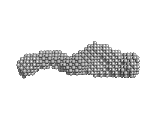
Nucleoporin POM152 monomer, 49 kDa Saccharomyces cerevisiae protein
Buffer: 10mM HEPES, 150mM NaCl, 10%(v/v) glycerol, 5mM DTT, pH: 7.5
Experiment: SAXS data collected at BL4-2, Stanford Synchrotron Radiation Lightsource (SSRL) on 2015 Apr 12
Molecular Architecture of the Major Membrane Ring Component of the Nuclear Pore Complex. Structure 25(3):434-445 (2017)
Upla P, Kim SJ, Sampathkumar P, Dutta K, Cahill SM, Chemmama IE, Williams R, Bonanno JB, Rice WJ, Stokes DL, Cowburn D, Almo SC, Sali A, Rout MP, Fernandez-Martinez J
RgGuinier 4.3 nm
Dmax 15.4 nm
VolumePorod 67 nm3