Search

 
Advanced search  

159 hits found for LI

SASDWP6 – RAD51 [F86E, A89E] in complex with BRCA2 truncation containing the first, second, third and fourth BRC repeats (BRC1-4) at 0.5 mg/mL

Breast cancer type 2 susceptibility protein (BRC repeats 1-4)DNA repair protein RAD51 homolog 1 (F86E A89E) experimental SAS data
Breast cancer type 2 susceptibility protein (BRC repeats 1-4) DNA repair protein RAD51 homolog 1 (F86E A89E) Kratky plot
Sample: ...lity protein (BRC repeats 1-4) monomer, 62 kDa Homo sapiens protein
DNA repair protein RAD51 homolog 1 (F86E A89E) tetramer, 148 kDa Homo sapiens protein
Buffer: ...Li₂SO₄, 5% glycerol, 1 mM DTT, pH: 8
Experiment: SAXS data collected at B21, ...Light Source on 2024 Dec 2
Breaking new ground into RAD51–BRC repeats interplay in Homologous Recombination (2025)
...li A
RgGuinier 8.1 nm
Dmax 33.0 nm
VolumePorod 422 nm3

SASDWQ6 – RAD51 [F86E, A89E] in complex with BRCA2 truncation containing the first, second, third and fourth BRC repeats (BRC1-4) at 0.98 mg/mL

Breast cancer type 2 susceptibility protein (BRC repeats 1-4)DNA repair protein RAD51 homolog 1 (F86E A89E) experimental SAS data
Breast cancer type 2 susceptibility protein (BRC repeats 1-4) DNA repair protein RAD51 homolog 1 (F86E A89E) Kratky plot
Sample: ...lity protein (BRC repeats 1-4) monomer, 62 kDa Homo sapiens protein
DNA repair protein RAD51 homolog 1 (F86E A89E) tetramer, 148 kDa Homo sapiens protein
Buffer: ...Li₂SO₄, 5% glycerol, 1 mM DTT, pH: 8
Experiment: SAXS data collected at B21, ...Light Source on 2024 Dec 2
Breaking new ground into RAD51–BRC repeats interplay in Homologous Recombination (2025)
...li A
RgGuinier 7.6 nm
Dmax 33.3 nm
VolumePorod 392 nm3

SASDWR6 – RAD51 [F86E, A89E] in complex with BRCA2 truncation containing the first, second, third and fourth BRC repeats (BRC1-4) at 2 mg/mL

Breast cancer type 2 susceptibility protein (BRC repeats 1-4)DNA repair protein RAD51 homolog 1 (F86E A89E) experimental SAS data
Breast cancer type 2 susceptibility protein (BRC repeats 1-4) DNA repair protein RAD51 homolog 1 (F86E A89E) Kratky plot
Sample: ...lity protein (BRC repeats 1-4) monomer, 62 kDa Homo sapiens protein
DNA repair protein RAD51 homolog 1 (F86E A89E) tetramer, 148 kDa Homo sapiens protein
Buffer: ...Li₂SO₄, 5% glycerol, 1 mM DTT, pH: 8
Experiment: SAXS data collected at B21, ...Light Source on 2024 Dec 2
Breaking new ground into RAD51–BRC repeats interplay in Homologous Recombination (2025)
...li A
RgGuinier 7.7 nm
Dmax 33.0 nm
VolumePorod 417 nm3

SASDVW6 – Monomeric Adhesion-associated domain in MUC4 and Other Proteins (AMOP-domain) of Isthmin-1

Isthmin-1 AMOP-domain experimental SAS data
Isthmin-1 AMOP-domain Kratky plot
Sample: Isthmin-1 AMOP-domain monomer, 25 kDa Homo sapiens protein
Buffer: 20 mM HEPES 100 mM NaCl, pH: 7.5
Experiment: SAXS data collected at Rigaku BioSAXS-2000, Pennsylvania State University on 2023 Jun 21
...ling. Nat Commun 16(1):3580 (2025)
Li T, Stayrook SE, Li W, Wang Y, Li H, Zhang J, Liu Y, Klein DE
RgGuinier 2.3 nm
Dmax 9.7 nm
VolumePorod 45264 nm3

SASDVX6 – Monomeric Adhesion-associated domain in MUC4 and Other Proteins (AMOP-domain) of Isthmin-1, I455E, F458E douple mutant

L-AMOP-IFEE experimental SAS data
L-AMOP-IFEE Kratky plot
Sample: L-AMOP-IFEE monomer, 25 kDa Homo sapiens protein
Buffer: 20 mM HEPES 100 mM NaCl, pH: 7.5
Experiment: SAXS data collected at Rigaku BioSAXS-2000, Pennsylvania State University on 2023 Jun 21
...ling. Nat Commun 16(1):3580 (2025)
Li T, Stayrook SE, Li W, Wang Y, Li H, Zhang J, Liu Y, Klein DE
RgGuinier 2.4 nm
Dmax 8.8 nm
VolumePorod 46874 nm3

SASDE47 – Aldehyde dehydrogenase 16 from Loktanella sp. (LsALDH16): 2 mg/ml

Aldehyde dehydrogenase 16 from Loktanella sp. experimental SAS data
PYMOL model
Sample: Aldehyde dehydrogenase 16 from Loktanella sp. dimer, 161 kDa Loktanella sp. 3ANDIMAR09 protein
Buffer: 20 mM Tris-HCl, 100 mM NaCl, 2.0% glycerol, 0.5 mM Tris(3-hydroxypropyl)phosphine, pH: 8
Experiment: SAXS data collected at 12.3.1 (SIBYLS), ...Light Source (ALS) on 2017 Dec 13
Crystal Structure of Aldehyde Dehydrogenase 16 Reveals Trans-Hierarchical Structural Similarity and a New Dimer. J Mol Biol (2018)
Liu LK, Tanner JJ
RgGuinier 3.6 nm
Dmax 10.9 nm
VolumePorod 202 nm3

SASDE57 – Aldehyde dehydrogenase 16 from Loktanella sp. (LsALDH16): 4 mg/ml

Aldehyde dehydrogenase 16 from Loktanella sp. experimental SAS data
PYMOL model
Sample: Aldehyde dehydrogenase 16 from Loktanella sp. dimer, 161 kDa Loktanella sp. 3ANDIMAR09 protein
Buffer: 20 mM Tris-HCl, 100 mM NaCl, 2.0% glycerol, 0.5 mM Tris(3-hydroxypropyl)phosphine, pH: 8
Experiment: SAXS data collected at 12.3.1 (SIBYLS), ...Light Source (ALS) on 2017 Dec 13
Crystal Structure of Aldehyde Dehydrogenase 16 Reveals Trans-Hierarchical Structural Similarity and a New Dimer. J Mol Biol (2018)
Liu LK, Tanner JJ
RgGuinier 3.6 nm
Dmax 11.2 nm
VolumePorod 204 nm3

SASDE67 – Aldehyde dehydrogenase 16 from Loktanella sp. (LsALDH16): 6 mg/ml

Aldehyde dehydrogenase 16 from Loktanella sp. experimental SAS data
PYMOL model
Sample: Aldehyde dehydrogenase 16 from Loktanella sp. dimer, 161 kDa Loktanella sp. 3ANDIMAR09 protein
Buffer: 20 mM Tris-HCl, 100 mM NaCl, 2.0% glycerol, 0.5 mM Tris(3-hydroxypropyl)phosphine, pH: 8
Experiment: SAXS data collected at 12.3.1 (SIBYLS), ...Light Source (ALS) on 2017 Dec 13
Crystal Structure of Aldehyde Dehydrogenase 16 Reveals Trans-Hierarchical Structural Similarity and a New Dimer. J Mol Biol (2018)
Liu LK, Tanner JJ
RgGuinier 3.5 nm
Dmax 10.6 nm
VolumePorod 207 nm3

SASDE77 – Aldehyde dehydrogenase 16 from Loktanella sp. (LsALDH16): 8 mg/ml

Aldehyde dehydrogenase 16 from Loktanella sp. experimental SAS data
PYMOL model
Sample: Aldehyde dehydrogenase 16 from Loktanella sp. dimer, 161 kDa Loktanella sp. 3ANDIMAR09 protein
Buffer: 20 mM Tris-HCl, 100 mM NaCl, 2.0% glycerol, 0.5 mM Tris(3-hydroxypropyl)phosphine, pH: 8
Experiment: SAXS data collected at 12.3.1 (SIBYLS), ...Light Source (ALS) on 2017 Dec 13
Crystal Structure of Aldehyde Dehydrogenase 16 Reveals Trans-Hierarchical Structural Similarity and a New Dimer. J Mol Biol (2018)
Liu LK, Tanner JJ
RgGuinier 3.6 nm
Dmax 10.8 nm
VolumePorod 205 nm3

SASDE87 – Aldehyde dehydrogenase family 16 member A1 from Homo sapiens (HsALDH16A1): 1 mg/ml

Aldehyde dehydrogenase family 16 member A1 from Homo sapiens experimental SAS data
PYMOL model
Sample: Aldehyde dehydrogenase family 16 member A1 from Homo sapiens dimer, 171 kDa Homo sapiens protein
Buffer: 20 mM Tris-HCl, 100 mM NaCl, 2.0% glycerol, 0.5 mM Tris(3-hydroxypropyl)phosphine, pH: 8
Experiment: SAXS data collected at 12.3.1 (SIBYLS), ...Light Source (ALS) on 2017 Dec 13
Crystal Structure of Aldehyde Dehydrogenase 16 Reveals Trans-Hierarchical Structural Similarity and a New Dimer. J Mol Biol (2018)
Liu LK, Tanner JJ
RgGuinier 3.6 nm
Dmax 10.9 nm
VolumePorod 230 nm3

SASDE97 – Aldehyde dehydrogenase family 16 member A1 from Homo sapiens (HsALDH16A1): 1.6 mg/ml

Aldehyde dehydrogenase family 16 member A1 from Homo sapiens experimental SAS data
PYMOL model
Sample: Aldehyde dehydrogenase family 16 member A1 from Homo sapiens dimer, 171 kDa Homo sapiens protein
Buffer: 20 mM Tris-HCl, 100 mM NaCl, 2.0% glycerol, 0.5 mM Tris(3-hydroxypropyl)phosphine, pH: 8
Experiment: SAXS data collected at 12.3.1 (SIBYLS), ...Light Source (ALS) on 2017 Dec 13
Crystal Structure of Aldehyde Dehydrogenase 16 Reveals Trans-Hierarchical Structural Similarity and a New Dimer. J Mol Biol (2018)
Liu LK, Tanner JJ
RgGuinier 3.8 nm
Dmax 11.2 nm
VolumePorod 236 nm3

SASDY97 – ...line phosphatase (ALP) under vis

Wild Alkaline Phosphatase under vis experimental SAS data
PYMOL model
Sample: ...line Phosphatase under vis dimer, 97 kDa ...li protein
Buffer: 25 mM Tris, 100 mM NaCl, 2 mM TCEP, 3% (w/v) glycerol, pH: 7.5
Experiment: SAXS data collected at BL19U2, ...lity (SSRF) on 2024 Dec 23
Single-engineered-residue solvation perturbations regulate global protein architecture and function. Nat Commun (2026)
Liu Y, Zhai J, Cao S, Guo H, Zhang H, Song B, Guo J, Men D, He X, Li D
RgGuinier 2.8 nm
Dmax 9.0 nm
VolumePorod 75 nm3

SASDEA7 – Aldehyde dehydrogenase family 16 member A1 from Homo sapiens (HsALDH16A1): 3.2 mg/ml

Aldehyde dehydrogenase family 16 member A1 from Homo sapiens experimental SAS data
PYMOL model
Sample: Aldehyde dehydrogenase family 16 member A1 from Homo sapiens dimer, 171 kDa Homo sapiens protein
Buffer: 20 mM Tris-HCl, 100 mM NaCl, 2.0% glycerol, 0.5 mM Tris(3-hydroxypropyl)phosphine, pH: 8
Experiment: SAXS data collected at 12.3.1 (SIBYLS), ...Light Source (ALS) on 2017 Dec 13
Crystal Structure of Aldehyde Dehydrogenase 16 Reveals Trans-Hierarchical Structural Similarity and a New Dimer. J Mol Biol (2018)
Liu LK, Tanner JJ
RgGuinier 3.8 nm
Dmax 11.5 nm
VolumePorod 237 nm3

SASDVS7 – Cadherin EGF LAG seven-pass G-type receptor 1 (CELSR1) ECR without calcium

Cadherin EGF LAG seven-pass G-type receptor 1 experimental SAS data
Cadherin EGF LAG seven-pass G-type receptor 1 Kratky plot
Sample: Cadherin EGF LAG seven-pass G-type receptor 1 monomer, 245 kDa Mus musculus protein
Buffer: 10 mM Tris, 150 mM NaCl, pH: 8.5
Experiment: SAXS data collected at BioCAT 18ID, Advanced Photon Source (APS), Argonne National Laboratory on 2022 Dec 8
Structural basis for regulation of CELSR1 by a compact module in its extracellular region. Nat Commun 16(1):3972 (2025)
...Li J, Shearer T, Sando RC, Araç D
RgGuinier 6.0 nm
Dmax 22.5 nm
VolumePorod 473 nm3

SASDVT7 – Cadherin EGF LAG seven-pass G-type receptor 1 (CELSR1) ECR with calcium

Cadherin EGF LAG seven-pass G-type receptor 1 experimental SAS data
Cadherin EGF LAG seven-pass G-type receptor 1 Kratky plot
Sample: Cadherin EGF LAG seven-pass G-type receptor 1 dimer, 491 kDa Mus musculus protein
Buffer: 10 mM Tris, 150 mM NaCl, 1 mM CaCl2, pH: 8.5
Experiment: SAXS data collected at BioCAT 18ID, Advanced Photon Source (APS), Argonne National Laboratory on 2022 Dec 8
Structural basis for regulation of CELSR1 by a compact module in its extracellular region. Nat Commun 16(1):3972 (2025)
...Li J, Shearer T, Sando RC, Araç D
RgGuinier 16.2 nm
Dmax 67.5 nm
VolumePorod 722 nm3

SASDJU7 – HIV-1 Primer Binding Site (PBS)-Segment RNA

Primer Binding Site-Segment experimental SAS data
DAMMIF model
Sample: Primer Binding Site-Segment monomer, 33 kDa HIV-1: pNL4-3 RNA
Buffer: 10 mM Tris, 140 mM KCl, 10 mM NaCl, 1 mM MgCl2, pH: 7.5
Experiment: SAXS data collected at BioCAT 18ID, Advanced Photon Source (APS), Argonne National Laboratory on 2015 Jul 29
The three-way junction structure of the HIV-1 PBS-segment binds host enzyme important for viral infectivity. Nucleic Acids Res (2021)
...Li J, Qiu L, Ji J, Lange MJ, Zuo X, Chen SJ, Zou X, Boris-Lawrie K, Heng X
RgGuinier 3.4 nm
Dmax 12.8 nm
VolumePorod 87 nm3

SASDQ38 – Nucleoside triphosphate pyrophosphohydrolase (full-length)

Nucleoside triphosphate pyrophosphohydrolase experimental SAS data
DAMMIF model
Sample: Nucleoside triphosphate pyrophosphohydrolase dimer, 71 kDa Mycobacterium tuberculosis (strain … protein
Buffer: 20 mM Tris-HCl, 150 mM NaCl, pH: 8
Experiment: SAXS data collected at BL19U2, ...lity (SSRF) on 2022 Sep 4
Structural analysis of the housecleaning nucleoside triphosphate pyrophosphohydrolase MazG from Mycobacterium tuberculosis Frontiers in Microbiology 14 (2023)
...Li J
RgGuinier 3.4 nm
Dmax 12.2 nm
VolumePorod 121 nm3

SASDPE8 – SMARCA2 bromodomain (isoform1):Protac (ACBI1):VCB (VHL-elongin C-elongin B)

Probable global transcription activator SNF2L2 (isoform 1)von Hippel-Lindau disease tumor suppressorElongin-BElongin-CACBI1 protac experimental SAS data
Probable global transcription activator SNF2L2 (isoform 1) von Hippel-Lindau disease tumor suppressor Elongin-B Elongin-C ACBI1 protac Kratky plot
Sample: Probable global transcription activator SNF2L2 (isoform 1) monomer, 16 kDa Homo sapiens protein
...Lindau disease tumor suppressor monomer, 19 kDa Homo sapiens protein
Elongin-B monomer, 12 kDa Homo sapiens protein
Elongin-C monomer, 11 kDa Homo sapiens protein
ACBI1 protac monomer, 1 kDa
Buffer: 20 mM HEPES, 150 mM NaCl, 1 mM DTT, pH: 7.5
Experiment: SAXS data collected at Xenocs BioXolver L with MetalJet, Département de Biochimie, Université de Montréal on 2021 Aug 11
Predicting the structural basis of targeted protein degradation by integrating molecular dynamics simulations with structural mass spectrometry. Nat Commun 13(1):5884 (2022)
...Li Y, Vinitsky A, Zhang X, Razavi AM, Kolossváry I, Imbriglio J, Evdokimov A, Bergeron L, Zhou W, Adhikari J, Ruprecht B, Dickson A, Xu H, Sherman W, Izaguirre JA
RgGuinier 3.3 nm
Dmax 12.5 nm
VolumePorod 83 nm3

SASDPF8 – SMARCA2 bromodomain (isoform2):Protac (ACBI1):VCB (VHL-elongin C-elongin B)

von Hippel-Lindau disease tumor suppressorElongin-BElongin-CACBI1 protacProbable global transcription activator SNF2L2 (isoform 2) experimental SAS data
von Hippel-Lindau disease tumor suppressor Elongin-B Elongin-C ACBI1 protac Probable global transcription activator SNF2L2 (isoform 2) Kratky plot
Sample: ...Lindau disease tumor suppressor monomer, 19 kDa Homo sapiens protein
Elongin-B monomer, 12 kDa Homo sapiens protein
Elongin-C monomer, 11 kDa Homo sapiens protein
ACBI1 protac monomer, 1 kDa
Probable global transcription activator SNF2L2 (isoform 2) monomer, 14 kDa Homo sapiens protein
Buffer: 20 mM HEPES, 150 mM NaCl, 1 mM DTT, pH: 7.5
Experiment: SAXS data collected at Xenocs BioXolver L with MetalJet, Département de Biochimie, Université de Montréal on 2021 Aug 11
Predicting the structural basis of targeted protein degradation by integrating molecular dynamics simulations with structural mass spectrometry. Nat Commun 13(1):5884 (2022)
...Li Y, Vinitsky A, Zhang X, Razavi AM, Kolossváry I, Imbriglio J, Evdokimov A, Bergeron L, Zhou W, Adhikari J, Ruprecht B, Dickson A, Xu H, Sherman W, Izaguirre JA
RgGuinier 3.2 nm
Dmax 11.1 nm
VolumePorod 75 nm3

SASDLM8 – Human apo Nocturnin - Extended

Nocturnin experimental SAS data
Human apo Nocturnin - Extended Rg histogram
Sample: Nocturnin monomer, 41 kDa Homo sapiens protein
Buffer: 50 mM HEPES, 150 mM KCl, 10% glycerol, 5 mM MgCl2, 1 mM TCEP, pH: 7.5
Experiment: SAXS data collected at BioCAT 18ID, Advanced Photon Source (APS), Argonne National Laboratory on 2021 Feb 7
The Disordered Amino Terminus of the Circadian Enzyme Nocturnin Modulates Its NADP(H) Phosphatase Activity by Changing Protein Dynamics. Biochemistry (2022)
...Li L, Hopkins JB, Joachimiak LA, Green CB
RgGuinier 2.9 nm
Dmax 12.3 nm
VolumePorod 85 nm3

SASDLN8 – Human apo Nocturnin - Compact

Nocturnin experimental SAS data
Human apo Nocturnin - Compact Rg histogram
Sample: Nocturnin monomer, 41 kDa Homo sapiens protein
Buffer: 50 mM HEPES, 150 mM KCl, 10% glycerol, 5 mM MgCl2, 1 mM TCEP, pH: 7.5
Experiment: SAXS data collected at BioCAT 18ID, Advanced Photon Source (APS), Argonne National Laboratory on 2021 Feb 7
The Disordered Amino Terminus of the Circadian Enzyme Nocturnin Modulates Its NADP(H) Phosphatase Activity by Changing Protein Dynamics. Biochemistry (2022)
...Li L, Hopkins JB, Joachimiak LA, Green CB
RgGuinier 2.4 nm
Dmax 9.6 nm
VolumePorod 76 nm3

SASDLP8 – Human apo Nocturnin - Deletion construct (Δ107-120) - Extended

Nocturnin - Deletion construct - Δ107-120 experimental SAS data
Human apo Nocturnin - Deletion construct (Δ107-120) - Extended Rg histogram
Sample: Nocturnin - Deletion construct - Δ107-120 monomer, 40 kDa protein
Buffer: 50 mM HEPES, 150 mM KCl, 10% glycerol, 5 mM MgCl2, 1 mM TCEP, pH: 7.5
Experiment: SAXS data collected at BioCAT 18ID, Advanced Photon Source (APS), Argonne National Laboratory on 2021 Feb 7
The Disordered Amino Terminus of the Circadian Enzyme Nocturnin Modulates Its NADP(H) Phosphatase Activity by Changing Protein Dynamics. Biochemistry (2022)
...Li L, Hopkins JB, Joachimiak LA, Green CB
RgGuinier 2.8 nm
Dmax 11.2 nm
VolumePorod 72 nm3

SASDLQ8 – Human apo Nocturnin - Deletion construct (Δ107-120) - Compact

Nocturnin - Deletion construct - Δ107-120 experimental SAS data
Human apo Nocturnin - Deletion construct (Δ107-120) - Compact Rg histogram
Sample: Nocturnin - Deletion construct - Δ107-120 monomer, 40 kDa protein
Buffer: 50 mM HEPES, 150 mM KCl, 10% glycerol, 5 mM MgCl2, 1 mM TCEP, pH: 7.5
Experiment: SAXS data collected at BioCAT 18ID, Advanced Photon Source (APS), Argonne National Laboratory on 2021 Feb 7
The Disordered Amino Terminus of the Circadian Enzyme Nocturnin Modulates Its NADP(H) Phosphatase Activity by Changing Protein Dynamics. Biochemistry (2022)
...Li L, Hopkins JB, Joachimiak LA, Green CB
RgGuinier 2.5 nm
Dmax 10.0 nm
VolumePorod 80 nm3

SASDQQ8 – Endo-D-arabinanase, 54 kDa, dimer, Microbacterium arabinogalactanolyticum

Endo-D-arabinanase experimental SAS data
REFMAC model
Sample: Endo-D-arabinanase dimer, 107 kDa Microbacterium arabinogalactanolyticum protein
Buffer: 20 mM Tris-HCl, 200 mM NaCl, pH: 7.5
Experiment: SAXS data collected at BL-10C, Photon Factory (PF), High Energy Accelerator Research Organization (KEK) on 2022 Nov 3
...lipoarabinomannan and arabinogalactan of mycobacteria Nature Communications 14(1) (2023)
...Li J, Fukushima R, Sawai N, Nakamori M, Tanaka Y, Kudo A, Morikami S, Iwanaga N, Akai G, Shimizu N, Arakawa T, Yamada C, Kitahara K, Tanaka K, Ito Y, Fushinobu S, Fujita K
RgGuinier 3.1 nm
Dmax 10.4 nm
VolumePorod 131 nm3

SASDLR8 – Human apo Nocturnin - N terminus truncated (118-NOCT)

Nocturnin - N terminus truncated (118-NOCT) experimental SAS data
EOM/RANCH model
Sample: Nocturnin - N terminus truncated (118-NOCT) monomer, 36 kDa Homo sapiens protein
Buffer: 50 mM HEPES, 150 mM KCl, 10% glycerol, 5 mM MgCl2, 1 mM TCEP, pH: 7.5
Experiment: SAXS data collected at BioCAT 18ID, Advanced Photon Source (APS), Argonne National Laboratory on 2021 Feb 7
The Disordered Amino Terminus of the Circadian Enzyme Nocturnin Modulates Its NADP(H) Phosphatase Activity by Changing Protein Dynamics. Biochemistry (2022)
...Li L, Hopkins JB, Joachimiak LA, Green CB
RgGuinier 2.3 nm
Dmax 10.3 nm
VolumePorod 63 nm3

SASDCW8 – Human Rev7 monomer (R124A mutant) in complex with Rev3 peptide @ 4mg/mL

Mitotic spindle assembly checkpoint protein MAD2BDNA polymerase zeta catalytic subunit experimental SAS data
PDB (PROTEIN DATA BANK) model
Sample: Mitotic spindle assembly checkpoint protein MAD2B monomer, 24 kDa Homo sapiens protein
DNA polymerase zeta catalytic subunit monomer, 3 kDa Homo sapiens protein
Buffer: 20 mM Tris, 150 mM NaCl, 1 mM EDTA, 10 mM DTT, 5% glycerol, pH: 8.4
Experiment: SAXS data collected at G1, Cornell High Energy Synchrotron Source (CHESS) on 2016 May 14
Rev7 dimerization is important for assembly and function of the Rev1/Polζ translesion synthesis complex. Proc Natl Acad Sci U S A 115(35):E8191-E8200 (2018)
...Li Y, Hao B, Hemann MT, Walker GC, Korzhnev DM
RgGuinier 2.1 nm
Dmax 6.5 nm
VolumePorod 46 nm3

SASDCX8 – Human Rev7 monomer (R124A mutant) in complex with Rev3 peptide @ 6mg/mL

Mitotic spindle assembly checkpoint protein MAD2BDNA polymerase zeta catalytic subunit experimental SAS data
PDB (PROTEIN DATA BANK) model
Sample: Mitotic spindle assembly checkpoint protein MAD2B monomer, 24 kDa Homo sapiens protein
DNA polymerase zeta catalytic subunit monomer, 3 kDa Homo sapiens protein
Buffer: 20 mM Tris, 150 mM NaCl, 1 mM EDTA, 10 mM DTT, 5% glycerol, pH: 8.4
Experiment: SAXS data collected at G1, Cornell High Energy Synchrotron Source (CHESS) on 2016 May 14
Rev7 dimerization is important for assembly and function of the Rev1/Polζ translesion synthesis complex. Proc Natl Acad Sci U S A 115(35):E8191-E8200 (2018)
...Li Y, Hao B, Hemann MT, Walker GC, Korzhnev DM
RgGuinier 2.1 nm
Dmax 7.6 nm
VolumePorod 47 nm3

SASDCY8 – Human Rev7 monomer (R124A mutant) in complex with Rev3 peptide @ 8mg/mL

Mitotic spindle assembly checkpoint protein MAD2BDNA polymerase zeta catalytic subunit experimental SAS data
PDB (PROTEIN DATA BANK) model
Sample: Mitotic spindle assembly checkpoint protein MAD2B monomer, 24 kDa Homo sapiens protein
DNA polymerase zeta catalytic subunit monomer, 3 kDa Homo sapiens protein
Buffer: 20 mM Tris, 150 mM NaCl, 1 mM EDTA, 10 mM DTT, 5% glycerol, pH: 8.4
Experiment: SAXS data collected at G1, Cornell High Energy Synchrotron Source (CHESS) on 2016 May 14
Rev7 dimerization is important for assembly and function of the Rev1/Polζ translesion synthesis complex. Proc Natl Acad Sci U S A 115(35):E8191-E8200 (2018)
...Li Y, Hao B, Hemann MT, Walker GC, Korzhnev DM
RgGuinier 2.1 nm
Dmax 7.5 nm
VolumePorod 46 nm3

SASDCZ8 – Human Rev7 dimer in complex with Rev3 peptide @ 12.2 mg/mL

Mitotic spindle assembly checkpoint protein MAD2BDNA polymerase zeta catalytic subunitDNA polymerase zeta catalytic subunit experimental SAS data
Mitotic spindle assembly checkpoint protein MAD2B DNA polymerase zeta catalytic subunit DNA polymerase zeta catalytic subunit Kratky plot
Sample: Mitotic spindle assembly checkpoint protein MAD2B dimer, 49 kDa Homo sapiens protein
DNA polymerase zeta catalytic subunit monomer, 3 kDa Homo sapiens protein
Buffer: 20 mM HEPES, 10 mM DTT, 5% glycerol, pH: 8
Experiment: SAXS data collected at G1, Cornell High Energy Synchrotron Source (CHESS) on 2016 May 14
Rev7 dimerization is important for assembly and function of the Rev1/Polζ translesion synthesis complex. Proc Natl Acad Sci U S A 115(35):E8191-E8200 (2018)
...Li Y, Hao B, Hemann MT, Walker GC, Korzhnev DM
RgGuinier 2.8 nm

SASDC29 – Human Rev7 dimer in complex with Rev3 peptide @ 10.6 mg/mL

Mitotic spindle assembly checkpoint protein MAD2BDNA polymerase zeta catalytic subunitDNA polymerase zeta catalytic subunit experimental SAS data
HADDOCK model
Sample: Mitotic spindle assembly checkpoint protein MAD2B dimer, 49 kDa Homo sapiens protein
DNA polymerase zeta catalytic subunit monomer, 3 kDa Homo sapiens protein
Buffer: 20 mM HEPES, 10 mM DTT, 5% glycerol, pH: 8
Experiment: SAXS data collected at G1, Cornell High Energy Synchrotron Source (CHESS) on 2016 May 14
Rev7 dimerization is important for assembly and function of the Rev1/Polζ translesion synthesis complex. Proc Natl Acad Sci U S A 115(35):E8191-E8200 (2018)
...Li Y, Hao B, Hemann MT, Walker GC, Korzhnev DM
RgGuinier 2.9 nm
Dmax 11.0 nm
VolumePorod 108 nm3

SASDC39 – Human Rev7 dimer in complex with Rev3 peptide @ 9.1mg/mL

Mitotic spindle assembly checkpoint protein MAD2BDNA polymerase zeta catalytic subunitDNA polymerase zeta catalytic subunit experimental SAS data
HADDOCK model
Sample: Mitotic spindle assembly checkpoint protein MAD2B dimer, 49 kDa Homo sapiens protein
DNA polymerase zeta catalytic subunit monomer, 3 kDa Homo sapiens protein
Buffer: 20 mM HEPES, 10 mM DTT, 5% glycerol, pH: 8
Experiment: SAXS data collected at G1, Cornell High Energy Synchrotron Source (CHESS) on 2016 May 14
Rev7 dimerization is important for assembly and function of the Rev1/Polζ translesion synthesis complex. Proc Natl Acad Sci U S A 115(35):E8191-E8200 (2018)
...Li Y, Hao B, Hemann MT, Walker GC, Korzhnev DM
RgGuinier 3.0 nm
Dmax 11.4 nm
VolumePorod 107 nm3

SASDC49 – Human Rev7 dimer in complex with Rev3 peptide @ 6.1mg/mL

Mitotic spindle assembly checkpoint protein MAD2BDNA polymerase zeta catalytic subunitDNA polymerase zeta catalytic subunit experimental SAS data
Mitotic spindle assembly checkpoint protein MAD2B DNA polymerase zeta catalytic subunit DNA polymerase zeta catalytic subunit Kratky plot
Sample: Mitotic spindle assembly checkpoint protein MAD2B dimer, 49 kDa Homo sapiens protein
DNA polymerase zeta catalytic subunit monomer, 3 kDa Homo sapiens protein
Buffer: 20 mM HEPES, 10 mM DTT, 5% glycerol, pH: 8
Experiment: SAXS data collected at G1, Cornell High Energy Synchrotron Source (CHESS) on 2016 May 14
Rev7 dimerization is important for assembly and function of the Rev1/Polζ translesion synthesis complex. Proc Natl Acad Sci U S A 115(35):E8191-E8200 (2018)
...Li Y, Hao B, Hemann MT, Walker GC, Korzhnev DM
RgGuinier 3.1 nm
Dmax 11.6 nm
VolumePorod 105 nm3

SASDC59 – Human Rev7 dimer in complex with Rev3 peptide @ 7.6mg/mL

Mitotic spindle assembly checkpoint protein MAD2BDNA polymerase zeta catalytic subunitDNA polymerase zeta catalytic subunit experimental SAS data
Mitotic spindle assembly checkpoint protein MAD2B DNA polymerase zeta catalytic subunit DNA polymerase zeta catalytic subunit Kratky plot
Sample: Mitotic spindle assembly checkpoint protein MAD2B dimer, 49 kDa Homo sapiens protein
DNA polymerase zeta catalytic subunit monomer, 3 kDa Homo sapiens protein
Buffer: 20 mM HEPES, 10 mM DTT, 5% glycerol, pH: 8
Experiment: SAXS data collected at G1, Cornell High Energy Synchrotron Source (CHESS) on 2016 May 14
Rev7 dimerization is important for assembly and function of the Rev1/Polζ translesion synthesis complex. Proc Natl Acad Sci U S A 115(35):E8191-E8200 (2018)
...Li Y, Hao B, Hemann MT, Walker GC, Korzhnev DM
RgGuinier 2.9 nm
Dmax 11.1 nm
VolumePorod 106 nm3

SASDC69 – Human Rev7 dimer in complex with Rev3 peptide @ 4.6mg/mL

Mitotic spindle assembly checkpoint protein MAD2BDNA polymerase zeta catalytic subunitDNA polymerase zeta catalytic subunit experimental SAS data
Mitotic spindle assembly checkpoint protein MAD2B DNA polymerase zeta catalytic subunit DNA polymerase zeta catalytic subunit Kratky plot
Sample: Mitotic spindle assembly checkpoint protein MAD2B dimer, 49 kDa Homo sapiens protein
DNA polymerase zeta catalytic subunit monomer, 3 kDa Homo sapiens protein
Buffer: 20 mM HEPES, 10 mM DTT, 5% glycerol, pH: 8
Experiment: SAXS data collected at G1, Cornell High Energy Synchrotron Source (CHESS) on 2016 May 14
Rev7 dimerization is important for assembly and function of the Rev1/Polζ translesion synthesis complex. Proc Natl Acad Sci U S A 115(35):E8191-E8200 (2018)
...Li Y, Hao B, Hemann MT, Walker GC, Korzhnev DM
RgGuinier 2.9 nm

SASDC79 – Human Rev7 dimer in complex with Rev3 peptide @ 3.0mg/mL

Mitotic spindle assembly checkpoint protein MAD2BDNA polymerase zeta catalytic subunitDNA polymerase zeta catalytic subunit experimental SAS data
Mitotic spindle assembly checkpoint protein MAD2B DNA polymerase zeta catalytic subunit DNA polymerase zeta catalytic subunit Kratky plot
Sample: Mitotic spindle assembly checkpoint protein MAD2B dimer, 49 kDa Homo sapiens protein
DNA polymerase zeta catalytic subunit monomer, 3 kDa Homo sapiens protein
Buffer: 20 mM HEPES, 10 mM DTT, 5% glycerol, pH: 8
Experiment: SAXS data collected at G1, Cornell High Energy Synchrotron Source (CHESS) on 2016 May 14
Rev7 dimerization is important for assembly and function of the Rev1/Polζ translesion synthesis complex. Proc Natl Acad Sci U S A 115(35):E8191-E8200 (2018)
...Li Y, Hao B, Hemann MT, Walker GC, Korzhnev DM
RgGuinier 2.8 nm

SASDC89 – Human Rev7 dimer in complex with Rev3 peptide @ 1.5 mg/mL

Mitotic spindle assembly checkpoint protein MAD2BDNA polymerase zeta catalytic subunitDNA polymerase zeta catalytic subunit experimental SAS data
Mitotic spindle assembly checkpoint protein MAD2B DNA polymerase zeta catalytic subunit DNA polymerase zeta catalytic subunit Kratky plot
Sample: Mitotic spindle assembly checkpoint protein MAD2B dimer, 49 kDa Homo sapiens protein
DNA polymerase zeta catalytic subunit monomer, 3 kDa Homo sapiens protein
Buffer: 20 mM HEPES, 10 mM DTT, 5% glycerol, pH: 8
Experiment: SAXS data collected at G1, Cornell High Energy Synchrotron Source (CHESS) on 2016 May 14
Rev7 dimerization is important for assembly and function of the Rev1/Polζ translesion synthesis complex. Proc Natl Acad Sci U S A 115(35):E8191-E8200 (2018)
...Li Y, Hao B, Hemann MT, Walker GC, Korzhnev DM
RgGuinier 2.7 nm

SASDEB9 – ...li

Flagellar brake protein YcgR experimental SAS data
SASREF model
Sample: Flagellar brake protein YcgR monomer, 29 kDa ...li protein
Buffer: 20 mM HEPES, 150mM NaCl, 10% glycerol,, pH: 7.5
Experiment: SAXS data collected at BL19U2, ...lity (SSRF) on 2016 Jan 4
...lity in Escherichia coli. J Biol Chem 295(3):808-821 (2020)
...Li DF
RgGuinier 2.6 nm
Dmax 9.1 nm
VolumePorod 44 nm3

SASDEC9 – ...li

Flagellar brake protein YcgR in complex with c-di-GMP experimental SAS data
PDB (PROTEIN DATA BANK) model
Sample: Flagellar brake protein YcgR in complex with c-di-GMP monomer, 29 kDa ...li protein
Buffer: 20 mM HEPES, 150mM NaCl, 10% glycerol,, pH: 7.5
Experiment: SAXS data collected at BL19U2, ...lity (SSRF) on 2016 Jan 4
...lity in Escherichia coli. J Biol Chem 295(3):808-821 (2020)
...Li DF
RgGuinier 2.2 nm
Dmax 7.3 nm
VolumePorod 44 nm3

SASDQF9 – GRP94 protein (Endoplasmin)

Endoplasmin experimental SAS data
Endoplasmin Kratky plot
Sample: Endoplasmin dimer, 169 kDa Mus musculus protein
Buffer: 25 mM HEPES, 200 mM NaCl, 1 mM TCEP, pH: 8
Experiment: SAXS data collected at BL19U2, ...lity (SSRF) on 2018 Jul 11
...lization of conformational transition of GRP94 in solution. Life Sci Alliance 7(2) (2024)
...Li N, Yang B
RgGuinier 5.6 nm
Dmax 24.0 nm
VolumePorod 260 nm3

SASDQG9 – GRP94 protein (Endoplasmin) bound to ADP

Endoplasmin experimental SAS data
Endoplasmin Kratky plot
Sample: Endoplasmin dimer, 169 kDa Mus musculus protein
Buffer: 25 mM HEPES, 200 mM NaCl, 1 mM TCEP, pH: 8
Experiment: SAXS data collected at BL19U2, ...lity (SSRF) on 2018 Jul 11
...lization of conformational transition of GRP94 in solution. Life Sci Alliance 7(2) (2024)
...Li N, Yang B
RgGuinier 5.7 nm
Dmax 24.0 nm
VolumePorod 274 nm3

SASDCH9 – Bifunctional enzyme responsible for the synthesis and hydrolysis of c-di-GMP (DcpA) with GDP

Sensory box/response regulator experimental SAS data
CORAL model
Sample: Sensory box/response regulator dimer, 136 kDa Mycobacterium smegmatis (strain … protein
Buffer: 20 mM HEPES, 100 mM NaCl, 5% glycerol, 2 mM MgCl2, pH: 7.5
Experiment: SAXS data collected at BL19U2, ...lity (SSRF) on 2016 Jun 26
The GDP-switched GAF domain of DcpA modulates the concerted synthesis/hydrolysis of c-di-GMP in Mycobacterium smegmatis. Biochem J 475(7):1295-1308 (2018)
...Li N, Luo Y, Jiang YL, Zhou CZ, Chen Y, Li Q
RgGuinier 5.0 nm
Dmax 20.0 nm
VolumePorod 299 nm3

SASDQH9 – GRP94 protein (Endoplasmin) bound to AMP-PNP

Endoplasmin experimental SAS data
Endoplasmin Kratky plot
Sample: Endoplasmin dimer, 169 kDa Mus musculus protein
Buffer: 25 mM HEPES, 200 mM NaCl, 1 mM TCEP, pH: 8
Experiment: SAXS data collected at BL19U2, ...lity (SSRF) on 2018 Jul 11
...lization of conformational transition of GRP94 in solution. Life Sci Alliance 7(2) (2024)
...Li N, Yang B
RgGuinier 5.5 nm
Dmax 24.5 nm
VolumePorod 260 nm3

SASDBJ9 – Geobacillus stearothermophilus DnaB1-300

Geobacillus stearothermophilus DnaB1-300 experimental SAS data
GASBOR model
Sample: Geobacillus stearothermophilus DnaB1-300 tetramer, 138 kDa Geobacillus stearothermophilus protein
Buffer: 20 mM Tris, 300 mM NaCl and 5 mM β-ME, pH: 8
Experiment: SAXS data collected at 23A, Taiwan Photon Source, NSRRC on 2015 Oct 18
Structural analyses of the bacterial primosomal protein DnaB reveal that it is a tetramer and forms a complex with a primosomal re-initiation protein. J Biol Chem 292(38):15744-15757 (2017)
Li YC, Naveen V, Lin MG, Hsiao CD
RgGuinier 3.5 nm
Dmax 11.0 nm
VolumePorod 315 nm3

SASDCJ9 – Bifunctional enzyme responsible for the synthesis and hydrolysis of c-di-GMP (DcpA) without GDP

Sensory box/response regulator experimental SAS data
CORAL model
Sample: Sensory box/response regulator dimer, 136 kDa Mycobacterium smegmatis (strain … protein
Buffer: 20 mM HEPES, 100 mM NaCl, 5% glycerol, 2 mM MgCl2, pH: 7.5
Experiment: SAXS data collected at BL19U2, ...lity (SSRF) on 2016 Jun 26
The GDP-switched GAF domain of DcpA modulates the concerted synthesis/hydrolysis of c-di-GMP in Mycobacterium smegmatis. Biochem J 475(7):1295-1308 (2018)
...Li N, Luo Y, Jiang YL, Zhou CZ, Chen Y, Li Q
RgGuinier 4.8 nm
Dmax 17.0 nm
VolumePorod 271 nm3

SASDBK9 – Geobacillus stearothermophilus DnaB full-length

Geobacillus stearothermophilus DnaB full-length experimental SAS data
GASBOR model
Sample: Geobacillus stearothermophilus DnaB full-length tetramer, 214 kDa Geobacillus stearothermophilus protein
Buffer: 20 mM Tris, 300 mM NaCl and 5 mM β-ME, pH: 8
Experiment: SAXS data collected at 23A, Taiwan Photon Source, NSRRC on 2015 Mar 12
Structural analyses of the bacterial primosomal protein DnaB reveal that it is a tetramer and forms a complex with a primosomal re-initiation protein. J Biol Chem 292(38):15744-15757 (2017)
Li YC, Naveen V, Lin MG, Hsiao CD
RgGuinier 5.7 nm
Dmax 20.0 nm
VolumePorod 432 nm3

SASDFT9 – Adhesion G-protein coupled receptor G6 - zfGpr126 S2 (-ss) ECR

Adhesion G-protein coupled receptor G6 S2 experimental SAS data
Adhesion G-protein coupled receptor G6 S2 Kratky plot
Sample: Adhesion G-protein coupled receptor G6 S2 monomer, 86 kDa Danio rerio protein
Buffer: 150 mM NaCl, 20 mM HEPES, pH: 7.5
Experiment: SAXS data collected at BioCAT 18ID, Advanced Photon Source (APS), Argonne National Laboratory on 2018 Jun 28
Structural basis for adhesion G protein-coupled receptor Gpr126 function Nature Communications 11(1) (2020)
...Li J, Sosnick T, Zhao M, Monk K, Araç D
RgGuinier 4.0 nm
Dmax 14.1 nm
VolumePorod 169 nm3

SASDFU9 – Adhesion G-protein coupled receptor G6 - zfGpr126 S1 (+ss) ECR

Adhesion G-protein coupled receptor G6 S1 experimental SAS data
Adhesion G-protein coupled receptor G6 S1 Kratky plot
Sample: Adhesion G-protein coupled receptor G6 S1 monomer, 89 kDa Danio rerio protein
Buffer: 150 mM NaCl, 20 mM HEPES, pH: 7.5
Experiment: SAXS data collected at BioCAT 18ID, Advanced Photon Source (APS), Argonne National Laboratory on 2018 Jun 28
Structural basis for adhesion G protein-coupled receptor Gpr126 function Nature Communications 11(1) (2020)
...Li J, Sosnick T, Zhao M, Monk K, Araç D
RgGuinier 4.2 nm
Dmax 14.8 nm
VolumePorod 191 nm3

SASDFV9 – Adhesion G-protein coupled receptor G6 - zfGpr126 S2 (-ss) D134A/F135A ECR

Adhesion G-protein coupled receptor G6 S2 D134A/F135A experimental SAS data
Adhesion G-protein coupled receptor G6 S2 D134A/F135A Kratky plot
Sample: Adhesion G-protein coupled receptor G6 S2 D134A/F135A monomer, 86 kDa Danio rerio protein
Buffer: 150 mM NaCl, 20 mM HEPES, pH: 7.5
Experiment: SAXS data collected at BioCAT 18ID, Advanced Photon Source (APS), Argonne National Laboratory on 2018 Jun 28
Structural basis for adhesion G protein-coupled receptor Gpr126 function Nature Communications 11(1) (2020)
...Li J, Sosnick T, Zhao M, Monk K, Araç D
RgGuinier 4.3 nm
Dmax 14.8 nm
VolumePorod 181 nm3

SASDQV9 – Nanobody1 in complex with the extracellular domain of human BCMA

Tumor necrosis factor receptor superfamily member 17Nanobody1 experimental SAS data
Tumor necrosis factor receptor superfamily member 17 Nanobody1 Kratky plot
Sample: Tumor necrosis factor receptor superfamily member 17 monomer, 6 kDa Homo sapiens protein
Nanobody1 monomer, 14 kDa protein
Buffer: 20 mM Tris, 100 mM NaCl, pH: 8
Experiment: SAXS data collected at BL19U2, ...lity (SSRF) on 2022 Aug 28
...lifies on-tumor cytotoxicity. Signal Transduct Target Ther 8(1):445 (2023)
...Liu KW, Li N, Cui JC, Huang GX, Liu CL, Xu J, Mi JQ, Chen Z, Fan XH, Chen SJ, Chen S
RgGuinier 3.0 nm
Dmax 9.8 nm
VolumePorod 52 nm3

SASDFW9 – Adhesion G-protein coupled receptor G6 - hGPR126 S2 (-ss) ECR

Adhesion G-protein coupled receptor G6 S2 experimental SAS data
Adhesion G-protein coupled receptor G6 S2 Kratky plot
Sample: Adhesion G-protein coupled receptor G6 S2 monomer, 88 kDa Homo sapiens protein
Buffer: 150 mM NaCl, 20 mM HEPES, pH: 7.5
Experiment: SAXS data collected at BioCAT 18ID, Advanced Photon Source (APS), Argonne National Laboratory on 2018 Jun 28
Structural basis for adhesion G protein-coupled receptor Gpr126 function Nature Communications 11(1) (2020)
...Li J, Sosnick T, Zhao M, Monk K, Araç D
RgGuinier 4.4 nm
Dmax 15.7 nm
VolumePorod 199 nm3

SASDQW9 – Nanobody2 in complex with the extracellular domain of human BCMA

Tumor necrosis factor receptor superfamily member 17Nanobody2 experimental SAS data
Tumor necrosis factor receptor superfamily member 17 Nanobody2 Kratky plot
Sample: Tumor necrosis factor receptor superfamily member 17 monomer, 6 kDa Homo sapiens protein
Nanobody2 monomer, 14 kDa protein
Buffer: 20 mM Tris, 100 mM NaCl, pH: 8
Experiment: SAXS data collected at BL19U2, ...lity (SSRF) on 2022 Aug 28
...lifies on-tumor cytotoxicity. Signal Transduct Target Ther 8(1):445 (2023)
...Liu KW, Li N, Cui JC, Huang GX, Liu CL, Xu J, Mi JQ, Chen Z, Fan XH, Chen SJ, Chen S
RgGuinier 2.0 nm
Dmax 9.0 nm
VolumePorod 24 nm3

SASDFX9 – Adhesion G-protein coupled receptor G6 - hGPR126 S1 (+ss) ECR

Adhesion G-protein coupled receptor G6 S1 experimental SAS data
Adhesion G-protein coupled receptor G6 S1 Kratky plot
Sample: Adhesion G-protein coupled receptor G6 S1 monomer, 91 kDa Homo sapiens protein
Buffer: 150 mM NaCl, 20 mM HEPES, pH: 7.5
Experiment: SAXS data collected at BioCAT 18ID, Advanced Photon Source (APS), Argonne National Laboratory on 2018 Jun 28
Structural basis for adhesion G protein-coupled receptor Gpr126 function Nature Communications 11(1) (2020)
...Li J, Sosnick T, Zhao M, Monk K, Araç D
RgGuinier 4.9 nm
Dmax 17.1 nm
VolumePorod 213 nm3

SASDQX9 – Tandem nanobody in complex with the extracellular domain of human BCMA

Tumor necrosis factor receptor superfamily member 17Tandem nanobody experimental SAS data
Tumor necrosis factor receptor superfamily member 17 Tandem nanobody Kratky plot
Sample: Tumor necrosis factor receptor superfamily member 17 monomer, 6 kDa Homo sapiens protein
Tandem nanobody monomer, 27 kDa protein
Buffer: 20 mM Tris, 100 mM NaCl, pH: 8
Experiment: SAXS data collected at BL19U2, ...lity (SSRF) on 2022 Aug 28
...lifies on-tumor cytotoxicity. Signal Transduct Target Ther 8(1):445 (2023)
...Liu KW, Li N, Cui JC, Huang GX, Liu CL, Xu J, Mi JQ, Chen Z, Fan XH, Chen SJ, Chen S
RgGuinier 5.1 nm
Dmax 23.3 nm
VolumePorod 247 nm3

SASDPA4 – SAXS data for the trimeric C-phycocyanin

C-phycocyanin alpha subunitC-phycocyanin beta subunit experimental SAS data
GASBOR model
Sample: C-phycocyanin alpha subunit trimer, 53 kDa Arthrospira platensis protein
C-phycocyanin beta subunit trimer, 54 kDa Arthrospira platensis protein
Buffer: 150 mM NaCl, 20 mM Tris, pH: 7.6
Experiment: SAXS data collected at Rigaku MicroMax 007-HF, Moscow Institute of Physics and Technology (MIPT) on 2022 Apr 14
...lity of the C-phycocyanin chromophores Structural Dynamics 9(5):054701 (2022)
...limanova E, Ryzhykau Y, Kuklin A, Semenov Y, Ge B, Li W, Qin S, Friedrich T, Sluchanko N, Maksimov E
RgGuinier 3.9 nm
Dmax 13.2 nm
VolumePorod 154 nm3

SASDWT6 – Frataxin bound to tailored nanobody 16C10

Frataxin, mitochondrialNanobody 16C10 experimental SAS data
COOT model
Sample: Frataxin, mitochondrial monomer, 14 kDa Homo sapiens protein
Nanobody 16C10 monomer, 15 kDa synthetic construct protein
Buffer: 20 mM Tris, 150 mM NaCl, pH: 7.5
Experiment: SAXS data collected at B21, ...Light Source on 2024 Sep 18
Nanobodies as tools for studying human frataxin biology. Commun Biol (2026)
...lina R, Muñoz IG, Grossi J, Noguera M, Vila A, García AE, Gentili HG, Rodríguez NA, Aran M, Parreño V, Bok M, Hermoso JA, Ibañez LI, Santos J
RgGuinier 2.4 nm
Dmax 8.0 nm
VolumePorod 37 nm3

SASDLD9 – 2-aminomuconic 6-semialdehyde dehydrogenase wild-type

2-aminomuconic 6-semialdehyde dehydrogenase experimental SAS data
PDB (PROTEIN DATA BANK) model
Sample: 2-aminomuconic 6-semialdehyde dehydrogenase tetramer, 215 kDa Pseudomonas sp. protein
Buffer: 20 mM HEPES, 150 mM NaCl, pH: 7
Experiment: SAXS data collected at BL19U2, ...lity (SSRF) on 2021 Feb 1
The tetrameric assembly of 2-aminomuconic 6-semialdehyde dehydrogenase is a functional requirement of cofactor NAD(+) binding. Environ Microbiol 24(7):2994-3012 (2022)
...Li X, Dong H, Chen C, Zhong Z, Yang C, Liu G, Su D
RgGuinier 3.9 nm
Dmax 11.6 nm
VolumePorod 326 nm3

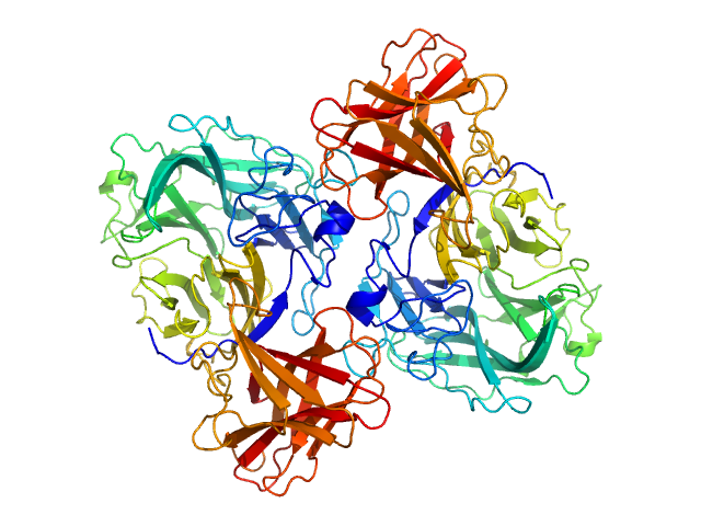
SASDPB4 – SAXS data for the C-phycocyanin in 8 M urea solution

C-phycocyanin alpha subunitC-phycocyanin beta subunit experimental SAS data
GASBOR model
Sample: C-phycocyanin alpha subunit monomer, 18 kDa Arthrospira platensis protein
C-phycocyanin beta subunit monomer, 18 kDa Arthrospira platensis protein
Buffer: 8 M urea, 30 mM βME, pH: 7
Experiment: SAXS data collected at Rigaku MicroMax 007-HF, Moscow Institute of Physics and Technology (MIPT) on 2022 Apr 14
...lity of the C-phycocyanin chromophores Structural Dynamics 9(5):054701 (2022)
...limanova E, Ryzhykau Y, Kuklin A, Semenov Y, Ge B, Li W, Qin S, Friedrich T, Sluchanko N, Maksimov E
RgGuinier 6.6 nm
Dmax 23.5 nm

SASDWS6 – Frataxin bound to tailored nanobody 28F6

Frataxin, mitochondrialNanobody 28F6 experimental SAS data
ALPHAFOLD model
Sample: Frataxin, mitochondrial monomer, 14 kDa Homo sapiens protein
Nanobody 28F6 monomer, 15 kDa synthetic construct protein
Buffer: 20 mM Tris, 150 mM NaCl, pH: 7.5
Experiment: SAXS data collected at B21, ...Light Source on 2024 Sep 18
Nanobodies as tools for studying human frataxin biology. Commun Biol (2026)
...lina R, Muñoz IG, Grossi J, Noguera M, Vila A, García AE, Gentili HG, Rodríguez NA, Aran M, Parreño V, Bok M, Hermoso JA, Ibañez LI, Santos J
RgGuinier 2.2 nm
Dmax 7.4 nm
VolumePorod 36 nm3

SASDLC9 – 2-aminomuconic 6-semialdehyde dehydrogenase double truncation mutant (Δ124-138; Δ477-491)

2-aminomuconic 6-semialdehyde dehydrogenase experimental SAS data
PDB (PROTEIN DATA BANK) model
Sample: 2-aminomuconic 6-semialdehyde dehydrogenase dimer, 101 kDa Pseudomonas sp. protein
Buffer: 20 mM HEPES, 150 mM NaCl, pH: 7
Experiment: SAXS data collected at BL19U2, ...lity (SSRF) on 2021 Feb 1
The tetrameric assembly of 2-aminomuconic 6-semialdehyde dehydrogenase is a functional requirement of cofactor NAD(+) binding. Environ Microbiol 24(7):2994-3012 (2022)
...Li X, Dong H, Chen C, Zhong Z, Yang C, Liu G, Su D
RgGuinier 2.9 nm
Dmax 9.0 nm
VolumePorod 117 nm3