Search

 
Advanced search  

67 hits found for Maxim Petoukhov

SASDKN2 – Matrix protein from Newcastle disease virus at neutral pH

Matrix protein experimental SAS data
DAMMIN model
Sample: Matrix protein , 40 kDa Newcastle disease virus … protein
Buffer: STE buffer 100 mM NaCl, 10 mM Tris-HCl, and 1 mM EDTA, pH: 4
Experiment: SAXS data collected at EMBL P12, PETRA III on 2013 Dec 11
Solution Structure, Self-Assembly, and Membrane Interactions of the Matrix Protein from Newcastle Disease Virus at Neutral and Acidic pH Journal of Virology 93(6) (2019)
...Petoukhov M, Dadinova L, Fedorova N, Tashkin V, Timofeeva T, Ksenofontov A, Loshkarev N, Baratova L, Jeffries C, Svergun D, Batishchev O, García-Sastre A
RgGuinier 3.5 nm

SASDKP2 – Matrix protein from Newcastle disease virus at acidic pH

Matrix protein experimental SAS data
PDB (PROTEIN DATA BANK) model
Sample: Matrix protein dimer, 79 kDa Newcastle disease virus … protein
Buffer: STE buffer 100 mM NaCl, 10 mM Tris-HCl, and 1 mM EDTA, pH: 4
Experiment: SAXS data collected at EMBL P12, PETRA III on 2013 Dec 11
Solution Structure, Self-Assembly, and Membrane Interactions of the Matrix Protein from Newcastle Disease Virus at Neutral and Acidic pH Journal of Virology 93(6) (2019)
...Petoukhov M, Dadinova L, Fedorova N, Tashkin V, Timofeeva T, Ksenofontov A, Loshkarev N, Baratova L, Jeffries C, Svergun D, Batishchev O, García-Sastre A
RgGuinier 3.3 nm

SASDAX2 – Pyruvate decarboxylase (PDC) from Z. mobilis

Pyruvate decarboxylase experimental SAS data
CRYSOL model
Sample: Pyruvate decarboxylase tetramer, 244 kDa Zymomonas mobilis protein
Buffer: 100 mM Sodium Citrate, 17% Glycerol, 22.5% PEG 1500, pH: 6
Experiment: SAXS data collected at EMBL X33, DORIS III, DESY on 1998 Nov 3
Crystal versus solution structures of thiamine diphosphate-dependent enzymes. J Biol Chem 275(1):297-302 (2000)
...Petoukhov MV, Koch MH, König S
RgGuinier 3.9 nm
Dmax 11.0 nm

SASDBC3 – RepB, the replication initiator protein of a promiscuous Streptococcal plasmid pMV158

Replication initiator protein of a promiscuous streptococcal plasmid pMV158. experimental SAS data
NONE model
Sample: Replication initiator protein of a promiscuous streptococcal plasmid pMV158. hexamer, Streptococcus sp. protein
Buffer: 10 mM TRIS 5 mM EDTA 1.0 M KCl, pH: 8.5
Experiment: SAXS data collected at EMBL X33, DORIS III, DESY on 2007 Jun 16
Conformational plasticity of RepB, the replication initiator protein of promiscuous streptococcal plasmid pMV158. Sci Rep 6:20915 (2016)
...Petoukhov MV, Machón C, Svergun DI, Orozco M, del Solar G, Coll M
RgGuinier 3.8 nm
Dmax 10.6 nm
VolumePorod 282 nm3

SASDAN3 – MutS dimer

DNA mismatch repair protein MutS experimental SAS data
DAMMIF model
Sample: DNA mismatch repair protein MutS dimer, 191 kDa Escherichia coli protein
Buffer: 50 mM HEPES 50 mM KCl, pH: 7.5
Experiment: SAXS data collected at EMBL P12, PETRA III on 2013 Feb 28
Using stable MutS dimers and tetramers to quantitatively analyze DNA mismatch recognition and sliding clamp formation. Nucleic Acids Res 41(17):8166-81 (2013)
...Petoukhov MV, Reumer A, Manelyte L, Winterwerp HH, Marinus MG, Lebbink JH, Svergun DI, Friedhoff P, Sixma TK
RgGuinier 4.7 nm
Dmax 15.5 nm
VolumePorod 307 nm3

SASDAQ3 – MutS tetramer

DNA mismatch repair protein MutS experimental SAS data
DAMMIF model
Sample: DNA mismatch repair protein MutS tetramer, 381 kDa Escherichia coli protein
Buffer: 50 mM HEPES 50 mM KCl, pH: 7.5
Experiment: SAXS data collected at EMBL X33, DORIS III, DESY on 2011 May 12
Using stable MutS dimers and tetramers to quantitatively analyze DNA mismatch recognition and sliding clamp formation. Nucleic Acids Res 41(17):8166-81 (2013)
...Petoukhov MV, Reumer A, Manelyte L, Winterwerp HH, Marinus MG, Lebbink JH, Svergun DI, Friedhoff P, Sixma TK
RgGuinier 7.8 nm
Dmax 28.0 nm
VolumePorod 700 nm3

SASDKS3 – The dimeric ectodomain of the alkali-sensing insulin receptor–related receptor (ectoIRR) at pH7

Insulin receptor-related protein experimental SAS data
CORAL model
Sample: Insulin receptor-related protein dimer, 201 kDa Homo sapiens protein
Buffer: 150 mM NaCl, 20 mM Tris, pH: 7
Experiment: SAXS data collected at EMBL P12, PETRA III on 2016 Oct 22
The dimeric ectodomain of the alkali-sensing insulin receptor-related receptor (ectoIRR) has a droplike shape. J Biol Chem 294(47):17790-17798 (2019)
...Petoukhov MV, Mozhaev AA, Deyev IE, Dadinova LA, Loshkarev NA, Goryashchenko AS, Bocharov EV, Jeffries CM, Svergun DI, Batishchev OV, Petrenko AG
RgGuinier 5.4 nm
Dmax 19.5 nm
VolumePorod 444 nm3

SASDKT3 – The dimeric ectodomain of the alkali-sensing insulin receptor–related receptor (ectoIRR) at pH9

Insulin receptor-related protein experimental SAS data
CORAL model
Sample: Insulin receptor-related protein dimer, 201 kDa Homo sapiens protein
Buffer: 150 mM NaCl, 20 mM Tris, pH: 9
Experiment: SAXS data collected at EMBL P12, PETRA III on 2016 Oct 22
The dimeric ectodomain of the alkali-sensing insulin receptor-related receptor (ectoIRR) has a droplike shape. J Biol Chem 294(47):17790-17798 (2019)
...Petoukhov MV, Mozhaev AA, Deyev IE, Dadinova LA, Loshkarev NA, Goryashchenko AS, Bocharov EV, Jeffries CM, Svergun DI, Batishchev OV, Petrenko AG
RgGuinier 5.3 nm
Dmax 19.0 nm
VolumePorod 430 nm3

SASDAV3 – Geminin:Cdt1 2:1 heterotrimer

GemininDNA replication factor Cdt1 experimental SAS data
CRYSOL model
Sample: Geminin dimer, 47 kDa Homo sapiens protein
DNA replication factor Cdt1 monomer, 60 kDa Homo sapiens protein
Buffer: 25 mM Tris75 200 mM NaCl, pH: 7.5
Experiment: SAXS data collected at EMBL X33, DORIS III, DESY on 2006 Aug 13
Quaternary structure of the human Cdt1-Geminin complex regulates DNA replication licensing. Proc Natl Acad Sci U S A 106(47):19807-12 (2009)
...Petoukhov MV, Iliou MS, Lygerou Z, Medema RH, Blow JJ, Svergun DI, Taraviras S, Perrakis A
RgGuinier 2.9 nm
Dmax 10.0 nm
VolumePorod 70 nm3

SASDAW3 – Geminin:Cdt1 4:2 heterohexamer

GemininDNA replication factor Cdt1 experimental SAS data
CRYSOL model
Sample: Geminin dimer, 47 kDa Homo sapiens protein
DNA replication factor Cdt1 monomer, 60 kDa Homo sapiens protein
Buffer: 25 mM Tris75 200 mM NaCl, pH: 7.5
Experiment: SAXS data collected at EMBL X33, DORIS III, DESY on 2006 Oct 5
Quaternary structure of the human Cdt1-Geminin complex regulates DNA replication licensing. Proc Natl Acad Sci U S A 106(47):19807-12 (2009)
...Petoukhov MV, Iliou MS, Lygerou Z, Medema RH, Blow JJ, Svergun DI, Taraviras S, Perrakis A
RgGuinier 3.8 nm
Dmax 14.0 nm
VolumePorod 120 nm3

SASDAX3 – MutS tetramer

DNA mismatch repair protein MutS experimental SAS data
DNA mismatch repair protein MutS Kratky plot
Sample: DNA mismatch repair protein MutS tetramer, 381 kDa Escherichia coli protein
Buffer: 50 mM HEPES 50 mM KCl, pH: 7.5
Experiment: SAXS data collected at EMBL X33, DORIS III, DESY on 2011 May 12
Using stable MutS dimers and tetramers to quantitatively analyze DNA mismatch recognition and sliding clamp formation. Nucleic Acids Res 41(17):8166-81 (2013)
...Petoukhov MV, Reumer A, Manelyte L, Winterwerp HH, Marinus MG, Lebbink JH, Svergun DI, Friedhoff P, Sixma TK
RgGuinier 8.5 nm
Dmax 29.0 nm
VolumePorod 750 nm3

SASDAY3 – MutS tetramer

DNA mismatch repair protein MutS experimental SAS data
DNA mismatch repair protein MutS Kratky plot
Sample: DNA mismatch repair protein MutS tetramer, 381 kDa Escherichia coli protein
Buffer: 50 mM HEPES 50 mM KCl, pH: 7.5
Experiment: SAXS data collected at EMBL X33, DORIS III, DESY on 2011 May 12
Using stable MutS dimers and tetramers to quantitatively analyze DNA mismatch recognition and sliding clamp formation. Nucleic Acids Res 41(17):8166-81 (2013)
...Petoukhov MV, Reumer A, Manelyte L, Winterwerp HH, Marinus MG, Lebbink JH, Svergun DI, Friedhoff P, Sixma TK
RgGuinier 8.3 nm
Dmax 29.0 nm
VolumePorod 720 nm3

SASDAZ3 – MutS tetramer

DNA mismatch repair protein MutS experimental SAS data
DNA mismatch repair protein MutS Kratky plot
Sample: DNA mismatch repair protein MutS tetramer, 381 kDa Escherichia coli protein
Buffer: 50 mM HEPES 50 mM KCl, pH: 7.5
Experiment: SAXS data collected at EMBL X33, DORIS III, DESY on 2011 May 12
Using stable MutS dimers and tetramers to quantitatively analyze DNA mismatch recognition and sliding clamp formation. Nucleic Acids Res 41(17):8166-81 (2013)
...Petoukhov MV, Reumer A, Manelyte L, Winterwerp HH, Marinus MG, Lebbink JH, Svergun DI, Friedhoff P, Sixma TK
RgGuinier 8.0 nm

SASDA24 – MutS tetramer

DNA mismatch repair protein MutS experimental SAS data
DNA mismatch repair protein MutS Kratky plot
Sample: DNA mismatch repair protein MutS tetramer, 381 kDa Escherichia coli protein
Buffer: 50 mM HEPES 50 mM KCl, pH: 7.5
Experiment: SAXS data collected at EMBL X33, DORIS III, DESY on 2011 May 12
Using stable MutS dimers and tetramers to quantitatively analyze DNA mismatch recognition and sliding clamp formation. Nucleic Acids Res 41(17):8166-81 (2013)
...Petoukhov MV, Reumer A, Manelyte L, Winterwerp HH, Marinus MG, Lebbink JH, Svergun DI, Friedhoff P, Sixma TK
RgGuinier 7.8 nm
Dmax 27.0 nm

SASDKF4 – DNA binding protein of starvation (DPS)

DNA protection during starvation protein experimental SAS data
DAMMIN model
Sample: DNA protection during starvation protein dodecamer, 225 kDa Escherichia coli protein
Buffer: 50 mM Tris-HCl, 50 mM NaCl, 0.5 mM EDTA, pH: 8
Experiment: SAXS data collected at EMBL P12, PETRA III on 2018 Nov 26
Spatial Organization of Dps and DNA-Dps Complexes. J Mol Biol :166930 (2021)
...Petoukhov MV, Yu Soshinskaya E, Mozhaev AA, Klinov DV, Schäffer TE, Shtykova EV, Batishchev OV
RgGuinier 3.9 nm
Dmax 14.0 nm
VolumePorod 314 nm3

SASDAH4 – DH-PH

DH-PH module of PDZRhoGEF experimental SAS data
DAMMIN model
Sample: DH-PH module of PDZRhoGEF monomer, 41 kDa Homo sapiens protein
Buffer: 50 mM Tris-HCL 150 mM NaCl 1.0 mM DTT, pH: 7.5
Experiment: SAXS data collected at EMBL X33, DORIS III, DESY on 2008 Oct 30
The solution structure and dynamics of the DH-PH module of PDZRhoGEF in isolation and in complex with nucleotide-free RhoA. Protein Sci 18(10):2067-79 (2009)
...Petoukhov M, Zhang A, Fernandez EJ, Svergun DI, Derewenda U, Bushweller JH, Derewenda ZS
RgGuinier 2.9 nm
Dmax 9.0 nm
VolumePorod 60 nm3

SASDLM4 – Prebiotic-producing GH10 xylanase (RmXyn10A, full length)

Prebiotic-producing GH10 xylanase (RmXyn10A, full length) experimental SAS data
BUNCH model
Sample: Prebiotic-producing GH10 xylanase (RmXyn10A, full length) monomer, 108 kDa Rhodothermus marinus protein
Buffer: 50 mM TRIS, 0.5 M NaCl, pH: 7.4
Experiment: SAXS data collected at EMBL X33, DORIS III, DESY on 2004 Mar 12
Structural insights of RmXyn10A - A prebiotic-producing GH10 xylanase with a non-conserved aglycone binding region. Biochim Biophys Acta Proteins Proteom 1866(2):292-306 (2018)
...Petoukhov MV, Crennell SJ, Svergun DI, Linares-Pastén JA, Nordberg Karlsson E
RgGuinier 3.9 nm
Dmax 12.5 nm
VolumePorod 170 nm3

SASDAN4 – Calmodulin:peptide complex

CalmodulinC-terminal region of human myelin basic protein experimental SAS data
SASREF model
Sample: Calmodulin monomer, 17 kDa Homo sapiens protein
C-terminal region of human myelin basic protein monomer, 2 kDa Homo sapiens protein
Buffer: 25 mM Tris75 200 mM NaCl, pH: 7.5
Experiment: SAXS data collected at EMBL X33, DORIS III, DESY on 2006 Nov 28
Interaction between the C-terminal region of human myelin basic protein and calmodulin: analysis of complex formation and solution structure. BMC Struct Biol 8:10 (2008)
...Petoukhov MV, Hayashi N, Pirilä P, Svergun DI, Kursula P
RgGuinier 2.1 nm
Dmax 7.0 nm
VolumePorod 36 nm3

SASDLN4 – Insomniac protein (INC) – cullin 3 complex

Cullin 3Insomniac protein experimental SAS data
SASREF model
Sample: Cullin 3 pentamer, 60 kDa Drosophila melanogaster protein
Insomniac protein pentamer, 210 kDa Drosophila melanogaster protein
Buffer: 50 mM TrisHCl, 300 mM NaCl, 2 mM DTT, pH: 8
Experiment: SAXS data collected at B21, Diamond Light Source on 2014 Nov 24
Proteins involved in sleep homeostasis: Biophysical characterization of INC and its partners Biochimie 131:106-114 (2016)
...Petoukhov M, Spilotros A, Svergun D, Di Gaetano S, Vitagliano L, Pedone E
RgGuinier 6.7 nm
Dmax 25.2 nm

SASDAP4 – Chitinase 60 from Moritella marina

Chitinase 60 experimental SAS data
Chitinase 60 from Moritella marina Rg histogram
Sample: Chitinase 60 monomer, 61 kDa Moritella marina protein
Buffer: 20 mM Tris 200 mM NaCl, pH: 8
Experiment: SAXS data collected at EMBL P12, PETRA III on 2013 Mar 18
Crystal structures of substrate-bound chitinase from the psychrophilic bacterium Moritella marina and its structure in solution. Acta Crystallogr D Biol Crystallogr 70(Pt 3):676-84 (2014)
...Petoukhov MV, Svergun DI, Rypniewski W
RgGuinier 3.2 nm
Dmax 11.3 nm
VolumePorod 75 nm3

SASDAQ4 – 6:3 complex of GAD:CaM

CalmodulinGlutamate decarboxylase experimental SAS data
BUNCH model
Sample: Calmodulin monomer, 17 kDa Homo sapiens protein
Glutamate decarboxylase hexamer, 340 kDa Petunia x hybrida protein
Buffer: 50 mM HEPES 50 mM KCl, pH: 7.5
Experiment: SAXS data collected at EMBL X33, DORIS III, DESY on 2004 Nov 9
A common structural basis for pH- and calmodulin-mediated regulation in plant glutamate decarboxylase. J Mol Biol 392(2):334-51 (2009)
...Petoukhov MV, Svergun DI, Grütter MG, Capitani G
RgGuinier 5.9 nm
Dmax 23.0 nm
VolumePorod 630 nm3

SASDAR4 – PTB full

Polypyrimidine tract-binding protein 1 experimental SAS data
BUNCH model
Sample: Polypyrimidine tract-binding protein 1 monomer, 57 kDa Homo sapiens protein
Buffer: 25 mM Tris 100 mM NaCl 5.0 mM DTT, pH: 7.2
Experiment: SAXS data collected at EMBL X33, DORIS III, DESY on 2004 Feb 11
Conformation of polypyrimidine tract binding protein in solution. Structure 14(6):1021-7 (2006)
Petoukhov MV, Monie TP, Allain FH, Matthews S, Curry S, Svergun DI
RgGuinier 4.0 nm
Dmax 16.5 nm

SASDMS4 – Listeria monocytogenes invasion protein internalin B

Internalin B experimental SAS data
PDB (PROTEIN DATA BANK) model
Sample: Internalin B monomer, 36 kDa Listeria monocytogenes serotype … protein
Buffer: 25mM Na-phosphate buffer, 150 mM NaCl, 1mM DTT, pH: 7.5
Experiment: SAXS data collected at EMBL X33, DORIS III, DESY on 2003 Nov 11
X-ray and Neutron Small-Angle Scattering Analysis of the Complex Formed by the Met Receptor and the Listeria monocytogenes Invasion Protein InlB Journal of Molecular Biology 377(2):489-500 (2008)
...Petoukhov M, Härtlein M, Moulin M, Gherardi E, Timmins P, Heinz D, Svergun D
RgGuinier 2.8 nm
Dmax 9.6 nm
VolumePorod 54 nm3

SASDMT4 – Complex of Met receptor with InlB

Hepatocyte growth factor receptorInternalin B experimental SAS data
SASREF CV model
Sample: Hepatocyte growth factor receptor monomer, 100 kDa Mus musculus protein
Internalin B monomer, 36 kDa Listeria monocytogenes serotype … protein
Buffer: 25mM Na-phosphate buffer, 150 mM NaCl, 1mM DTT, pH: 7.5
Experiment: SAXS data collected at EMBL X33, DORIS III, DESY on 2004 Mar 1
X-ray and Neutron Small-Angle Scattering Analysis of the Complex Formed by the Met Receptor and the Listeria monocytogenes Invasion Protein InlB Journal of Molecular Biology 377(2):489-500 (2008)
...Petoukhov M, Härtlein M, Moulin M, Gherardi E, Timmins P, Heinz D, Svergun D
RgGuinier 4.6 nm
Dmax 16.0 nm
VolumePorod 270 nm3

SASDMU4 – Retinoic acid receptor RXR-alpha ligand binding domain (LBD)

Retinoic acid receptor RXR-alpha experimental SAS data
CUSTOM IN-HOUSE model
Sample: Retinoic acid receptor RXR-alpha dimer, 51 kDa Homo sapiens protein
Buffer: 20 mM Tris, 100 mM NaCl, 100 mM KCl, 5% glycerol, 2 mM Chaps, and 5 mM DTT, pH: 7.5
Experiment: SAXS data collected at EMBL X33, DORIS III, DESY on 2010 Oct 7
Solution Behavior of the Intrinsically Disordered N-Terminal Domain of Retinoid X Receptor α in the Context of the Full-Length Protein Biochemistry 55(12):1741-1748 (2016)
...Petoukhov M, Peluso-Iltis C, Kieffer B, Svergun D, Rochel N
RgGuinier 2.5 nm
Dmax 8.3 nm
VolumePorod 60 nm3

SASDLV4 – Ceruloplasmin ternary complex with lactoferrin and myeloperoxidase

CeruloplasminLactotransferrinMyeloperoxidase experimental SAS data
OTHER [STATIC IMAGE] model
Sample: Ceruloplasmin dimer, 244 kDa Homo sapiens protein
Lactotransferrin dimer, 156 kDa Homo sapiens protein
Myeloperoxidase dimer, 132 kDa Homo sapiens protein
Buffer: 0.1 M sodium-acetate buffer, pH: 5.6
Experiment: SAXS data collected at EMBL X33, DORIS III, DESY on 2007 Nov 24
Ceruloplasmin: Macromolecular Assemblies with Iron-Containing Acute Phase Proteins PLoS ONE 8(7):e67145 (2013)
...Petoukhov M, Pulina M, Zakharova E, Vasilyev V, Bartunik H, Svergun D, Tuma R
RgGuinier 6.9 nm
Dmax 22.6 nm
VolumePorod 726 nm3

SASDMV4 – Full-length Retinoic acid receptor RXR-alpha in complex with Ramp2 DNA

Retinoic acid receptor RXR-alphaRamp2 DNA experimental SAS data
EOM/RANCH model
Sample: Retinoic acid receptor RXR-alpha dimer, 102 kDa Homo sapiens protein
Ramp2 DNA monomer, 11 kDa DNA
Buffer: 20 mM Tris, 50 mM NaCl, 50 mM KCl, 5% glycerol, 2 mM Chaps, and 5 mM DTT, pH: 7.5
Experiment: SAXS data collected at EMBL X33, DORIS III, DESY on 2010 May 7
Solution Behavior of the Intrinsically Disordered N-Terminal Domain of Retinoid X Receptor α in the Context of the Full-Length Protein Biochemistry 55(12):1741-1748 (2016)
...Petoukhov M, Peluso-Iltis C, Kieffer B, Svergun D, Rochel N
RgGuinier 4.4 nm
Dmax 13.9 nm
VolumePorod 161 nm3

SASDLW4 – Human Protein S100-A4

Protein S100-A4 experimental SAS data
EOM/RANCH model
Sample: Protein S100-A4 dimer, 23 kDa Homo sapiens protein
Buffer: 20 mM HEPES, 20 mM NaCl, 0.1 mM TCEP, pH: 7.5
Experiment: SAXS data collected at BM29, ESRF on 2013 Mar 2
The C-Terminal Random Coil Region Tunes the Ca2+-Binding Affinity of S100A4 through Conformational Activation PLoS ONE 9(5):e97654 (2014)
...Petoukhov M, Svergun D, Nyitray L, Katona G, Csernoch L
RgGuinier 2.1 nm
Dmax 7.0 nm
VolumePorod 48 nm3

SASDMW4 – Retinoic acid receptor RXR-alpha N-terminal and DNA binding domains (DBD) in complex with Ramp2 DNA

Ramp2 DNARetinoic acid receptor RXR-alpha experimental SAS data
EOM/RANCH model
Sample: Ramp2 DNA monomer, 11 kDa DNA
Retinoic acid receptor RXR-alpha dimer, 44 kDa Homo sapiens protein
Buffer: 20 mM Tris, 50 mM NaCl, 50 mM KCl, 5% glycerol, 2 mM Chaps, and 5 mM DTT, pH: 7.5
Experiment: SAXS data collected at EMBL X33, DORIS III, DESY on 2009 Jul 17
Solution Behavior of the Intrinsically Disordered N-Terminal Domain of Retinoid X Receptor α in the Context of the Full-Length Protein Biochemistry 55(12):1741-1748 (2016)
...Petoukhov M, Peluso-Iltis C, Kieffer B, Svergun D, Rochel N
RgGuinier 4.3 nm
Dmax 14.3 nm
VolumePorod 89 nm3

SASDLX4 – Full-length flavorubredoxin

Anaerobic nitric oxide reductase flavorubredoxin experimental SAS data
Full-length flavorubredoxin Rg histogram
Sample: Anaerobic nitric oxide reductase flavorubredoxin tetramer, 217 kDa Escherichia coli (strain … protein
Buffer: 50 mM Tris-HCl, 18% glycerol, pH: 7.5
Experiment: SAXS data collected at EMBL X33, DORIS III, DESY on 2006 Nov 19
Quaternary Structure of Flavorubredoxin as Revealed by Synchrotron Radiation Small-Angle X-Ray Scattering Structure 16(9):1428-1436 (2008)
Petoukhov M, Vicente J, Crowley P, Carrondo M, Teixeira M, Svergun D
RgGuinier 4.1 nm
Dmax 14.2 nm
VolumePorod 297 nm3

SASDMX4 – Retinoic acid receptor RXR-alpha DNA binding (DBD) and ligand binding (LBD) domains in complex with Ramp2 DNA

Ramp2 DNARetinoic acid receptor RXR-alpha experimental SAS data
CORAL model
Sample: Ramp2 DNA monomer, 11 kDa DNA
Retinoic acid receptor RXR-alpha dimer, 74 kDa Homo sapiens protein
Buffer: 20 mM Tris, 50 mM NaCl, 50 mM KCl, 5% glycerol, 2 mM Chaps, and 5 mM DTT, pH: 7.5
Experiment: SAXS data collected at EMBL X33, DORIS III, DESY on 2010 Oct 7
Solution Behavior of the Intrinsically Disordered N-Terminal Domain of Retinoid X Receptor α in the Context of the Full-Length Protein Biochemistry 55(12):1741-1748 (2016)
...Petoukhov M, Peluso-Iltis C, Kieffer B, Svergun D, Rochel N
RgGuinier 3.6 nm
Dmax 12.2 nm

SASDKY4 – DNA binding protein of starvation (no EDTA)

DNA protection during starvation protein experimental SAS data
DAMMIN model
Sample: DNA protection during starvation protein dodecamer, 225 kDa Escherichia coli protein
Buffer: 20мМ Tris-HCl, 100 мМ NaCl, pH: 7.5
Experiment: SAXS data collected at EMBL P12, PETRA III on 2018 Nov 19
Spatial Organization of Dps and DNA-Dps Complexes. J Mol Biol :166930 (2021)
...Petoukhov MV, Yu Soshinskaya E, Mozhaev AA, Klinov DV, Schäffer TE, Shtykova EV, Batishchev OV
RgGuinier 3.8 nm
Dmax 11.9 nm
VolumePorod 277 nm3

SASDMY4 – Retinoic acid receptor RXR-alpha DNA binding domain (DBD) in complex with Ramp2

Ramp2 DNARetinoic acid receptor RXR-alpha experimental SAS data
CUSTOM IN-HOUSE model
Sample: Ramp2 DNA monomer, 11 kDa DNA
Retinoic acid receptor RXR-alpha dimer, 20 kDa Homo sapiens protein
Buffer: 20 mM Tris, 50 mM NaCl, 50 mM KCl, 5% glycerol, 2 mM Chaps, and 5 mM DTT, pH: 7.5
Experiment: SAXS data collected at EMBL X33, DORIS III, DESY on 2010 Oct 7
Solution Behavior of the Intrinsically Disordered N-Terminal Domain of Retinoid X Receptor α in the Context of the Full-Length Protein Biochemistry 55(12):1741-1748 (2016)
...Petoukhov M, Peluso-Iltis C, Kieffer B, Svergun D, Rochel N
RgGuinier 1.9 nm
Dmax 5.8 nm

SASDMZ4 – Mixture of estrogen-related receptor gamma:Inverse repeat IR3 DNA - 6:3 and 2:1 complexes - at 2.9 mg/ml

Estrogen-related receptor gammaInverse repeat IR3 DNA experimental SAS data
SASREF MX model
Sample: Estrogen-related receptor gamma hexamer, 230 kDa Homo sapiens protein
Inverse repeat IR3 DNA trimer, 46 kDa DNA
Buffer: 20 mM Tris-HCl, 100 mM NaCl, 100 mM KCl, 5 mM MgCl2, 1% (v/v) glycerol, and 1 mM CHAPS, pH: 8
Experiment: SAXS data collected at EMBL X33, DORIS III, DESY on 2009 Mar 3
Reconstruction of Quaternary Structure from X-ray Scattering by Equilibrium Mixtures of Biological Macromolecules Biochemistry 52(39):6844-6855 (2013)
Petoukhov M, Billas I, Takacs M, Graewert M, Moras D, Svergun D
RgGuinier 4.7 nm
Dmax 14.6 nm

SASDM25 – Mixture of estrogen-related receptor gamma:Inverse repeat IR3 DNA - 6:3 and 2:1 complexes - at 1.9 mg/ml

Estrogen-related receptor gammaInverse repeat IR3 DNA experimental SAS data
SASREF MX model
Sample: Estrogen-related receptor gamma hexamer, 230 kDa Homo sapiens protein
Inverse repeat IR3 DNA trimer, 46 kDa DNA
Buffer: 20 mM Tris-HCl, 100 mM NaCl, 100 mM KCl, 5 mM MgCl2, 1% (v/v) glycerol, and 1 mM CHAPS, pH: 8
Experiment: SAXS data collected at EMBL X33, DORIS III, DESY on 2009 Mar 3
Reconstruction of Quaternary Structure from X-ray Scattering by Equilibrium Mixtures of Biological Macromolecules Biochemistry 52(39):6844-6855 (2013)
Petoukhov M, Billas I, Takacs M, Graewert M, Moras D, Svergun D
RgGuinier 4.3 nm
Dmax 13.2 nm

SASDM35 – Mixture of estrogen-related receptor gamma:Inverse repeat IR3 DNA - 6:3 and 2:1 complexes - at 1.0 mg/ml

Estrogen-related receptor gammaInverse repeat IR3 DNA experimental SAS data
SASREF MX model
Sample: Estrogen-related receptor gamma hexamer, 230 kDa Homo sapiens protein
Inverse repeat IR3 DNA trimer, 46 kDa DNA
Buffer: 20 mM Tris-HCl, 100 mM NaCl, 100 mM KCl, 5 mM MgCl2, 1% (v/v) glycerol, and 1 mM CHAPS, pH: 8
Experiment: SAXS data collected at EMBL X33, DORIS III, DESY on 2009 Mar 3
Reconstruction of Quaternary Structure from X-ray Scattering by Equilibrium Mixtures of Biological Macromolecules Biochemistry 52(39):6844-6855 (2013)
Petoukhov M, Billas I, Takacs M, Graewert M, Moras D, Svergun D
RgGuinier 4.1 nm
Dmax 12.8 nm

SASDB45 – Trimeric periplasmic holdase chaperone protein Skp

Periplasmic holdase chaperone protein Skp experimental SAS data
Trimeric periplasmic holdase chaperone protein Skp Rg histogram
Sample: Periplasmic holdase chaperone protein Skp trimer, 47 kDa Escherichia coli protein
Buffer: 25 mM HEPES 150 mM NaCl 1 mM DTT, pH: 7.5
Experiment: SAXS data collected at EMBL P12, PETRA III on 2013 Sep 24
A Spring-Loaded Mechanism Governs the Clamp-like Dynamics of the Skp Chaperone. Structure 25(7):1079-1088.e3 (2017)
...Petoukhov MV, Svergun DI, Hiller S, Bond PJ
RgGuinier 3.6 nm
Dmax 12.8 nm
VolumePorod 168 nm3

SASDAX5 – Endophilin-CoA complex

Endophilin-A1 BAR domainarachidonyl-CoA experimental SAS data
SASREF model
Sample: Endophilin-A1 BAR domain dimer, 58 kDa Mus musculus protein
arachidonyl-CoA , 60 kDa
Buffer: 50 mM TRIS-HCL 300 mM NaCl, pH: 8
Experiment: SAXS data collected at EMBL X33, DORIS III, DESY on 2006 Nov 27
Endophilin-A1 BAR domain interaction with arachidonyl CoA. Front Mol Biosci 1:20 (2014)
Petoukhov MV, Weissenhorn W, Svergun DI
RgGuinier 5.9 nm
Dmax 19.0 nm
VolumePorod 480 nm3

SASDLX5 – Wild-type human mitochondrial deoxyuridine 5'-triphosphate nucleotidohydrolase (dUTPase)

Deoxyuridine 5'-triphosphate nucleotidohydrolase, mitochondrial experimental SAS data
BUNCH model
Sample: Deoxyuridine 5'-triphosphate nucleotidohydrolase, mitochondrial trimer, 80 kDa Homo sapiens protein
Buffer: 20 mM citrate buffer, 1 mM DTT, 0.1 mM PMSF and 1 mM MgCl2, pH: 5
Experiment: SAXS data collected at EMBL X33, DORIS III, DESY on 2005 Feb 1
Molecular shape and prominent role of β-strand swapping in organization of dUTPase oligomers FEBS Letters 583(5):865-871 (2009)
...Petoukhov M, Svergun D, Vértessy B
RgGuinier 2.6 nm
Dmax 7.0 nm
VolumePorod 101 nm3

SASDAY5 – Free endophilin

Endophilin-A1 BAR domain experimental SAS data
CORAL model
Sample: Endophilin-A1 BAR domain dimer, 58 kDa Mus musculus protein
Buffer: 50 mM TRIS-HCL 300 mM NaCl, pH: 8
Experiment: SAXS data collected at EMBL X33, DORIS III, DESY on 2004 Feb 13
Endophilin-A1 BAR domain interaction with arachidonyl CoA. Front Mol Biosci 1:20 (2014)
Petoukhov MV, Weissenhorn W, Svergun DI
RgGuinier 3.3 nm
Dmax 13.5 nm
VolumePorod 90 nm3

SASDLY5 – Monomer-dimer equilibrium of glutamyl-tRNA synthetase

Glutamate--tRNA ligase experimental SAS data
PDB (PROTEIN DATA BANK) model
Sample: Glutamate--tRNA ligase dimer, 108 kDa Mycobacterium tuberculosis (strain … protein
Buffer: 35 mM HEPES⁄NaOH, pH: 6.5
Experiment: SAXS data collected at EMBL X33, DORIS III, DESY on 2007 Feb 19
Kinetic and mechanistic characterization of Mycobacterium tuberculosis glutamyl-tRNA synthetase and determination of its oligomeric structure in solution FEBS Journal 276(5):1398-1417 (2009)
...Petoukhov M, Bernier S, Chênevert R, Svergun D, Curti B, Vanoni M
RgGuinier 3.5 nm
Dmax 11.0 nm
VolumePorod 123 nm3

SASDLZ5 – Hepatocyte growth factor/Hepatocyte growth factor receptor (NK1-Met567) complex

Hepatocyte growth factorHepatocyte growth factor receptor experimental SAS data
MONSA model
Sample: Hepatocyte growth factor dimer, 39 kDa Homo sapiens protein
Hepatocyte growth factor receptor dimer, 122 kDa Homo sapiens protein
Buffer: Phosphate-buffered saline containing EDTA, surfactant P20 and 5 M NaCl, pH: 6
Experiment: SAXS data collected at EMBL X33, DORIS III, DESY on 2007 Jun 26
Engineering the NK1 Fragment of Hepatocyte Growth Factor/Scatter Factor as a MET Receptor Antagonist Journal of Molecular Biology 377(3):616-622 (2008)
...Petoukhov M, Nessen M, Stivala S, Svergun D, Gherardi E
RgGuinier 5.3 nm
Dmax 20.0 nm
VolumePorod 290 nm3

SASDLZ6 – Hexamer - monomer equilibrium of Glutamate Synthase

Glutamate synthase [NADPH] large chainGlutamate synthase [NADPH] small chain experimental SAS data
PDB (PROTEIN DATA BANK) model
Sample: Glutamate synthase [NADPH] large chain hexamer, 968 kDa Azospirillum brasilense protein
Glutamate synthase [NADPH] small chain hexamer, 296 kDa Azospirillum brasilense protein
Buffer: 25 mM Hepes/KOH, 1 mM EDTA, 1 mM dithiothreitol, pH: 7.5
Experiment: SAXS data collected at EMBL X33, DORIS III, DESY on 2007 Feb 19
The Subnanometer Resolution Structure of the Glutamate Synthase 1.2-MDa Hexamer by Cryoelectron Microscopy and Its Oligomerization Behavior in Solution Journal of Biological Chemistry 283(13):8237-8249 (2008)
...Petoukhov M, Caprini G, Paravisi S, Svergun D, Vanoni M, Boisset N
RgGuinier 7.7 nm
Dmax 22.4 nm

SASDL27 – N-Oct-3 POU transcription factor domain in complex with rat CRH DNA

POU domain, class 3, transcription factor 2Rat CRH DNA experimental SAS data
CUSTOM IN-HOUSE model
Sample: POU domain, class 3, transcription factor 2 monomer, 19 kDa Homo sapiens protein
Rat CRH DNA monomer, 15 kDa Rattus norvegicus DNA
Buffer: 50 mM Tris, 0.4 M NaCl, 2% glycerol, 2 mM DTT, pH: 7.5
Experiment: SAXS data collected at EMBL X33, DORIS III, DESY on 2003 Oct 9
Fine-tuning of intrinsic N-Oct-3 POU domain allostery by regulatory DNA targets Nucleic Acids Research 35(13):4420-4432 (2007)
...Petoukhov M, Svergun D, Erard M
RgGuinier 2.9 nm
Dmax 11.0 nm
VolumePorod 41 nm3

SASDL37 – N-Oct-3 POU transcription factor domain in complex with human DR-alpha DNA

POU domain, class 3, transcription factor 2Human DR-alpha DNA experimental SAS data
CUSTOM IN-HOUSE model
Sample: POU domain, class 3, transcription factor 2 monomer, 19 kDa Homo sapiens protein
Human DR-alpha DNA monomer, 15 kDa Homo sapiens DNA
Buffer: 50 mM Tris, 0.4 M NaCl, 2% glycerol, 2 mM DTT, pH: 7.5
Experiment: SAXS data collected at EMBL X33, DORIS III, DESY on 2004 May 26
Fine-tuning of intrinsic N-Oct-3 POU domain allostery by regulatory DNA targets Nucleic Acids Research 35(13):4420-4432 (2007)
...Petoukhov M, Svergun D, Erard M
RgGuinier 2.9 nm
Dmax 11.0 nm
VolumePorod 44 nm3

SASDLB7 – Procollagen C-proteinase enhancer

Procollagen C-endopeptidase enhancer 1 experimental SAS data
GASBOR model
Sample: Procollagen C-endopeptidase enhancer 1 monomer, 46 kDa Homo sapiens protein
Buffer: 20 mM Hepes, 500 mM NaCl, pH: 7.4
Experiment: SAXS data collected at EMBL X33, DORIS III, DESY on 2002 Jul 9
Low Resolution Structure Determination Shows Procollagen C-Proteinase Enhancer to be an Elongated Multidomain Glycoprotein Journal of Biological Chemistry 278(9):7199-7205 (2003)
...Petoukhov M, Ruggiero F, Ricard-Blum S, Ebel C, Geourjon C, Deléage G, Font B, Eichenberger D, Greenspan D, Hulmes D
RgGuinier 4.1 nm
Dmax 15.0 nm
VolumePorod 117 nm3

SASDLN7 – 1:1 complex of Ferredoxin-dependent glutamate synthase 2 (FdGlts) with ferredoxin

Ferredoxin-dependent glutamate synthase 2Ferredoxin-1 experimental SAS data
OTHER model
Sample: Ferredoxin-dependent glutamate synthase 2 monomer, 169 kDa Synechocystis sp. (strain … protein
Ferredoxin-1 monomer, 11 kDa Nostoc sp. (strain … protein
Buffer: Hepes– KOH buffer, pH: 7.5
Experiment: SAXS data collected at EMBL X33, DORIS III, DESY on 2002 Jun 4
The Active Conformation of Glutamate Synthase and its Binding to Ferredoxin Journal of Molecular Biology 330(1):113-128 (2003)
...Petoukhov M, Coda A, Curti B, Ravasio S, Vanoni M, Mattevi A
RgGuinier 3.6 nm
Dmax 12.3 nm
VolumePorod 262 nm3

SASDLP7 – Two-chain hepatocyte growth factor/scatter factor in complex with receptor tyrosine kinase MET

Hepatocyte growth factorHepatocyte growth factor receptor experimental SAS data
SASREF model
Sample: Hepatocyte growth factor monomer, 79 kDa Mus musculus protein
Hepatocyte growth factor receptor monomer, 100 kDa Mus musculus protein
Buffer: 50 mM MES, 150 mM NaCl, pH: 6.7
Experiment: SAXS data collected at EMBL X33, DORIS III, DESY on 2004 Feb 9
Structural basis of hepatocyte growth factor/scatter factor and MET signalling Proceedings of the National Academy of Sciences 103(11):4046-4051 (2006)
...Petoukhov M, Finch J, Youles M, Ofverstedt L, Miguel R, Blundell T, Vande Woude G, Skoglund U, Svergun D
RgGuinier 6.7 nm
Dmax 20.0 nm
VolumePorod 370 nm3

SASDLQ7 – receptor tyrosine kinase MET

Hepatocyte growth factor receptor experimental SAS data
SASREF model
Sample: Hepatocyte growth factor receptor monomer, 100 kDa Mus musculus protein
Buffer: 50 mM MES, 150 mM NaCl, pH: 6.7
Experiment: SAXS data collected at EMBL X33, DORIS III, DESY on 2004 Feb 7
Structural basis of hepatocyte growth factor/scatter factor and MET signalling Proceedings of the National Academy of Sciences 103(11):4046-4051 (2006)
...Petoukhov M, Finch J, Youles M, Ofverstedt L, Miguel R, Blundell T, Vande Woude G, Skoglund U, Svergun D
RgGuinier 4.8 nm
Dmax 16.0 nm
VolumePorod 190 nm3

SASDCD8 – Horse spleen apoferritin (WAXS)

Horse spleen apoferritin experimental SAS data
PDB (PROTEIN DATA BANK) model
Sample: Horse spleen apoferritin 24-mer, 476 kDa Equus caballus protein
Buffer: tbs, pH: 7.5
Experiment: SAXS data collected at EMBL P12, PETRA III on 2015 Jul 15
WAXS benchmark on standard proteins
Maxim Petoukhov
RgGuinier 6.3 nm

SASDCE8 – Beta amylase from sweet potato (WAXS)

Beta-amylase experimental SAS data
PDB (PROTEIN DATA BANK) model
Sample: Beta-amylase tetramer, 224 kDa Ipomoea batatas protein
Buffer: tbs, pH: 7.5
Experiment: SAXS data collected at EMBL P12, PETRA III on 2015 Jul 15
WAXS benchmark on standard proteins
Maxim Petoukhov
RgGuinier 4.0 nm

SASDCF8 – Bovine serum albumin (WAXS)

Serum albumin experimental SAS data
PDB (PROTEIN DATA BANK) model
Sample: Serum albumin monomer, 66 kDa Bos taurus protein
Buffer: Hepes, pH: 7.5
Experiment: SAXS data collected at EMBL P12, PETRA III on 2015 Jun 25
WAXS benchmark on standard proteins
Maxim Petoukhov
RgGuinier 2.7 nm

SASDCG8 – Bovine carbonic anhydrase (WAXS)

Carbonic anhydrase 2 experimental SAS data
PDB (PROTEIN DATA BANK) model
Sample: Carbonic anhydrase 2 monomer, 29 kDa Bos taurus protein
Buffer: Tbs, pH: 7.5
Experiment: SAXS data collected at EMBL P12, PETRA III on 2015 Jul 16
WAXS benchmark on standard proteins
Maxim Petoukhov
RgGuinier 1.9 nm

SASDCH8 – Horse heart cytochrome C (WAXS)

Cytochrome c experimental SAS data
PDB (PROTEIN DATA BANK) model
Sample: Cytochrome c monomer, 12 kDa Equus caballus protein
Buffer: tbs, pH: 7.5
Experiment: SAXS data collected at EMBL P12, PETRA III on 2015 Jul 16
WAXS benchmark on standard proteins
Maxim Petoukhov
RgGuinier 1.2 nm

SASDCJ8 – Glucose isomerase (WAXS)

Xylose isomerase experimental SAS data
PDB (PROTEIN DATA BANK) model
Sample: Xylose isomerase tetramer, 172 kDa Streptomyces rubiginosus protein
Buffer: 100 mM tris pH 8.0, 100 mM NaCl, 1 mM MgCl2, pH: 8
Experiment: SAXS data collected at EMBL P12, PETRA III on 2016 Oct 17
WAXS benchmark on standard proteins
Maxim Petoukhov
RgGuinier 3.2 nm

SASDCK8 – Lysozyme (WAXS)

Lysozyme C experimental SAS data
PDB (PROTEIN DATA BANK) model
Sample: Lysozyme C monomer, 14 kDa Gallus gallus protein
Buffer: 40 mM Sodium Acetate pH 4.0, 50 mM NaCl, pH: 4
Experiment: SAXS data collected at EMBL P12, PETRA III on 2015 May 31
WAXS benchmark on standard proteins
Maxim Petoukhov
RgGuinier 1.4 nm

SASDCL8 – Horse myoglobin (WAXS)

Myoglobin experimental SAS data
PDB (PROTEIN DATA BANK) model
Sample: Myoglobin monomer, 17 kDa Equus caballus protein
Buffer: 100 mM tris pH 7.5, 100 mM NaCl, pH: 7.5
Experiment: SAXS data collected at EMBL P12, PETRA III on 2015 Jun 2
WAXS benchmark on standard proteins
Maxim Petoukhov
RgGuinier 1.6 nm

SASDAM8 – MHV-68 LANA

Latency-associated nuclear antigen experimental SAS data
MHV-68 LANA Rg histogram
Sample: Latency-associated nuclear antigen tetramer, 87 kDa Murid herpesvirus 4 protein
Buffer: 25 mM Na/K Phosphate, pH: 7.5
Experiment: SAXS data collected at EMBL P12, PETRA III on 2013 Apr 27
KSHV but not MHV-68 LANA induces a strong bend upon binding to terminal repeat viral DNA. Nucleic Acids Res 43(20):10039-54 (2015)
...Petoukhov MV, Correia B, Custodio TF, Juillard F, Tan M, Pires de Miranda M, Carrondo MA, Simas JP, Kaye KM, Svergun DI, McVey CE
RgGuinier 4.2 nm
Dmax 16.0 nm
VolumePorod 117 nm3

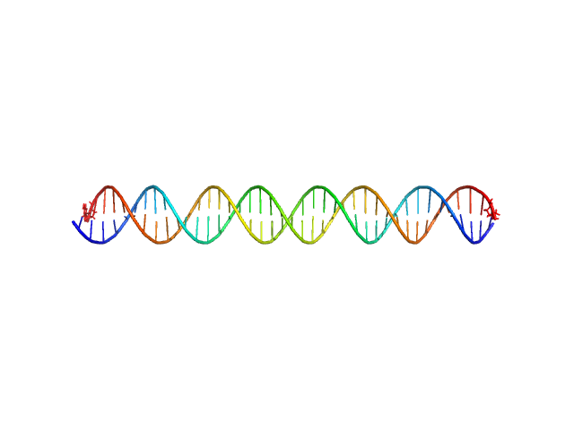
SASDAN8 – mLBS1-2 DNA

MHV-68 TR DNA experimental SAS data
CRYSOL model
Sample: MHV-68 TR DNA monomer, 30 kDa unidentified herpesvirus DNA
Buffer: 10 mM TRIS 150 mM NaCl, pH: 7.6
Experiment: SAXS data collected at EMBL P12, PETRA III on 2013 Apr 27
KSHV but not MHV-68 LANA induces a strong bend upon binding to terminal repeat viral DNA. Nucleic Acids Res 43(20):10039-54 (2015)
...Petoukhov MV, Correia B, Custodio TF, Juillard F, Tan M, Pires de Miranda M, Carrondo MA, Simas JP, Kaye KM, Svergun DI, McVey CE
RgGuinier 4.0 nm
Dmax 16.0 nm
VolumePorod 50 nm3

SASDAP8 – kLBS1-2 DNA

kLBS1-2 DNA experimental SAS data
CRYSOL model
Sample: kLBS1-2 DNA monomer, 24 kDa unidentified herpesvirus DNA
Buffer: Tris, pH: 7.6
Experiment: SAXS data collected at BM29, ESRF on 2013 Apr 27
KSHV but not MHV-68 LANA induces a strong bend upon binding to terminal repeat viral DNA. Nucleic Acids Res 43(20):10039-54 (2015)
...Petoukhov MV, Correia B, Custodio TF, Juillard F, Tan M, Pires de Miranda M, Carrondo MA, Simas JP, Kaye KM, Svergun DI, McVey CE
RgGuinier 4.0 nm
Dmax 16.0 nm
VolumePorod 50 nm3

SASDAQ8 – kLANA mutant dimer-tetramer mixture

ORF73 tetramerORF73 dimer experimental SAS data
NONE model
Sample: ORF73 tetramer tetramer, 63 kDa Human herpesvirus 8 protein
ORF73 dimer dimer, 32 kDa Human herpesvirus 8 protein
Buffer: 25 mM Na/K Phosphate, pH: 7.5
Experiment: SAXS data collected at BM29, ESRF on 2014 Jun 21
KSHV but not MHV-68 LANA induces a strong bend upon binding to terminal repeat viral DNA. Nucleic Acids Res 43(20):10039-54 (2015)
...Petoukhov MV, Correia B, Custodio TF, Juillard F, Tan M, Pires de Miranda M, Carrondo MA, Simas JP, Kaye KM, Svergun DI, McVey CE
RgGuinier 2.4 nm
Dmax 9.5 nm
VolumePorod 50 nm3

SASDAR8 – mLANA 124-316 mLBS1-2 8:1 complex

MHV-68 TR DNALatency-associated nuclear antigen experimental SAS data
CRYSOL model
Sample: MHV-68 TR DNA monomer, 30 kDa unidentified herpesvirus DNA
Latency-associated nuclear antigen octamer, 269 kDa Murid herpesvirus 4 protein
Buffer: 25 mM Na/K Phosphate, pH: 7.5
Experiment: SAXS data collected at EMBL P12, PETRA III on 2013 Apr 27
KSHV but not MHV-68 LANA induces a strong bend upon binding to terminal repeat viral DNA. Nucleic Acids Res 43(20):10039-54 (2015)
...Petoukhov MV, Correia B, Custodio TF, Juillard F, Tan M, Pires de Miranda M, Carrondo MA, Simas JP, Kaye KM, Svergun DI, McVey CE
RgGuinier 5.8 nm
Dmax 20.0 nm
VolumePorod 475 nm3

SASDAS8 – kLANA 1008-1150 -- kLBS1-2 complex 8:2 (partially dissociated)

kLBS1-2 DNAORF73 tetramerORF73 octamerkLBS1-2 DNA two monomers experimental SAS data
NONE model
Sample: kLBS1-2 DNA monomer, 24 kDa unidentified herpesvirus DNA
ORF73 tetramer tetramer, 63 kDa Human herpesvirus 8 protein
ORF73 octamer octamer, 126 kDa Human herpesvirus 8 protein
kLBS1-2 DNA two monomers dimer, 48 kDa unidentified herpesvirus RNA
Buffer: 25 mM Na/K Phosphate, pH: 7.5
Experiment: SAXS data collected at EMBL P12, PETRA III on 2013 Apr 27
KSHV but not MHV-68 LANA induces a strong bend upon binding to terminal repeat viral DNA. Nucleic Acids Res 43(20):10039-54 (2015)
...Petoukhov MV, Correia B, Custodio TF, Juillard F, Tan M, Pires de Miranda M, Carrondo MA, Simas JP, Kaye KM, Svergun DI, McVey CE
RgGuinier 4.8 nm
Dmax 16.0 nm
VolumePorod 250 nm3

SASDDR9 – NADPH oxidase (H2O2 producing and [F-actin] oxidizing) MICAL1 (monomer)

[F-actin]-monooxygenase MICAL1 (monomer) experimental SAS data
CORAL model
Sample: [F-actin]-monooxygenase MICAL1 (monomer) monomer, 118 kDa Homo sapiens protein
Buffer: 50 mM sodium phosphate buffer, pH 7.5, 5 % glycerol, 100 mM NaCl, 1 mM EDTA, 1 mM DTT, pH: 7.5
Experiment: SAXS data collected at EMBL P12, PETRA III on 2017 Jun 20
Human MICAL1: activation by the small GTPase Rab8 and small-angle X-ray scattering studies on the oligomerization state of MICAL1 and its complex with Rab8. Protein Sci (2018)
...Petoukhov MV, Rai A, Svergun DI, Vanoni MA
RgGuinier 3.7 nm
Dmax 12.1 nm
VolumePorod 212 nm3

SASDDS9 – NADPH oxidase (H2O2 producing and [F-actin] oxidizing) MICAL1 (monomer) (Truncated MOCHLIM construct)

[F-actin]-monooxygenase MICAL1 (MoChLim) experimental SAS data
CORAL model
Sample: [F-actin]-monooxygenase MICAL1 (MoChLim) monomer, 85 kDa Homo sapiens protein
Buffer: 20 mM Hepes/NaOH, pH 7.5, 50 mM NaCl, 2 mM MgCl2, 2 mM DTT, pH: 7.5
Experiment: SAXS data collected at EMBL P12, PETRA III on 2017 Jun 20
Human MICAL1: activation by the small GTPase Rab8 and small-angle X-ray scattering studies on the oligomerization state of MICAL1 and its complex with Rab8. Protein Sci (2018)
...Petoukhov MV, Rai A, Svergun DI, Vanoni MA
RgGuinier 4.0 nm
Dmax 18.0 nm
VolumePorod 145 nm3

SASDDT9 – NADPH oxidase (H2O2 producing and [F-actin] oxidizing) MICAL1 (monomer) in complex with Ras-related protein Rab 8 (MICAL1-Rab8 complex)

[F-actin]-monooxygenase MICAL1 (monomer)Ras-related protein 8 experimental SAS data
CORAL model
Sample: [F-actin]-monooxygenase MICAL1 (monomer) monomer, 118 kDa Homo sapiens protein
Ras-related protein 8 monomer, 20 kDa protein
Buffer: 20 mM Hepes/NaOH, pH 7.5, 50 mM NaCl, 2 mM MgCl2, 2 mM DTT, pH: 7.5
Experiment: SAXS data collected at EMBL P12, PETRA III on 2017 Jun 20
Human MICAL1: activation by the small GTPase Rab8 and small-angle X-ray scattering studies on the oligomerization state of MICAL1 and its complex with Rab8. Protein Sci (2018)
...Petoukhov MV, Rai A, Svergun DI, Vanoni MA
RgGuinier 3.7 nm
Dmax 11.8 nm
VolumePorod 234 nm3

SASDDU9 – NADPH oxidase (H2O2 producing and [F-actin] oxidizing) MICAL1 (monomer) (Truncated MOCH construct)

[F-actin]-monooxygenase MICAL1 (MoCh) experimental SAS data
PDB (PROTEIN DATA BANK) model
Sample: [F-actin]-monooxygenase MICAL1 (MoCh) monomer, 67 kDa Homo sapiens protein
Buffer: 50 mM sodium phosphate buffer, 5 % glycerol, 100 mM NaCl, 1 mM EDTA, 1 mM DTT, pH: 7.5
Experiment: SAXS data collected at EMBL P12, PETRA III on 2016 Jun 6
Human MICAL1: activation by the small GTPase Rab8 and small-angle X-ray scattering studies on the oligomerization state of MICAL1 and its complex with Rab8. Protein Sci (2018)
...Petoukhov MV, Rai A, Svergun DI, Vanoni MA
RgGuinier 3.4 nm
Dmax 12.0 nm
VolumePorod 100 nm3