Search

 
Advanced search  

352 hits found for X33

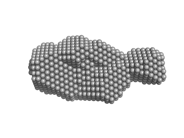
SASDMV4 – Full-length Retinoic acid receptor RXR-alpha in complex with Ramp2 DNA

Retinoic acid receptor RXR-alphaRamp2 DNA experimental SAS data
EOM/RANCH model
Sample: Retinoic acid receptor RXR-alpha dimer, 102 kDa Homo sapiens protein
Ramp2 DNA monomer, 11 kDa DNA
Buffer: 20 mM Tris, 50 mM NaCl, 50 mM KCl, 5% glycerol, 2 mM Chaps, and 5 mM DTT, pH: 7.5
Experiment: SAXS data collected at ...X33, DORIS III, DESY on 2010 May 7
Solution Behavior of the Intrinsically Disordered N-Terminal Domain of Retinoid X Receptor α in the Context of the Full-Length Protein Biochemistry 55(12):1741-1748 (2016)
Belorusova A, Osz J, Petoukhov M, Peluso-Iltis C, Kieffer B, Svergun D, Rochel N
RgGuinier 4.4 nm
Dmax 13.9 nm
VolumePorod 161 nm3

SASDAW4 – uPAR wild-type AE234 complex

Urokinase plasminogen activator surface receptorsynthetic peptide AE234 experimental SAS data
Urokinase plasminogen activator surface receptor synthetic peptide AE234 Kratky plot
Sample: Urokinase plasminogen activator surface receptor monomer, 37 kDa Homo sapiens protein
synthetic peptide AE234 monomer, 1 kDa protein
Buffer: 25 mM Sodium Phosphate 5 % Glycerol 50 mM NaSO4, pH: 7.2
Experiment: SAXS data collected at ...X33, DORIS III, DESY on 2010 Jun 10
A flexible multidomain structure drives the function of the urokinase-type plasminogen activator receptor (uPAR). J Biol Chem 287(41):34304-15 (2012)
Mertens HD, Kjaergaard M, Mysling S, Gårdsvoll H, Jørgensen TJ, Svergun DI, Ploug M
RgGuinier 2.4 nm
Dmax 8.3 nm
VolumePorod 57 nm3

SASDMW4 – Retinoic acid receptor RXR-alpha N-terminal and DNA binding domains (DBD) in complex with Ramp2 DNA

Ramp2 DNARetinoic acid receptor RXR-alpha experimental SAS data
EOM/RANCH model
Sample: Ramp2 DNA monomer, 11 kDa DNA
Retinoic acid receptor RXR-alpha dimer, 44 kDa Homo sapiens protein
Buffer: 20 mM Tris, 50 mM NaCl, 50 mM KCl, 5% glycerol, 2 mM Chaps, and 5 mM DTT, pH: 7.5
Experiment: SAXS data collected at ...X33, DORIS III, DESY on 2009 Jul 17
Solution Behavior of the Intrinsically Disordered N-Terminal Domain of Retinoid X Receptor α in the Context of the Full-Length Protein Biochemistry 55(12):1741-1748 (2016)
Belorusova A, Osz J, Petoukhov M, Peluso-Iltis C, Kieffer B, Svergun D, Rochel N
RgGuinier 4.3 nm
Dmax 14.3 nm
VolumePorod 89 nm3

SASDAX4 – uPAR wild-type ATF complex

Urokinase plasminogen activator surface receptorUrokinase-type plasminogen activator experimental SAS data
Urokinase plasminogen activator surface receptor Urokinase-type plasminogen activator Kratky plot
Sample: Urokinase plasminogen activator surface receptor monomer, 37 kDa Homo sapiens protein
Urokinase-type plasminogen activator monomer, 49 kDa Homo sapiens protein
Buffer: 25 mM Sodium Phosphate 5 % Glycerol 50 mM NaSO4, pH: 7.2
Experiment: SAXS data collected at ...X33, DORIS III, DESY on 2010 Jun 10
A flexible multidomain structure drives the function of the urokinase-type plasminogen activator receptor (uPAR). J Biol Chem 287(41):34304-15 (2012)
Mertens HD, Kjaergaard M, Mysling S, Gårdsvoll H, Jørgensen TJ, Svergun DI, Ploug M
RgGuinier 2.8 nm
Dmax 9.4 nm
VolumePorod 102 nm3

SASDLX4 – Full-length flavorubredoxin

Anaerobic nitric oxide reductase flavorubredoxin experimental SAS data
Full-length flavorubredoxin Rg histogram
Sample: Anaerobic nitric oxide reductase flavorubredoxin tetramer, 217 kDa Escherichia coli (strain … protein
Buffer: 50 mM Tris-HCl, 18% glycerol, pH: 7.5
Experiment: SAXS data collected at ...X33, DORIS III, DESY on 2006 Nov 19
Quaternary Structure of Flavorubredoxin as Revealed by Synchrotron Radiation Small-Angle X-Ray Scattering Structure 16(9):1428-1436 (2008)
Petoukhov M, Vicente J, Crowley P, Carrondo M, Teixeira M, Svergun D
RgGuinier 4.1 nm
Dmax 14.2 nm
VolumePorod 297 nm3

SASDMX4 – Retinoic acid receptor RXR-alpha DNA binding (DBD) and ligand binding (LBD) domains in complex with Ramp2 DNA

Ramp2 DNARetinoic acid receptor RXR-alpha experimental SAS data
CORAL model
Sample: Ramp2 DNA monomer, 11 kDa DNA
Retinoic acid receptor RXR-alpha dimer, 74 kDa Homo sapiens protein
Buffer: 20 mM Tris, 50 mM NaCl, 50 mM KCl, 5% glycerol, 2 mM Chaps, and 5 mM DTT, pH: 7.5
Experiment: SAXS data collected at ...X33, DORIS III, DESY on 2010 Oct 7
Solution Behavior of the Intrinsically Disordered N-Terminal Domain of Retinoid X Receptor α in the Context of the Full-Length Protein Biochemistry 55(12):1741-1748 (2016)
Belorusova A, Osz J, Petoukhov M, Peluso-Iltis C, Kieffer B, Svergun D, Rochel N
RgGuinier 3.6 nm
Dmax 12.2 nm

SASDAY4 – fH1015

Factor H CCP modules 10 to 15 experimental SAS data
DAMMIF model
Sample: Factor H CCP modules 10 to 15 monomer, 41 kDa Homo sapiens protein
Buffer: 50 mM Potassium Phosphate, pH: 7.4
Experiment: SAXS data collected at ...X33, DORIS III, DESY on 2008 Dec 2
The central portion of factor H (modules 10-15) is compact and contains a structurally deviant CCP module. J Mol Biol 395(1):105-22 (2010)
Schmidt CQ, Herbert AP, Mertens HD, Guariento M, Soares DC, Uhrin D, Rowe AJ, Svergun DI, Barlow PN
RgGuinier 3.0 nm
Dmax 10.4 nm
VolumePorod 68 nm3

SASDMY4 – Retinoic acid receptor RXR-alpha DNA binding domain (DBD) in complex with Ramp2

Ramp2 DNARetinoic acid receptor RXR-alpha experimental SAS data
CUSTOM IN-HOUSE model
Sample: Ramp2 DNA monomer, 11 kDa DNA
Retinoic acid receptor RXR-alpha dimer, 20 kDa Homo sapiens protein
Buffer: 20 mM Tris, 50 mM NaCl, 50 mM KCl, 5% glycerol, 2 mM Chaps, and 5 mM DTT, pH: 7.5
Experiment: SAXS data collected at ...X33, DORIS III, DESY on 2010 Oct 7
Solution Behavior of the Intrinsically Disordered N-Terminal Domain of Retinoid X Receptor α in the Context of the Full-Length Protein Biochemistry 55(12):1741-1748 (2016)
Belorusova A, Osz J, Petoukhov M, Peluso-Iltis C, Kieffer B, Svergun D, Rochel N
RgGuinier 1.9 nm
Dmax 5.8 nm

SASDAZ4 – fH1114

Factor H CCP modules 11 to 14 experimental SAS data
DAMMIF model
Sample: Factor H CCP modules 11 to 14 monomer, 28 kDa Homo sapiens protein
Buffer: 50 mM Potassium Phosphate, pH: 7.4
Experiment: SAXS data collected at ...X33, DORIS III, DESY on 2008 Dec 2
The central portion of factor H (modules 10-15) is compact and contains a structurally deviant CCP module. J Mol Biol 395(1):105-22 (2010)
Schmidt CQ, Herbert AP, Mertens HD, Guariento M, Soares DC, Uhrin D, Rowe AJ, Svergun DI, Barlow PN
RgGuinier 3.0 nm
Dmax 10.5 nm
VolumePorod 38 nm3

SASDMZ4 – Mixture of estrogen-related receptor gamma:Inverse repeat IR3 DNA - 6:3 and 2:1 complexes - at 2.9 mg/ml

Estrogen-related receptor gammaInverse repeat IR3 DNA experimental SAS data
SASREF MX model
Sample: Estrogen-related receptor gamma hexamer, 230 kDa Homo sapiens protein
Inverse repeat IR3 DNA trimer, 46 kDa DNA
Buffer: 20 mM Tris-HCl, 100 mM NaCl, 100 mM KCl, 5 mM MgCl2, 1% (v/v) glycerol, and 1 mM CHAPS, pH: 8
Experiment: SAXS data collected at ...X33, DORIS III, DESY on 2009 Mar 3
Reconstruction of Quaternary Structure from X-ray Scattering by Equilibrium Mixtures of Biological Macromolecules Biochemistry 52(39):6844-6855 (2013)
Petoukhov M, Billas I, Takacs M, Graewert M, Moras D, Svergun D
RgGuinier 4.7 nm
Dmax 14.6 nm

SASDA25 – fh1213

Factor H CCP modules 12 to 13 experimental SAS data
CRYSOL model
Sample: Factor H CCP modules 12 to 13 monomer, 14 kDa Homo sapiens protein
Buffer: 50 mM Potassium Phosphate, pH: 7.4
Experiment: SAXS data collected at ...X33, DORIS III, DESY on 2008 Dec 2
The central portion of factor H (modules 10-15) is compact and contains a structurally deviant CCP module. J Mol Biol 395(1):105-22 (2010)
Schmidt CQ, Herbert AP, Mertens HD, Guariento M, Soares DC, Uhrin D, Rowe AJ, Svergun DI, Barlow PN
RgGuinier 2.0 nm
Dmax 7.1 nm
VolumePorod 20 nm3

SASDM25 – Mixture of estrogen-related receptor gamma:Inverse repeat IR3 DNA - 6:3 and 2:1 complexes - at 1.9 mg/ml

Estrogen-related receptor gammaInverse repeat IR3 DNA experimental SAS data
SASREF MX model
Sample: Estrogen-related receptor gamma hexamer, 230 kDa Homo sapiens protein
Inverse repeat IR3 DNA trimer, 46 kDa DNA
Buffer: 20 mM Tris-HCl, 100 mM NaCl, 100 mM KCl, 5 mM MgCl2, 1% (v/v) glycerol, and 1 mM CHAPS, pH: 8
Experiment: SAXS data collected at ...X33, DORIS III, DESY on 2009 Mar 3
Reconstruction of Quaternary Structure from X-ray Scattering by Equilibrium Mixtures of Biological Macromolecules Biochemistry 52(39):6844-6855 (2013)
Petoukhov M, Billas I, Takacs M, Graewert M, Moras D, Svergun D
RgGuinier 4.3 nm
Dmax 13.2 nm

SASDB35 – Staphylococcus aureus thiaminase II

Thiaminase type II enzyme experimental SAS data
PDB (PROTEIN DATA BANK) model
Sample: Thiaminase type II enzyme tetramer, 107 kDa Staphylococcus aureus protein
Buffer: 100 mM Tris-HCl, pH: 7.5
Experiment: SAXS data collected at ...X33, DORIS III, DESY on 2011 May 11
Staphylococcus aureus thiaminase II: oligomerization warrants proteolytic protection against serine proteases. Acta Crystallogr D Biol Crystallogr 69(Pt 12):2320-9 (2013)
Begum A, Drebes J, Kikhney A, Müller IB, Perbandt M, Svergun D, Wrenger C, Betzel C
RgGuinier 3.4 nm
Dmax 11.0 nm
VolumePorod 168 nm3

SASDM35 – Mixture of estrogen-related receptor gamma:Inverse repeat IR3 DNA - 6:3 and 2:1 complexes - at 1.0 mg/ml

Estrogen-related receptor gammaInverse repeat IR3 DNA experimental SAS data
SASREF MX model
Sample: Estrogen-related receptor gamma hexamer, 230 kDa Homo sapiens protein
Inverse repeat IR3 DNA trimer, 46 kDa DNA
Buffer: 20 mM Tris-HCl, 100 mM NaCl, 100 mM KCl, 5 mM MgCl2, 1% (v/v) glycerol, and 1 mM CHAPS, pH: 8
Experiment: SAXS data collected at ...X33, DORIS III, DESY on 2009 Mar 3
Reconstruction of Quaternary Structure from X-ray Scattering by Equilibrium Mixtures of Biological Macromolecules Biochemistry 52(39):6844-6855 (2013)
Petoukhov M, Billas I, Takacs M, Graewert M, Moras D, Svergun D
RgGuinier 4.1 nm
Dmax 12.8 nm

SASDA45 – PpoA wild type enzyme

Psi-producing oxygenase A experimental SAS data
SASREF model
Sample: Psi-producing oxygenase A trimer, 362 kDa Aspergillus nidulans protein
Buffer: 20 Mm HEPES, pH: 7.5
Experiment: SAXS data collected at ...X33, DORIS III, DESY on 2011 Jun 24
A structural model of PpoA derived from SAXS-analysis-implications for substrate conversion. Biochim Biophys Acta 1831(9):1449-57 (2013)
Koch C, Tria G, Fielding AJ, Brodhun F, Valerius O, Feussner K, Braus GH, Svergun DI, Bennati M, Feussner I
RgGuinier 5.4 nm
Dmax 16.5 nm
VolumePorod 540 nm3

SASDA55 – Nucleoplasmin

Nucleoplasmin experimental SAS data
BUNCH model
Sample: Nucleoplasmin pentamer, 110 kDa Escherichia coli protein
Buffer: 20 mM Pipes buffer 150 mM NaCl, pH: 7.5
Experiment: SAXS data collected at ...X33, DORIS III, DESY on 2007 Dec 3
A mechanism for histone chaperoning activity of nucleoplasmin: thermodynamic and structural models. J Mol Biol 393(2):448-63 (2009)
Taneva SG, Bañuelos S, Falces J, Arregi I, Muga A, Konarev PV, Svergun DI, Velázquez-Campoy A, Urbaneja MA
RgGuinier 4.0 nm
Dmax 12.6 nm
VolumePorod 210 nm3

SASDA65 – Nucleoplasmin-H5 complex

NP-H5 experimental SAS data
MONSA model
Sample: NP-H5 pentamer, 200 kDa Escherichia coli protein
Buffer: 20 mM Pipes buffer 150 mM NaCl, pH: 7.5
Experiment: SAXS data collected at ...X33, DORIS III, DESY on 2007 Dec 3
A mechanism for histone chaperoning activity of nucleoplasmin: thermodynamic and structural models. J Mol Biol 393(2):448-63 (2009)
Taneva SG, Bañuelos S, Falces J, Arregi I, Muga A, Konarev PV, Svergun DI, Velázquez-Campoy A, Urbaneja MA
RgGuinier 5.2 nm
Dmax 16.7 nm
VolumePorod 410 nm3

SASDA75 – Nucleoplasmin-H2AH2B complex

NP-H2AH2B experimental SAS data
MONSA model
Sample: NP-H2AH2B pentamer, 250 kDa Escherichia coli protein
Buffer: 20 mM Pipes buffer 150 mM NaCl, pH: 7.5
Experiment: SAXS data collected at ...X33, DORIS III, DESY on 2014 Dec 3
A mechanism for histone chaperoning activity of nucleoplasmin: thermodynamic and structural models. J Mol Biol 393(2):448-63 (2009)
Taneva SG, Bañuelos S, Falces J, Arregi I, Muga A, Konarev PV, Svergun DI, Velázquez-Campoy A, Urbaneja MA
RgGuinier 4.7 nm
Dmax 14.5 nm
VolumePorod 470 nm3

SASDA85 – CHD4 (PP-CC-AH-D)

Human Chromatin Remodeler CHD4 (363-1353) experimental SAS data
DAMMIF model
Sample: Human Chromatin Remodeler CHD4 (363-1353) monomer, 117 kDa Homo sapiens protein
Buffer: 50 mM HEPES 5% Glycerol 300 mM NaCl, pH: 7.5
Experiment: SAXS data collected at ...X33, DORIS III, DESY on 2010 Nov 13
The PHD and chromo domains regulate the ATPase activity of the human chromatin remodeler CHD4. J Mol Biol 422(1):3-17 (2012)
Watson AA, Mahajan P, Mertens HD, Deery MJ, Zhang W, Pham P, Du X, Bartke T, Zhang W, Edlich C, Berridge G, Chen Y, Burgess-Brown NA, Kouzarides T, Wiechens N, Owen-Hughes T, Svergun DI, Gileadi O, La...
RgGuinier 4.9 nm
Dmax 17.4 nm

SASDA95 – CHD4 (CC-AH-D)

Human Chromatin Remodeler CHD4 (494-1353) experimental SAS data
DAMMIF model
Sample: Human Chromatin Remodeler CHD4 (494-1353) monomer, 101 kDa Homo sapiens protein
Buffer: 50 mM HEPES, 50 mM KCl, pH: 7.5
Experiment: SAXS data collected at ...X33, DORIS III, DESY on 2010 Nov 13
The PHD and chromo domains regulate the ATPase activity of the human chromatin remodeler CHD4. J Mol Biol 422(1):3-17 (2012)
Watson AA, Mahajan P, Mertens HD, Deery MJ, Zhang W, Pham P, Du X, Bartke T, Zhang W, Edlich C, Berridge G, Chen Y, Burgess-Brown NA, Kouzarides T, Wiechens N, Owen-Hughes T, Svergun DI, Gileadi O, La...
RgGuinier 4.0 nm
Dmax 14.5 nm

SASDAA5 – CHD4 (AH)

Human Chromatin Remodeler CHD4 (685-1233) experimental SAS data
DAMMIF model
Sample: Human Chromatin Remodeler CHD4 (685-1233) monomer, 63 kDa Homo sapiens protein
Buffer: 50 mM HEPES, 50 mM KCl, pH: 7.5
Experiment: SAXS data collected at ...X33, DORIS III, DESY on 2010 Nov 13
The PHD and chromo domains regulate the ATPase activity of the human chromatin remodeler CHD4. J Mol Biol 422(1):3-17 (2012)
Watson AA, Mahajan P, Mertens HD, Deery MJ, Zhang W, Pham P, Du X, Bartke T, Zhang W, Edlich C, Berridge G, Chen Y, Burgess-Brown NA, Kouzarides T, Wiechens N, Owen-Hughes T, Svergun DI, Gileadi O, La...
RgGuinier 4.0 nm
Dmax 14.5 nm

SASDAB5 – CHD4 (PP-CC)

Human Chromatin Remodeler CHD4 (363-682) experimental SAS data
DAMMIF model
Sample: Human Chromatin Remodeler CHD4 (363-682) monomer, 38 kDa Homo sapiens protein
Buffer: 50 mM HEPES, 50 mM KCl, pH: 7.5
Experiment: SAXS data collected at ...X33, DORIS III, DESY on 2010 Nov 13
The PHD and chromo domains regulate the ATPase activity of the human chromatin remodeler CHD4. J Mol Biol 422(1):3-17 (2012)
Watson AA, Mahajan P, Mertens HD, Deery MJ, Zhang W, Pham P, Du X, Bartke T, Zhang W, Edlich C, Berridge G, Chen Y, Burgess-Brown NA, Kouzarides T, Wiechens N, Owen-Hughes T, Svergun DI, Gileadi O, La...
RgGuinier 3.0 nm
Dmax 10.5 nm

SASDAC5 – ImportinA_ImportinB

ImportinA_ImportinB experimental SAS data
DAMMIF model
Sample: ImportinA_ImportinB monomer, 160 kDa Escherichia coli protein
Buffer: 50 mM Tris-HCL 150 mM NaCl 1.0 mM DTT, pH: 7.5
Experiment: SAXS data collected at ...X33, DORIS III, DESY on 2009 Oct 29
Recognition of nucleoplasmin by its nuclear transport receptor importin α/β: insights into a complete import complex. Biochemistry 49(45):9756-69 (2010)
Falces J, Arregi I, Konarev PV, Urbaneja MA, Svergun DI, Taneva SG, Bañuelos S
RgGuinier 5.7 nm
Dmax 19.0 nm
VolumePorod 390 nm3

SASDAD5 – Nucleoplasmin_ImpA_ImpB

Nucleoplasmin_importinA_importinB experimental SAS data
DAMMIF model
Sample: Nucleoplasmin_importinA_importinB , 900 kDa Escherichia coli protein
Buffer: 50 mM Tris-HCL 150 mM NaCl 1.0 mM DTT, pH: 7.5
Experiment: SAXS data collected at ...X33, DORIS III, DESY on 2008 May 23
Recognition of nucleoplasmin by its nuclear transport receptor importin α/β: insights into a complete import complex. Biochemistry 49(45):9756-69 (2010)
Falces J, Arregi I, Konarev PV, Urbaneja MA, Svergun DI, Taneva SG, Bañuelos S
RgGuinier 8.6 nm
Dmax 28.0 nm
VolumePorod 1800 nm3

SASDAE5 – CYNEX4

CYNEX4 FRET probe, (eYFP-AnnexinA4-eCFP) experimental SAS data
DAMMIF model
Sample: CYNEX4 FRET probe, (eYFP-AnnexinA4-eCFP) monomer, 93 kDa Homo sapiens protein
Buffer: 50 mM HEPES 150 mM NaCl 1 mM EGTA, pH: 7.5
Experiment: SAXS data collected at ...X33, DORIS III, DESY on 2010 Sep 11
Conformational analysis of a genetically encoded FRET biosensor by SAXS. Biophys J 102(12):2866-75 (2012)
Mertens HD, Piljić A, Schultz C, Svergun DI
RgGuinier 3.7 nm
Dmax 12.8 nm
VolumePorod 135 nm3

SASDAF5 – CYNEX4-T266D

CYNEX4 FRET probe, (eYFP-AnnexinA4-eCFP) T266D mutant experimental SAS data
DAMMIF model
Sample: CYNEX4 FRET probe, (eYFP-AnnexinA4-eCFP) T266D mutant monomer, 93 kDa Homo sapiens protein
Buffer: 50 mM HEPES, 50 mM KCl, pH: 7.5
Experiment: SAXS data collected at ...X33, DORIS III, DESY on 2010 Sep 11
Conformational analysis of a genetically encoded FRET biosensor by SAXS. Biophys J 102(12):2866-75 (2012)
Mertens HD, Piljić A, Schultz C, Svergun DI
RgGuinier 4.1 nm
Dmax 14.4 nm
VolumePorod 146 nm3

SASDAG5 – RNA shaperone Hfq

RNA chaperone Hfq experimental SAS data
DAMMIN model
Sample: RNA chaperone Hfq hexamer, 67 kDa Escherichia coli protein
Buffer: 50 mM Tris-HCL 150 mM NaCl 1.0 mM DTT, pH: 7.5
Experiment: SAXS data collected at ...X33, DORIS III, DESY on 2008 May 2
Structural insights into the dynamics and function of the C-terminus of the E. coli RNA chaperone Hfq. Nucleic Acids Res 39(11):4900-15 (2011)
Beich-Frandsen M, Vecerek B, Konarev PV, Sjöblom B, Kloiber K, Hämmerle H, Rajkowitsch L, Miles AJ, Kontaxis G, Wallace BA, Svergun DI, Konrat R, Bläsi U, Djinovic-Carugo K
RgGuinier 3.2 nm
Dmax 11.2 nm
VolumePorod 110 nm3

SASDLG5 – Human nerve growth factor

Beta-nerve growth factor experimental SAS data
PDB (PROTEIN DATA BANK) model
Sample: Beta-nerve growth factor dimer, 24 kDa Homo sapiens protein
Buffer: 50 mM Na-phosphate, 1 mM EDTA, pH: 7
Experiment: SAXS data collected at ...X33, DORIS III, DESY on 2007 Jul 2
The conundrum of the high-affinity NGF binding site formation unveiled? Biophys J 108(3):687-97 (2015)
Covaceuszach S, Konarev PV, Cassetta A, Paoletti F, Svergun DI, Lamba D, Cattaneo A
RgGuinier 2.5 nm

SASDAH5 – Complex of Hfq with DsrA

RNA chaperone HfqRNA DsrA experimental SAS data
SASREF model
Sample: RNA chaperone Hfq hexamer, 67 kDa Escherichia coli protein
RNA DsrA monomer, 12 kDa RNA
Buffer: 50 mM Tris-HCL 150 mM NaCl 1.0 mM DTT, pH: 7.5
Experiment: SAXS data collected at ...X33, DORIS III, DESY on 2010 Nov 16
Structural flexibility of RNA as molecular basis for Hfq chaperone function. Nucleic Acids Res 40(16):8072-84 (2012)
Ribeiro Ede A Jr, Beich-Frandsen M, Konarev PV, Shang W, Vecerek B, Kontaxis G, Hämmerle H, Peterlik H, Svergun DI, Bläsi U, Djinović-Carugo K
RgGuinier 4.3 nm
Dmax 14.5 nm
VolumePorod 210 nm3

SASDLH5 – Aggregation state of Ataxin-3 protein

Ataxin-3 experimental SAS data
DAMMIN model
Sample: Ataxin-3 monomer, 41 kDa Homo sapiens protein
Buffer: phosphate buffered saline (PBS), pH: 7.4
Experiment: SAXS data collected at ...X33, DORIS III, DESY on 2009 Dec 3
Interactions of ataxin-3 with its molecular partners in the protein machinery that sorts protein aggregates to the aggresome. Int J Biochem Cell Biol 51:58-64 (2014)
Bonanomi M, Mazzucchelli S, D'Urzo A, Nardini M, Konarev PV, Invernizzi G, Svergun DI, Vanoni M, Regonesi ME, Tortora P
RgGuinier 6.9 nm
Dmax 25.0 nm
VolumePorod 425 nm3

SASDAJ5 – Annexin-A4

Annexin-A4 experimental SAS data
CRYSOL model
Sample: Annexin-A4 monomer, 36 kDa Homo sapiens protein
Buffer: 50 mM HEPES, 50 mM KCl, pH: 7.5
Experiment: SAXS data collected at ...X33, DORIS III, DESY on 2010 Sep 11
Conformational analysis of a genetically encoded FRET biosensor by SAXS. Biophys J 102(12):2866-75 (2012)
Mertens HD, Piljić A, Schultz C, Svergun DI
RgGuinier 2.4 nm
Dmax 7.6 nm
VolumePorod 54 nm3

SASDLJ5 – Aggregation state of tubulin protein

Tubulin alpha-1A chain experimental SAS data
DAMMIN model
Sample: Tubulin alpha-1A chain monomer, 50 kDa Homo sapiens protein
Buffer: phosphate buffered saline (PBS), pH: 7.4
Experiment: SAXS data collected at ...X33, DORIS III, DESY on 2009 Dec 3
Interactions of ataxin-3 with its molecular partners in the protein machinery that sorts protein aggregates to the aggresome. Int J Biochem Cell Biol 51:58-64 (2014)
Bonanomi M, Mazzucchelli S, D'Urzo A, Nardini M, Konarev PV, Invernizzi G, Svergun DI, Vanoni M, Regonesi ME, Tortora P
RgGuinier 7.0 nm
Dmax 25.0 nm
VolumePorod 340 nm3

SASDAK5 – Myomesin-1 My12-My13

Myomesin-1 experimental SAS data
CRYSOL model
Sample: Myomesin-1 dimer, 46 kDa Homo sapiens protein
Buffer: 25 mM Tris/HCl 150 mM NaCl, pH: 7.5
Experiment: SAXS data collected at ...X33, DORIS III, DESY on 2005 May 7
Molecular basis of the C-terminal tail-to-tail assembly of the sarcomeric filament protein myomesin. EMBO J 27(1):253-64 (2008)
Pinotsis N, Lange S, Perriard JC, Svergun DI, Wilmanns M
RgGuinier 4.0 nm
Dmax 14.5 nm
VolumePorod 56 nm3

SASDLK5 – Aggregation state of ataxin-3-tubulin complex

Ataxin-3Tubulin alpha-1A chain experimental SAS data
DAMMIN model
Sample: Ataxin-3 monomer, 41 kDa Homo sapiens protein
Tubulin alpha-1A chain monomer, 50 kDa Homo sapiens protein
Buffer: phosphate buffered saline (PBS), pH: 7.4
Experiment: SAXS data collected at ...X33, DORIS III, DESY on 2009 Dec 3
Interactions of ataxin-3 with its molecular partners in the protein machinery that sorts protein aggregates to the aggresome. Int J Biochem Cell Biol 51:58-64 (2014)
Bonanomi M, Mazzucchelli S, D'Urzo A, Nardini M, Konarev PV, Invernizzi G, Svergun DI, Vanoni M, Regonesi ME, Tortora P
RgGuinier 8.4 nm
Dmax 30.0 nm
VolumePorod 900 nm3

SASDAL5 – Clostridium difficile bacteriophage 27 endolysin dimer, CD27L

Clostridium difficile bacteriophage 27 endolysin experimental SAS data
NONE model
Sample: Clostridium difficile bacteriophage 27 endolysin dimer, 64 kDa Clostridioides difficile protein
Buffer: 20 mM HEPES 150 mM NaCl, pH: 7.5
Experiment: SAXS data collected at ...X33, DORIS III, DESY on 2011 Mar 17
The CD27L and CTP1L endolysins targeting Clostridia contain a built-in trigger and release factor. PLoS Pathog 10(7):e1004228 (2014)
Dunne M, Mertens HD, Garefalaki V, Jeffries CM, Thompson A, Lemke EA, Svergun DI, Mayer MJ, Narbad A, Meijers R
RgGuinier 3.3 nm
Dmax 10.6 nm
VolumePorod 72 nm3

SASDLL5 – frataxin homolog, Yfh1, at 0 °C

Frataxin homolog, mitochondrial experimental SAS data
frataxin homolog, Yfh1, at 0 °C Rg histogram
Sample: Frataxin homolog, mitochondrial monomer, 14 kDa Saccharomyces cerevisiae (strain … protein
Buffer: 20 mM HEPES, pH: 7
Experiment: SAXS data collected at ...X33, DORIS III, DESY on 2008 Nov 28
The role of hydration in protein stability: comparison of the cold and heat unfolded states of Yfh1. J Mol Biol 417(5):413-24 (2012)
Adrover M, Martorell G, Martin SR, Urosev D, Konarev PV, Svergun DI, Daura X, Temussi P, Pastore A
RgGuinier 2.2 nm

SASDLM5 – frataxin homolog, Yfh1, at 20 °C

Frataxin homolog, mitochondrial experimental SAS data
frataxin homolog, Yfh1, at 20 °C Rg histogram
Sample: Frataxin homolog, mitochondrial monomer, 14 kDa Saccharomyces cerevisiae (strain … protein
Buffer: 20 mM HEPES, pH: 7
Experiment: SAXS data collected at ...X33, DORIS III, DESY on 2008 Nov 28
The role of hydration in protein stability: comparison of the cold and heat unfolded states of Yfh1. J Mol Biol 417(5):413-24 (2012)
Adrover M, Martorell G, Martin SR, Urosev D, Konarev PV, Svergun DI, Daura X, Temussi P, Pastore A
RgGuinier 2.1 nm

SASDAN5 – Cross-linked complex CytC_Adr

Cytochrome CAdrenodoxin experimental SAS data
DAMMIN model
Sample: Cytochrome C monomer, 11 kDa Escherichia coli protein
Adrenodoxin monomer, 11 kDa Escherichia coli protein
Buffer: 20 mM HEPES 2 mM DTT, pH: 7.4
Experiment: SAXS data collected at ...X33, DORIS III, DESY on 2006 Jun 15
Dynamics in a pure encounter complex of two proteins studied by solution scattering and paramagnetic NMR spectroscopy. J Am Chem Soc 130(20):6395-403 (2008)
Xu X, Reinle W, Hannemann F, Konarev PV, Svergun DI, Bernhardt R, Ubbink M
RgGuinier 2.1 nm
Dmax 8.5 nm
VolumePorod 42 nm3

SASDLN5 – frataxin homolog, Yfh1, at 50 °C

Frataxin homolog, mitochondrial experimental SAS data
frataxin homolog, Yfh1, at 50 °C Rg histogram
Sample: Frataxin homolog, mitochondrial monomer, 14 kDa Saccharomyces cerevisiae (strain … protein
Buffer: 20 mM HEPES, pH: 7
Experiment: SAXS data collected at ...X33, DORIS III, DESY on 2008 Nov 28
The role of hydration in protein stability: comparison of the cold and heat unfolded states of Yfh1. J Mol Biol 417(5):413-24 (2012)
Adrover M, Martorell G, Martin SR, Urosev D, Konarev PV, Svergun DI, Daura X, Temussi P, Pastore A
RgGuinier 2.5 nm

SASDAP5 – Native complex CytC_Adr

Cytochrome C dimerAdrenodoxin dimer experimental SAS data
DAMMIN model
Sample: Cytochrome C dimer dimer, 22 kDa Escherichia coli protein
Adrenodoxin dimer dimer, 22 kDa Escherichia coli protein
Buffer: 20 mM HEPES 2 mM DTT, pH: 7.4
Experiment: SAXS data collected at ...X33, DORIS III, DESY on 2006 Jun 15
Dynamics in a pure encounter complex of two proteins studied by solution scattering and paramagnetic NMR spectroscopy. J Am Chem Soc 130(20):6395-403 (2008)
Xu X, Reinle W, Hannemann F, Konarev PV, Svergun DI, Bernhardt R, Ubbink M
RgGuinier 2.9 nm
Dmax 9.5 nm
VolumePorod 64 nm3

SASDAQ5 – Lumazine Synthase

Lumazine Synthase experimental SAS data
DAMMIN model
Sample: Lumazine Synthase , 960 kDa Bacillus subtilis protein
Buffer: Borate buffer, pH: 7
Experiment: SAXS data collected at ...X33, DORIS III, DESY on 2004 Nov 25
Multiple assembly states of lumazine synthase: a model relating catalytic function and molecular assembly. J Mol Biol 362(4):753-70 (2006)
Zhang X, Konarev PV, Petoukhov MV, Svergun DI, Xing L, Cheng RH, Haase I, Fischer M, Bacher A, Ladenstein R, Meining W
RgGuinier 6.2 nm
Dmax 15.5 nm
VolumePorod 1450 nm3

SASDLQ5 – Tricorn protease oligomers

Tricorn protease experimental SAS data
PDB (PROTEIN DATA BANK) model
Sample: Tricorn protease hexamer, 730 kDa Thermoplasma acidophilum (strain … protein
Buffer: 20mM Tris-HCl, 100 mM NaCl, pH: 7.5
Experiment: SAXS data collected at ...X33, DORIS III, DESY on 2003 Jan 14
X-ray snapshots of peptide processing in mutants of tricorn-interacting factor F1 from Thermoplasma acidophilum. J Biol Chem 280(39):33387-96 (2005)
Goettig P, Brandstetter H, Groll M, Göhring W, Konarev PV, Svergun DI, Huber R, Kim JS
RgGuinier 5.9 nm

SASDAR5 – ProNGF

ProNGF experimental SAS data
DAMMIN model
Sample: ProNGF dimer, 45 kDa Mouse submandibulary glands protein
Buffer: 50 mM Naphosphat 0.5 M Ammonium Sulfate(NH4)2SO4, pH: 7
Experiment: SAXS data collected at ...X33, DORIS III, DESY on 2007 Oct 25
Intrinsic structural disorder of mouse proNGF. Proteins 75(4):990-1009 (2009)
Paoletti F, Covaceuszach S, Konarev PV, Gonfloni S, Malerba F, Schwarz E, Svergun DI, Cattaneo A, Lamba D
RgGuinier 2.6 nm
Dmax 8.0 nm
VolumePorod 92 nm3

SASDLR5 – Multidomain Regulatory Protein Rv1364c (monomer)

Multidomain regulatory protein Rv1364c experimental SAS data
GASBOR model
Sample: Multidomain regulatory protein Rv1364c monomer, 70 kDa Mycobacterium tuberculosis (strain … protein
Buffer: 25 mM Tris-HCl, 50 mM NaCl, pH: 7.5
Experiment: SAXS data collected at ...X33, DORIS III, DESY on 2007 Feb 14
Structural characterization of the multidomain regulatory protein Rv1364c from Mycobacterium tuberculosis. Structure 19(1):56-69 (2011)
King-Scott J, Konarev PV, Panjikar S, Jordanova R, Svergun DI, Tucker PA
RgGuinier 3.8 nm
Dmax 14.0 nm
VolumePorod 130 nm3

SASDAS5 – ad11 Fab

aD11 Fab experimental SAS data
DAMMIN model
Sample: aD11 Fab dimer, 50 kDa Mus musculus protein
Buffer: 50 mM Na-phosphate 1mM ethylenediaminetetraacetic aci, pH: 7
Experiment: SAXS data collected at ...X33, DORIS III, DESY on 2005 Oct 20
Dissecting NGF interactions with TrkA and p75 receptors by structural and functional studies of an anti-NGF neutralizing antibody. J Mol Biol 381(4):881-96 (2008)
Covaceuszach S, Cassetta A, Konarev PV, Gonfloni S, Rudolph R, Svergun DI, Lamba D, Cattaneo A
RgGuinier 2.6 nm
Dmax 8.0 nm
VolumePorod 95 nm3

SASDLS5 – Multidomain Regulatory Protein Rv1364c (dimer)

Multidomain regulatory protein Rv1364c experimental SAS data
GASBOR model
Sample: Multidomain regulatory protein Rv1364c dimer, 139 kDa Mycobacterium tuberculosis (strain … protein
Buffer: 25 mM Tris-HCl, 50 mM NaCl, pH: 7.5
Experiment: SAXS data collected at ...X33, DORIS III, DESY on 2007 Feb 14
Structural characterization of the multidomain regulatory protein Rv1364c from Mycobacterium tuberculosis. Structure 19(1):56-69 (2011)
King-Scott J, Konarev PV, Panjikar S, Jordanova R, Svergun DI, Tucker PA
RgGuinier 4.5 nm
Dmax 18.0 nm
VolumePorod 289 nm3

SASDAT5 – NGF

NGF experimental SAS data
DAMMIN model
Sample: NGF dimer, 50 kDa Mus musculus protein
Buffer: 10 mM Na-phosphate 150 mM NaCl, pH: 7.8
Experiment: SAXS data collected at ...X33, DORIS III, DESY on 2005 Oct 20
Dissecting NGF interactions with TrkA and p75 receptors by structural and functional studies of an anti-NGF neutralizing antibody. J Mol Biol 381(4):881-96 (2008)
Covaceuszach S, Cassetta A, Konarev PV, Gonfloni S, Rudolph R, Svergun DI, Lamba D, Cattaneo A
RgGuinier 3.1 nm
Dmax 12.0 nm
VolumePorod 102 nm3

SASDLT5 – Modification methylase SsoII (M.SsoII) protein bound to 12-bp DNA

Modification methylase SsoII12-bp DNA experimental SAS data
Modification methylase SsoII (M.SsoII) protein bound to 12-bp DNA Rg histogram
Sample: Modification methylase SsoII monomer, 43 kDa Shigella sonnei protein
12-bp DNA monomer, 8 kDa DNA
Buffer: 50 mM Na-phosphate buffer, pH: 7
Experiment: SAXS data collected at ...X33, DORIS III, DESY on 2007 Mar 13
Flexibility of the linker between the domains of DNA methyltransferase SsoII revealed by small-angle X-ray scattering: implications for transcription regulation in SsoII restriction-modification syste... PLoS One 9(4):e93453 (2014)
Konarev PV, Kachalova GS, Ryazanova AY, Kubareva EA, Karyagina AS, Bartunik HD, Svergun DI
RgGuinier 2.8 nm
Dmax 11.0 nm
VolumePorod 85 nm3

SASDAU5 – ad11 Fab + NGF

aD11 FabNGF experimental SAS data
DAMMIN model
Sample: aD11 Fab dimer, 50 kDa Mus musculus protein
NGF dimer, 50 kDa Mus musculus protein
Buffer: 30 mM Na-phosphate 75 mM NaCl, pH: 7
Experiment: SAXS data collected at ...X33, DORIS III, DESY on 2005 Dec 20
Dissecting NGF interactions with TrkA and p75 receptors by structural and functional studies of an anti-NGF neutralizing antibody. J Mol Biol 381(4):881-96 (2008)
Covaceuszach S, Cassetta A, Konarev PV, Gonfloni S, Rudolph R, Svergun DI, Lamba D, Cattaneo A
RgGuinier 4.3 nm
Dmax 16.0 nm
VolumePorod 255 nm3

SASDLU5 – Complex formed between the replication origin-binding protein UL9 (C-terminal domains) with 15-bp DNA

Replication origin-binding protein15-bp DNA experimental SAS data
MONSA model
Sample: Replication origin-binding protein dimer, 81 kDa Human herpesvirus 1 … protein
15-bp DNA monomer, 9 kDa DNA
Buffer: 20 mM Tris-HCl, 50 mM NaCl, 5 mM MgCl2, pH: 7.5
Experiment: SAXS data collected at ...X33, DORIS III, DESY on 2006 May 27
Structural and biophysical characterization of the proteins interacting with the herpes simplex virus 1 origin of replication. J Biol Chem 284(24):16343-16353 (2009)
Manolaridis I, Mumtsidu E, Konarev P, Makhov AM, Fullerton SW, Sinz A, Kalkhof S, McGeehan JE, Cary PD, Griffith JD, Svergun D, Kneale GG, Tucker PA
RgGuinier 3.6 nm
Dmax 15.0 nm

SASDAV5 – apo XMRV RT

apo XMRV RT experimental SAS data
CRYSOL model
Sample: apo XMRV RT monomer, 75 kDa Escherichia coli protein
Buffer: 10 mM HEPES 100 mM KCl 5% Glycerol, pH: 6.5
Experiment: SAXS data collected at ...X33, DORIS III, DESY on 2011 Dec 8
Structural analysis of monomeric retroviral reverse transcriptase in complex with an RNA/DNA hybrid. Nucleic Acids Res 41(6):3874-87 (2013)
Nowak E, Potrzebowski W, Konarev PV, Rausch JW, Bona MK, Svergun DI, Bujnicki JM, Le Grice SF, Nowotny M
RgGuinier 4.0 nm
Dmax 13.5 nm
VolumePorod 160 nm3

SASDLV5 – Myosin Filament-Linking Protein Myomesin-1 (domains My9-My13, dimer)

Myomesin-1 experimental SAS data
DAMMIN model
Sample: Myomesin-1 dimer, 121 kDa Homo sapiens protein
Buffer: 25 mMTris-HCl, 150 mM NaCl, pH: 7.5
Experiment: SAXS data collected at ...X33, DORIS III, DESY on 2005 May 10
Superhelical architecture of the myosin filament-linking protein myomesin with unusual elastic properties. PLoS Biol 10(2):e1001261 (2012)
Pinotsis N, Chatziefthimiou SD, Berkemeier F, Beuron F, Mavridis IM, Konarev PV, Svergun DI, Morris E, Rief M, Wilmanns M
RgGuinier 9.6 nm
Dmax 37.0 nm

SASDAW5 – XMRV RT + DNA/RNA hybrid

apo XMRV RTRNA_DNA hybrid substrate experimental SAS data
CRYSOL model
Sample: apo XMRV RT monomer, 75 kDa Escherichia coli protein
RNA_DNA hybrid substrate monomer, 15 kDa
Buffer: 10 mM HEPES 100 mM KCl 5% Glycerol, pH: 6.5
Experiment: SAXS data collected at ...X33, DORIS III, DESY on 2011 Dec 8
Structural analysis of monomeric retroviral reverse transcriptase in complex with an RNA/DNA hybrid. Nucleic Acids Res 41(6):3874-87 (2013)
Nowak E, Potrzebowski W, Konarev PV, Rausch JW, Bona MK, Svergun DI, Bujnicki JM, Le Grice SF, Nowotny M
RgGuinier 3.5 nm
Dmax 11.5 nm
VolumePorod 155 nm3

SASDLW5 – Cytochrome c nitrite reductase from Thioalkalivibrio nitratireducens

Cytochrome c-552 experimental SAS data
PDB (PROTEIN DATA BANK) model
Sample: Cytochrome c-552 hexamer, 356 kDa Thioalkalivibrio nitratireducens (strain … protein
Buffer: Tris-borate buffer, pH: 8.5
Experiment: SAXS data collected at ...X33, DORIS III, DESY on 2005 May 8
Isolation and oligomeric composition of cytochrome c nitrite reductase from the haloalkaliphilic bacterium Thioalkalivibrio nitratireducens. Biochemistry (Mosc) 73(2):164-70 (2008)
Tikhonova TV, Slutskaya ES, Filimonenkov AA, Boyko KM, Kleimenov SY, Konarev PV, Polyakov KM, Svergun DI, Trofimov AA, Khomenkov VG, Zvyagilskaya RA, Popov VO
RgGuinier 4.8 nm

SASDAX5 – Endophilin-CoA complex

Endophilin-A1 BAR domainarachidonyl-CoA experimental SAS data
SASREF model
Sample: Endophilin-A1 BAR domain dimer, 58 kDa Mus musculus protein
arachidonyl-CoA , 60 kDa
Buffer: 50 mM TRIS-HCL 300 mM NaCl, pH: 8
Experiment: SAXS data collected at ...X33, DORIS III, DESY on 2006 Nov 27
Endophilin-A1 BAR domain interaction with arachidonyl CoA. Front Mol Biosci 1:20 (2014)
Petoukhov MV, Weissenhorn W, Svergun DI
RgGuinier 5.9 nm
Dmax 19.0 nm
VolumePorod 480 nm3

SASDLX5 – Wild-type human mitochondrial deoxyuridine 5'-triphosphate nucleotidohydrolase (dUTPase)

Deoxyuridine 5'-triphosphate nucleotidohydrolase, mitochondrial experimental SAS data
BUNCH model
Sample: Deoxyuridine 5'-triphosphate nucleotidohydrolase, mitochondrial trimer, 80 kDa Homo sapiens protein
Buffer: 20 mM citrate buffer, 1 mM DTT, 0.1 mM PMSF and 1 mM MgCl2, pH: 5
Experiment: SAXS data collected at ...X33, DORIS III, DESY on 2005 Feb 1
Molecular shape and prominent role of β-strand swapping in organization of dUTPase oligomers FEBS Letters 583(5):865-871 (2009)
Takács E, Barabás O, Petoukhov M, Svergun D, Vértessy B
RgGuinier 2.6 nm
Dmax 7.0 nm
VolumePorod 101 nm3

SASDAY5 – Free endophilin

Endophilin-A1 BAR domain experimental SAS data
CORAL model
Sample: Endophilin-A1 BAR domain dimer, 58 kDa Mus musculus protein
Buffer: 50 mM TRIS-HCL 300 mM NaCl, pH: 8
Experiment: SAXS data collected at ...X33, DORIS III, DESY on 2004 Feb 13
Endophilin-A1 BAR domain interaction with arachidonyl CoA. Front Mol Biosci 1:20 (2014)
Petoukhov MV, Weissenhorn W, Svergun DI
RgGuinier 3.3 nm
Dmax 13.5 nm
VolumePorod 90 nm3

SASDLY5 – Monomer-dimer equilibrium of glutamyl-tRNA synthetase

Glutamate--tRNA ligase experimental SAS data
PDB (PROTEIN DATA BANK) model
Sample: Glutamate--tRNA ligase dimer, 108 kDa Mycobacterium tuberculosis (strain … protein
Buffer: 35 mM HEPES⁄NaOH, pH: 6.5
Experiment: SAXS data collected at ...X33, DORIS III, DESY on 2007 Feb 19
Kinetic and mechanistic characterization of Mycobacterium tuberculosis glutamyl-tRNA synthetase and determination of its oligomeric structure in solution FEBS Journal 276(5):1398-1417 (2009)
Paravisi S, Fumagalli G, Riva M, Morandi P, Morosi R, Konarev P, Petoukhov M, Bernier S, Chênevert R, Svergun D, Curti B, Vanoni M
RgGuinier 3.5 nm
Dmax 11.0 nm
VolumePorod 123 nm3

SASDLZ5 – Hepatocyte growth factor/Hepatocyte growth factor receptor (NK1-Met567) complex

Hepatocyte growth factorHepatocyte growth factor receptor experimental SAS data
MONSA model
Sample: Hepatocyte growth factor dimer, 39 kDa Homo sapiens protein
Hepatocyte growth factor receptor dimer, 122 kDa Homo sapiens protein
Buffer: Phosphate-buffered saline containing EDTA, surfactant P20 and 5 M NaCl, pH: 6
Experiment: SAXS data collected at ...X33, DORIS III, DESY on 2007 Jun 26
Engineering the NK1 Fragment of Hepatocyte Growth Factor/Scatter Factor as a MET Receptor Antagonist Journal of Molecular Biology 377(3):616-622 (2008)
Youles M, Holmes O, Petoukhov M, Nessen M, Stivala S, Svergun D, Gherardi E
RgGuinier 5.3 nm
Dmax 20.0 nm
VolumePorod 290 nm3

SASDA56 – BAZ2B in Tris

Bromodomain adjacent to zinc finger domain protein 2B, C-terminal experimental SAS data
EOM/RANCH model
Sample: Bromodomain adjacent to zinc finger domain protein 2B, C-terminal monomer, 28 kDa Homo sapiens protein
Buffer: 20 mM Tris 500 mM NaCl 2 mM DTT 10 microM ZnCl2, pH: 8
Experiment: SAXS data collected at ...X33, DORIS III, DESY on 2012 Sep 24
Molecular basis of histone tail recognition by human TIP5 PHD finger and bromodomain of the chromatin remodeling complex NoRC. Structure 23(1):80-92 (2015)
Tallant C, Valentini E, Fedorov O, Overvoorde L, Ferguson FM, Filippakopoulos P, Svergun DI, Knapp S, Ciulli A
RgGuinier 4.0 nm
Dmax 14.5 nm
VolumePorod 43 nm3

SASDC56 – Blue light receptor YtvA, dark/inactive state

pfyP - Blue light photoreceptor experimental SAS data
GASBOR model
Sample: pfyP - Blue light photoreceptor dimer, 58 kDa Bacillus subtilis protein
Buffer: PBS + 5 mM DTT, pH: 7.4
Experiment: SAXS data collected at ...X33, DORIS III, DESY on 2009 Oct 24
The switch that does not flip: the blue-light receptor YtvA from Bacillus subtilis adopts an elongated dimer conformation independent of the activation state as revealed by a combined AUC and SAXS stu... J Mol Biol 403(1):78-87 (2010)
Jurk M, Dorn M, Kikhney A, Svergun D, Gärtner W, Schmieder P
RgGuinier 3.2 nm
Dmax 10.1 nm
VolumePorod 75 nm3

SASDA66 – BAZ2B-18mer complex in Tris

Bromodomain adjacent to zinc finger domain protein 2B, C-terminalH3Kac9Kac14 experimental SAS data
Bromodomain adjacent to zinc finger domain protein 2B, C-terminal H3Kac9Kac14 Kratky plot
Sample: Bromodomain adjacent to zinc finger domain protein 2B, C-terminal monomer, 28 kDa Homo sapiens protein
H3Kac9Kac14 monomer, 5 kDa Homo sapiens protein
Buffer: 20 mM Tris 500 mM NaCl 2 mM DTT 10 microM ZnCl2, pH: 8
Experiment: SAXS data collected at ...X33, DORIS III, DESY on 2012 Sep 24
Molecular basis of histone tail recognition by human TIP5 PHD finger and bromodomain of the chromatin remodeling complex NoRC. Structure 23(1):80-92 (2015)
Tallant C, Valentini E, Fedorov O, Overvoorde L, Ferguson FM, Filippakopoulos P, Svergun DI, Knapp S, Ciulli A
RgGuinier 4.2 nm
Dmax 16.0 nm
VolumePorod 52 nm3

SASDC66 – Blue light receptor YtvA, lit/active state

pfyP - Blue light photoreceptor experimental SAS data
GASBOR model
Sample: pfyP - Blue light photoreceptor dimer, 58 kDa Bacillus subtilis protein
Buffer: PBS + 5 mM DTT, pH: 7.4
Experiment: SAXS data collected at ...X33, DORIS III, DESY on 2009 Oct 24
The switch that does not flip: the blue-light receptor YtvA from Bacillus subtilis adopts an elongated dimer conformation independent of the activation state as revealed by a combined AUC and SAXS stu... J Mol Biol 403(1):78-87 (2010)
Jurk M, Dorn M, Kikhney A, Svergun D, Gärtner W, Schmieder P
RgGuinier 3.2 nm
Dmax 10.1 nm
VolumePorod 74 nm3

SASDAH6 – WbdD(1-459)

bifunctional kinase- methyltransferase WbdD experimental SAS data
CORAL model
Sample: bifunctional kinase- methyltransferase WbdD monomer, 59 kDa Escherichia coli protein
Buffer: 20 mM BisTris 50 mM NaCl 5 mM DTT, pH: 7
Experiment: SAXS data collected at ...X33, DORIS III, DESY on 2011 Sep 23
A coiled-coil domain acts as a molecular ruler to regulate O-antigen chain length in lipopolysaccharide. Nat Struct Mol Biol 22(1):50-56 (2015)
Hagelueken G, Clarke BR, Huang H, Tuukkanen A, Danciu I, Svergun DI, Hussain R, Liu H, Whitfield C, Naismith JH
RgGuinier 3.1 nm
Dmax 10.0 nm
VolumePorod 90 nm3

SASDAJ6 – WbdD(1-556)

bifunctional kinase- methyltransferase WbdD experimental SAS data
CORAL model
Sample: bifunctional kinase- methyltransferase WbdD trimer, 190 kDa protein
Buffer: 20 mM BisTris 50 mM NaCl 5 mM DTT, pH: 7
Experiment: SAXS data collected at ...X33, DORIS III, DESY on 2011 Dec 2
A coiled-coil domain acts as a molecular ruler to regulate O-antigen chain length in lipopolysaccharide. Nat Struct Mol Biol 22(1):50-56 (2015)
Hagelueken G, Clarke BR, Huang H, Tuukkanen A, Danciu I, Svergun DI, Hussain R, Liu H, Whitfield C, Naismith JH
RgGuinier 5.2 nm
Dmax 17.0 nm
VolumePorod 380 nm3

SASDAK6 – Glucose Isomerase

Xylose isomerase experimental SAS data
DAMMIF model
Sample: Xylose isomerase tetramer, 172 kDa Streptomyces rubiginosus protein
Buffer: PBS, 50% Glycerol, 0.076 M NaCl, pH: 7.4
Experiment: SAXS data collected at ...X33, DORIS III, DESY on 2012 Apr 24
Standard proteins
Erica Valentini
RgGuinier 3.4 nm
Dmax 9.7 nm
VolumePorod 293 nm3

SASDAR6 – human CSF-1:CSF-1R extracellular signalling complex

Macrophage colony-stimulating factor 1Macrophage colony-stimulating factor 1 receptor experimental SAS data
SASREF model
Sample: Macrophage colony-stimulating factor 1 dimer, 35 kDa Homo sapiens protein
Macrophage colony-stimulating factor 1 receptor dimer, 107 kDa Homo sapiens protein
Buffer: 50 mM NaH2PO4, 100 m, pH: 7.4
Experiment: SAXS data collected at ...X33, DORIS III, DESY on 2009 Mar 13
Structure and Assembly Mechanism of the Signaling Complex Mediated by Human CSF-1. Structure 23(9):1621-1631 (2015)
Felix J, De Munck S, Verstraete K, Meuris L, Callewaert N, Elegheert J, Savvides SN
RgGuinier 5.7 nm
Dmax 17.9 nm
VolumePorod 299 nm3

SASDET6 – Polyglutamine-binding protein 1 p.Lys192Serfs*7 (PQBP-1 XLID mutant K192Sfs*7)

Polyglutamine-binding protein 1 p.Lys192Serfs*7 experimental SAS data
Polyglutamine-binding protein 1 p.Lys192Serfs*7 (PQBP-1 XLID mutant K192Sfs*7) Rg histogram
Sample: Polyglutamine-binding protein 1 p.Lys192Serfs*7 dimer, 47 kDa Homo sapiens protein
Buffer: Phosphate-buffered saline, pH: 7.4
Experiment: SAXS data collected at ...X33, DORIS III, DESY on 2013 Feb 15
Frameshift PQBP-1 mutants K192Sfs*7 and R153Sfs*41 implicated in X-linked intellectual disability form stable dimers. J Struct Biol (2019)
Rahman SK, Okazawa H, Chen YW
RgGuinier 3.5 nm
Dmax 13.0 nm
VolumePorod 114 nm3

SASDEU6 – Polyglutamine-binding protein 1 p.Arg153Serfs*41 (PQBP-1 XLID mutant R153Sfs*41)

Polyglutamine-binding protein 1 p.Arg153Serfs*41 experimental SAS data
Polyglutamine-binding protein 1 p.Arg153Serfs*41 (PQBP-1 XLID mutant R153Sfs*41) Rg histogram
Sample: Polyglutamine-binding protein 1 p.Arg153Serfs*41 dimer, 44 kDa Homo sapiens protein
Buffer: Phosphate-buffered saline, pH: 7.4
Experiment: SAXS data collected at ...X33, DORIS III, DESY on 2013 Feb 15
Frameshift PQBP-1 mutants K192Sfs*7 and R153Sfs*41 implicated in X-linked intellectual disability form stable dimers. J Struct Biol (2019)
Rahman SK, Okazawa H, Chen YW
RgGuinier 3.6 nm
Dmax 13.0 nm
VolumePorod 100 nm3

SASDAV6 – Cysteine desulfurase IscS dimer

Cysteine desulfurase IscS experimental SAS data
PDB (PROTEIN DATA BANK) model
Sample: Cysteine desulfurase IscS dimer, 87 kDa Escherichia coli protein
Buffer: 20 mM Tris-HCl 150 mM NaCl 10 mM β-mercaptoethanol, pH: 8
Experiment: SAXS data collected at ...X33, DORIS III, DESY on 2009 Nov 5
Structural bases for the interaction of frataxin with the central components of iron-sulphur cluster assembly. Nat Commun 1:95 (2010)
Prischi F, Konarev PV, Iannuzzi C, Pastore C, Adinolfi S, Martin SR, Svergun DI, Pastore A
RgGuinier 3.2 nm
Dmax 10.9 nm

SASDAW6 – Iron-sulfur cluster assembly scaffold IscU monomer

Iron-sulfur cluster assembly scaffold protein IscU experimental SAS data
PDB (PROTEIN DATA BANK) model
Sample: Iron-sulfur cluster assembly scaffold protein IscU monomer, 14 kDa Escherichia coli protein
Buffer: 20 mM Tris-HCl 150 mM NaCl 10 mM β-mercaptoethanol, pH: 8
Experiment: SAXS data collected at ...X33, DORIS III, DESY on 2009 Nov 5
Structural bases for the interaction of frataxin with the central components of iron-sulphur cluster assembly. Nat Commun 1:95 (2010)
Prischi F, Konarev PV, Iannuzzi C, Pastore C, Adinolfi S, Martin SR, Svergun DI, Pastore A
RgGuinier 1.9 nm
Dmax 6.4 nm

SASDAX6 – Protein CyaY monomer

Protein CyaY experimental SAS data
PDB (PROTEIN DATA BANK) model
Sample: Protein CyaY monomer, 12 kDa Escherichia coli protein
Buffer: 20 mM Tris-HCl 150 mM NaCl 10 mM β-mercaptoethanol, pH: 8
Experiment: SAXS data collected at ...X33, DORIS III, DESY on 2009 Nov 5
Structural bases for the interaction of frataxin with the central components of iron-sulphur cluster assembly. Nat Commun 1:95 (2010)
Prischi F, Konarev PV, Iannuzzi C, Pastore C, Adinolfi S, Martin SR, Svergun DI, Pastore A
RgGuinier 1.5 nm
Dmax 5.0 nm

SASDAY6 – IcsS-dimer and IscU-dimer complex

Cysteine desulfurase IscSIron-sulfur cluster assembly scaffold protein IscU experimental SAS data
DAMMIF model
Sample: Cysteine desulfurase IscS dimer, 87 kDa Escherichia coli protein
Iron-sulfur cluster assembly scaffold protein IscU dimer, 29 kDa Escherichia coli protein
Buffer: 20 mM Tris-HCl 150 mM NaCl 10 mM β-mercaptoethanol, pH: 8
Experiment: SAXS data collected at ...X33, DORIS III, DESY on 2009 Nov 5
Structural bases for the interaction of frataxin with the central components of iron-sulphur cluster assembly. Nat Commun 1:95 (2010)
Prischi F, Konarev PV, Iannuzzi C, Pastore C, Adinolfi S, Martin SR, Svergun DI, Pastore A
RgGuinier 3.6 nm
Dmax 12.1 nm

SASDAZ6 – CyaY-dimer and IscS-dimer complex

Cysteine desulfurase IscSProtein CyaY experimental SAS data
SASREF model
Sample: Cysteine desulfurase IscS dimer, 87 kDa Escherichia coli protein
Protein CyaY dimer, 24 kDa Escherichia coli protein
Buffer: 20 mM Tris-HCl, 50 mM NaCl, 10 mM β-mercaptoethanol and ferrous ammonium sulphate, pH: 8
Experiment: SAXS data collected at ...X33, DORIS III, DESY on 2009 Nov 5
Structural bases for the interaction of frataxin with the central components of iron-sulphur cluster assembly. Nat Commun 1:95 (2010)
Prischi F, Konarev PV, Iannuzzi C, Pastore C, Adinolfi S, Martin SR, Svergun DI, Pastore A
RgGuinier 3.1 nm
Dmax 10.9 nm

SASDLZ6 – Hexamer - monomer equilibrium of Glutamate Synthase

Glutamate synthase [NADPH] large chainGlutamate synthase [NADPH] small chain experimental SAS data
PDB (PROTEIN DATA BANK) model
Sample: Glutamate synthase [NADPH] large chain hexamer, 968 kDa Azospirillum brasilense protein
Glutamate synthase [NADPH] small chain hexamer, 296 kDa Azospirillum brasilense protein
Buffer: 25 mM Hepes/KOH, 1 mM EDTA, 1 mM dithiothreitol, pH: 7.5
Experiment: SAXS data collected at ...X33, DORIS III, DESY on 2007 Feb 19
The Subnanometer Resolution Structure of the Glutamate Synthase 1.2-MDa Hexamer by Cryoelectron Microscopy and Its Oligomerization Behavior in Solution Journal of Biological Chemistry 283(13):8237-8249 (2008)
Cottevieille M, Larquet E, Jonic S, Petoukhov M, Caprini G, Paravisi S, Svergun D, Vanoni M, Boisset N
RgGuinier 7.7 nm
Dmax 22.4 nm

SASDA27 – IcsS, IscU and CyaY dimeric complex

Cysteine desulfurase IscSIron-sulfur cluster assembly scaffold protein IscUProtein CyaY experimental SAS data
SASREF model
Sample: Cysteine desulfurase IscS dimer, 87 kDa Escherichia coli protein
Iron-sulfur cluster assembly scaffold protein IscU dimer, 29 kDa Escherichia coli protein
Protein CyaY dimer, 24 kDa Escherichia coli protein
Buffer: 20 mM Tris-HCl 150 mM NaCl 10 mM β-mercaptoethanol, pH: 8
Experiment: SAXS data collected at ...X33, DORIS III, DESY on 2009 Nov 5
Structural bases for the interaction of frataxin with the central components of iron-sulphur cluster assembly. Nat Commun 1:95 (2010)
Prischi F, Konarev PV, Iannuzzi C, Pastore C, Adinolfi S, Martin SR, Svergun DI, Pastore A
RgGuinier 3.5 nm
Dmax 11.9 nm

SASDL27 – N-Oct-3 POU transcription factor domain in complex with rat CRH DNA

POU domain, class 3, transcription factor 2Rat CRH DNA experimental SAS data
CUSTOM IN-HOUSE model
Sample: POU domain, class 3, transcription factor 2 monomer, 19 kDa Homo sapiens protein
Rat CRH DNA monomer, 15 kDa Rattus norvegicus DNA
Buffer: 50 mM Tris, 0.4 M NaCl, 2% glycerol, 2 mM DTT, pH: 7.5
Experiment: SAXS data collected at ...X33, DORIS III, DESY on 2003 Oct 9
Fine-tuning of intrinsic N-Oct-3 POU domain allostery by regulatory DNA targets Nucleic Acids Research 35(13):4420-4432 (2007)
Alazard R, Mourey L, Ebel C, Konarev P, Petoukhov M, Svergun D, Erard M
RgGuinier 2.9 nm
Dmax 11.0 nm
VolumePorod 41 nm3

SASDL37 – N-Oct-3 POU transcription factor domain in complex with human DR-alpha DNA

POU domain, class 3, transcription factor 2Human DR-alpha DNA experimental SAS data
CUSTOM IN-HOUSE model
Sample: POU domain, class 3, transcription factor 2 monomer, 19 kDa Homo sapiens protein
Human DR-alpha DNA monomer, 15 kDa Homo sapiens DNA
Buffer: 50 mM Tris, 0.4 M NaCl, 2% glycerol, 2 mM DTT, pH: 7.5
Experiment: SAXS data collected at ...X33, DORIS III, DESY on 2004 May 26
Fine-tuning of intrinsic N-Oct-3 POU domain allostery by regulatory DNA targets Nucleic Acids Research 35(13):4420-4432 (2007)
Alazard R, Mourey L, Ebel C, Konarev P, Petoukhov M, Svergun D, Erard M
RgGuinier 2.9 nm
Dmax 11.0 nm
VolumePorod 44 nm3

SASDL47 – Oligomeric composition of AXH Domain of Ataxin-1 (wild type and A567G, I580A mutants)

Ataxin-1 experimental SAS data
PDB (PROTEIN DATA BANK) model
Sample: Ataxin-1 monomer, 14 kDa Homo sapiens protein
Buffer: 20 mM Tris-HCl, pH: 7
Experiment: SAXS data collected at ...X33, DORIS III, DESY on 2010 Oct 21
Self-assembly and conformational heterogeneity of the AXH domain of ataxin-1: an unusual example of a chameleon fold. Biophys J 104(6):1304-13 (2013)
de Chiara C, Rees M, Menon RP, Pauwels K, Lawrence C, Konarev PV, Svergun DI, Martin SR, Chen YW, Pastore A
RgGuinier 5.5 nm

SASDM77 – Full-length polypyrimidine tract binding protein (PTB)

Polypyrimidine tract-binding protein 2 experimental SAS data
DAMMIN model
Sample: Polypyrimidine tract-binding protein 2 monomer, 57 kDa Homo sapiens protein
Buffer: 25 mM Tris, 250 mM NaCl, 2 mM DTT, pH: 7.2
Experiment: SAXS data collected at ...X33, DORIS III, DESY on 2004 Feb 11
Structure and RNA interactions of the N-terminal RRM domains of PTB. Structure 12(9):1631-43 (2004)
Simpson PJ, Monie TP, Szendröi A, Davydova N, Tyzack JK, Conte MR, Read CM, Cary PD, Svergun DI, Konarev PV, Curry S, Matthews S
RgGuinier 4.0 nm
Dmax 14.0 nm
VolumePorod 117 nm3

SASDM87 – Mouse ProNGF - pro-form of the NGF (nerve growth factor)

Mouse ProNGF - pro-form of the NGF (nerve growth factor) experimental SAS data
DAMMIN model
Sample: Mouse ProNGF - pro-form of the NGF (nerve growth factor) dimer, 100 kDa Mus musculus protein
Buffer: 50 mM Na-phosphate, pH: 7
Experiment: SAXS data collected at ...X33, DORIS III, DESY on 2005 Jul 18
Structural and functional properties of mouse proNGF. Biochem Soc Trans 34(Pt 4):605-6 (2006)
Paoletti F, Konarev PV, Covaceuszach S, Schwarz E, Cattaneo A, Lamba D, Svergun DI
RgGuinier 3.0 nm
Dmax 11.0 nm
VolumePorod 97 nm3

SASDC97 – Ethylene Receptor 1 (DHp + CA + RD domains)

Ethylene receptor 1 dimerization histidine phosphotransfer + catalytic ATP-binding + receiver domains experimental SAS data
Ethylene receptor 1 dimerization histidine phosphotransfer + catalytic ATP-binding + receiver domains Kratky plot
Sample: Ethylene receptor 1 dimerization histidine phosphotransfer + catalytic ATP-binding + receiver domains dimer, 88 kDa Arabidopsis thaliana protein
Buffer: 20 mM Tris 150 mM NaCl 1 mM DTT 5 mM ADP, pH: 8.8
Experiment: SAXS data collected at ...X33, DORIS III, DESY on 2010 Oct 11
Structural model of the cytosolic domain of the plant ethylene receptor 1 (ETR1). J Biol Chem 290(5):2644-58 (2015)
Mayerhofer H, Panneerselvam S, Kaljunen H, Tuukkanen A, Mertens HD, Mueller-Dieckmann J
RgGuinier 4.0 nm
Dmax 13.9 nm
VolumePorod 144 nm3

SASDM97 – Recombinant monoclonal anti-proNGF antibody in single chain Fv fragment (scFv)

Recombinant monoclonal anti-proNGF antibody in single chain Fv fragment (scFv) experimental SAS data
BUNCH model
Sample: Recombinant monoclonal anti-proNGF antibody in single chain Fv fragment (scFv) monomer, 29 kDa protein
Buffer: phosphate buffered saline, pH: 7
Experiment: SAXS data collected at ...X33, DORIS III, DESY on 2005 Oct 21
Direct intracellular selection and biochemical characterization of a recombinant anti-proNGF single chain antibody fragment. Arch Biochem Biophys 522(1):26-36 (2012)
Paoletti F, Malerba F, Konarev PV, Visintin M, Scardigli R, Fasulo L, Lamba D, Svergun DI, Cattaneo A
RgGuinier 2.7 nm
Dmax 8.0 nm
VolumePorod 39 nm3

SASDCA7 – Ethylene Receptor 1 (DHp + CA domains)

Ethylene receptor 1 dimerization histidine phosphotransfer + catalytic ATP-binding domains experimental SAS data
Ethylene receptor 1 dimerization histidine phosphotransfer + catalytic ATP-binding domains Kratky plot
Sample: Ethylene receptor 1 dimerization histidine phosphotransfer + catalytic ATP-binding domains dimer, 55 kDa Arabidopsis thaliana protein
Buffer: 20 mM Tris 150 mM NaCl 1 mM DTT 5 mM ADP, pH: 8.8
Experiment: SAXS data collected at ...X33, DORIS III, DESY on 2010 Oct 11
Structural model of the cytosolic domain of the plant ethylene receptor 1 (ETR1). J Biol Chem 290(5):2644-58 (2015)
Mayerhofer H, Panneerselvam S, Kaljunen H, Tuukkanen A, Mertens HD, Mueller-Dieckmann J
RgGuinier 2.7 nm
Dmax 8.7 nm
VolumePorod 74 nm3

SASDMA7 – Prion protein aggregate in solution

Major prion protein experimental SAS data
DAMMIN model
Sample: Major prion protein 24-mer, 664 kDa Homo sapiens protein
Buffer: 5 mM sodium acetate, pH: 5
Experiment: SAXS data collected at ...X33, DORIS III, DESY on 2005 Jun 2
Structural characterization of beta-sheeted oligomers formed on the pathway of oxidative prion protein aggregation in vitro. J Struct Biol 157(2):308-20 (2007)
Redecke L, von Bergen M, Clos J, Konarev PV, Svergun DI, Fittschen UE, Broekaert JA, Bruns O, Georgieva D, Mandelkow E, Genov N, Betzel C
RgGuinier 9.8 nm
Dmax 32.0 nm
VolumePorod 3320 nm3

SASDLB7 – Procollagen C-proteinase enhancer

Procollagen C-endopeptidase enhancer 1 experimental SAS data
GASBOR model
Sample: Procollagen C-endopeptidase enhancer 1 monomer, 46 kDa Homo sapiens protein
Buffer: 20 mM Hepes, 500 mM NaCl, pH: 7.4
Experiment: SAXS data collected at ...X33, DORIS III, DESY on 2002 Jul 9
Low Resolution Structure Determination Shows Procollagen C-Proteinase Enhancer to be an Elongated Multidomain Glycoprotein Journal of Biological Chemistry 278(9):7199-7205 (2003)
Bernocco S, Steiglitz B, Svergun D, Petoukhov M, Ruggiero F, Ricard-Blum S, Ebel C, Geourjon C, Deléage G, Font B, Eichenberger D, Greenspan D, Hulmes D
RgGuinier 4.1 nm
Dmax 15.0 nm
VolumePorod 117 nm3

SASDMB7 – Glucosamine-6-phosphate Synthase from Candida albicans

N-acetylglucosamine kinase 1 experimental SAS data
DAMMIN model
Sample: N-acetylglucosamine kinase 1 tetramer, 219 kDa Candida albicans (strain … protein
Buffer: 0.2 M magnesium acetate, 0.1 M sodium cacodylate, pH: 6.5
Experiment: SAXS data collected at ...X33, DORIS III, DESY on 2005 Apr 28
The crystal and solution studies of glucosamine-6-phosphate synthase from Candida albicans. J Mol Biol 372(3):672-88 (2007)
Raczynska J, Olchowy J, Konariev PV, Svergun DI, Milewski S, Rypniewski W
RgGuinier 5.1 nm
Dmax 16.0 nm
VolumePorod 421 nm3

SASDMC7 – Nuclear fragile X mental retardation-interacting protein 2 (FRMP)

Nuclear fragile X mental retardation-interacting protein 2 experimental SAS data
DAMMIN model
Sample: Nuclear fragile X mental retardation-interacting protein 2 monomer, 59 kDa Homo sapiens protein
Buffer: 50 mM Tris-HCl pH 8.0, 5 mM TCEP, pH:
Experiment: SAXS data collected at ...X33, DORIS III, DESY on 2007 Oct 11
A study of the ultrastructure of fragile-X-related proteins. Biochem J 419(2):347-57 (2009)
Sjekloća L, Konarev PV, Eccleston J, Taylor IA, Svergun DI, Pastore A
RgGuinier 4.1 nm
Dmax 15.0 nm
VolumePorod 92 nm3

SASDAD7 – Structure of a complex between full length and truncated CTP1L endolysin

Endolysin  experimental SAS data
CRYSOL model
Sample: Endolysin , 33 kDa Clostridium phage phiCTP1 protein
Buffer: 20 mM HEPES, pH: 7.4
Experiment: SAXS data collected at ...X33, DORIS III, DESY on 2011 Mar 17
Crystal Structure of the CTP1L Endolysin Reveals How Its Activity Is Regulated by a Secondary Translation Product. J Biol Chem 291(10):4882-93 (2016)
Dunne M, Leicht S, Krichel B, Mertens HD, Thompson A, Krijgsveld J, Svergun DI, Gómez-Torres N, Garde S, Uetrecht C, Narbad A, Mayer MJ, Meijers R
RgGuinier 3.7 nm
Dmax 13.8 nm
VolumePorod 95 nm3

SASDAE7 – Structure of a complex between full length and truncated CS74L endolysin

Endolysin CS74L  experimental SAS data
DAMMIF model
Sample: Endolysin CS74L , 31 kDa Clostridium phage phi8074-B1 protein
Buffer: 20 mM HEPES, pH: 7.4
Experiment: SAXS data collected at ...X33, DORIS III, DESY on 2011 Mar 17
Crystal Structure of the CTP1L Endolysin Reveals How Its Activity Is Regulated by a Secondary Translation Product. J Biol Chem 291(10):4882-93 (2016)
Dunne M, Leicht S, Krichel B, Mertens HD, Thompson A, Krijgsveld J, Svergun DI, Gómez-Torres N, Garde S, Uetrecht C, Narbad A, Mayer MJ, Meijers R
RgGuinier 3.6 nm
Dmax 14.0 nm
VolumePorod 79 nm3

SASDAF7 – Metal-bound hETHE1

Persulfide dioxygenase ETHE1, mitochondrial experimental SAS data
PDB (PROTEIN DATA BANK) model
Sample: Persulfide dioxygenase ETHE1, mitochondrial dimer, 56 kDa Homo sapiens protein
Buffer: 50 mM Tris 150 mM NaCl 2 mM TCEP, pH: 8
Experiment: SAXS data collected at ...X33, DORIS III, DESY on 2007 Oct 21
Distinctive features and structural significance of the Homo sapiens ethylmalonic encephalopathy protein iron binding site
Al Kikhney, Marco Salomone-Stagni
RgGuinier 2.5 nm
Dmax 7.5 nm
VolumePorod 70 nm3

SASDAH7 – Metal-free hETHE1, 0.5 mg/ml

Persulfide dioxygenase ETHE1, mitochondrial experimental SAS data
DAMMIN model
Sample: Persulfide dioxygenase ETHE1, mitochondrial dimer, 56 kDa Homo sapiens protein
Buffer: 50 mM Tris 150 mM NaCl 2 mM TCEP, pH: 8
Experiment: SAXS data collected at ...X33, DORIS III, DESY on 2009 Nov 23
Distinctive features and structural significance of the Homo sapiens ethylmalonic encephalopathy protein iron binding site
Al Kikhney, Marco Salomone-Stagni
RgGuinier 9.0 nm
Dmax 32.3 nm
VolumePorod 400 nm3

SASDAJ7 – Metal-free hETHE1, 1 mg/ml

Persulfide dioxygenase ETHE1, mitochondrial experimental SAS data
DAMMIN model
Sample: Persulfide dioxygenase ETHE1, mitochondrial dimer, 56 kDa Homo sapiens protein
Buffer: 50 mM Tris 150 mM NaCl 2 mM TCEP, pH: 8
Experiment: SAXS data collected at ...X33, DORIS III, DESY on 2009 Nov 23
Distinctive features and structural significance of the Homo sapiens ethylmalonic encephalopathy protein iron binding site
Al Kikhney, Marco Salomone-Stagni
RgGuinier 8.3 nm
Dmax 38.0 nm
VolumePorod 602 nm3

SASDAK7 – Metal-free hETHE1, 2 mg/ml

Persulfide dioxygenase ETHE1, mitochondrial experimental SAS data
DAMMIN model
Sample: Persulfide dioxygenase ETHE1, mitochondrial dimer, 56 kDa Homo sapiens protein
Buffer: 50 mM Tris 150 mM NaCl 2 mM TCEP, pH: 8
Experiment: SAXS data collected at ...X33, DORIS III, DESY on 2009 Nov 23
Distinctive features and structural significance of the Homo sapiens ethylmalonic encephalopathy protein iron binding site
Al Kikhney, Marco Salomone-Stagni
RgGuinier 8.8 nm
Dmax 42.5 nm
VolumePorod 647 nm3

SASDAL7 – Metal-free hETHE1, 3 mg/ml

Persulfide dioxygenase ETHE1, mitochondrial experimental SAS data
DAMMIN model
Sample: Persulfide dioxygenase ETHE1, mitochondrial dimer, 56 kDa Homo sapiens protein
Buffer: 50 mM Tris 150 mM NaCl 2 mM TCEP, pH: 8
Experiment: SAXS data collected at ...X33, DORIS III, DESY on 2009 Nov 23
Distinctive features and structural significance of the Homo sapiens ethylmalonic encephalopathy protein iron binding site
Al Kikhney, Marco Salomone-Stagni
RgGuinier 11.2 nm
Dmax 56.0 nm
VolumePorod 1445 nm3

SASDAM7 – Metal-free hETHE1, 5 mg/ml

Persulfide dioxygenase ETHE1, mitochondrial experimental SAS data
DAMMIN model
Sample: Persulfide dioxygenase ETHE1, mitochondrial dimer, 56 kDa Homo sapiens protein
Buffer: 50 mM Tris 150 mM NaCl 2 mM TCEP, pH: 8
Experiment: SAXS data collected at ...X33, DORIS III, DESY on 2009 Nov 23
Distinctive features and structural significance of the Homo sapiens ethylmalonic encephalopathy protein iron binding site
Al Kikhney, Marco Salomone-Stagni
RgGuinier 12.4 nm
Dmax 59.0 nm
VolumePorod 1820 nm3

SASDAN7 – Metal-free hETHE1, 10 mg/ml

Persulfide dioxygenase ETHE1, mitochondrial experimental SAS data
DAMMIN model
Sample: Persulfide dioxygenase ETHE1, mitochondrial dimer, 56 kDa Homo sapiens protein
Buffer: 50 mM Tris 150 mM NaCl 2 mM TCEP, pH: 8
Experiment: SAXS data collected at ...X33, DORIS III, DESY on 2009 Nov 23
Distinctive features and structural significance of the Homo sapiens ethylmalonic encephalopathy protein iron binding site
Al Kikhney, Marco Salomone-Stagni
RgGuinier 14.2 nm
Dmax 63.5 nm
VolumePorod 2317 nm3

SASDLN7 – 1:1 complex of Ferredoxin-dependent glutamate synthase 2 (FdGlts) with ferredoxin

Ferredoxin-dependent glutamate synthase 2Ferredoxin-1 experimental SAS data
OTHER model
Sample: Ferredoxin-dependent glutamate synthase 2 monomer, 169 kDa Synechocystis sp. (strain … protein
Ferredoxin-1 monomer, 11 kDa Nostoc sp. (strain … protein
Buffer: Hepes– KOH buffer, pH: 7.5
Experiment: SAXS data collected at ...X33, DORIS III, DESY on 2002 Jun 4
The Active Conformation of Glutamate Synthase and its Binding to Ferredoxin Journal of Molecular Biology 330(1):113-128 (2003)
van den Heuvel R, Svergun D, Petoukhov M, Coda A, Curti B, Ravasio S, Vanoni M, Mattevi A
RgGuinier 3.6 nm
Dmax 12.3 nm
VolumePorod 262 nm3

SASDLP7 – Two-chain hepatocyte growth factor/scatter factor in complex with receptor tyrosine kinase MET

Hepatocyte growth factorHepatocyte growth factor receptor experimental SAS data
SASREF model
Sample: Hepatocyte growth factor monomer, 79 kDa Mus musculus protein
Hepatocyte growth factor receptor monomer, 100 kDa Mus musculus protein
Buffer: 50 mM MES, 150 mM NaCl, pH: 6.7
Experiment: SAXS data collected at ...X33, DORIS III, DESY on 2004 Feb 9
Structural basis of hepatocyte growth factor/scatter factor and MET signalling Proceedings of the National Academy of Sciences 103(11):4046-4051 (2006)
Gherardi E, Sandin S, Petoukhov M, Finch J, Youles M, Ofverstedt L, Miguel R, Blundell T, Vande Woude G, Skoglund U, Svergun D
RgGuinier 6.7 nm
Dmax 20.0 nm
VolumePorod 370 nm3

SASDLQ7 – receptor tyrosine kinase MET

Hepatocyte growth factor receptor experimental SAS data
SASREF model
Sample: Hepatocyte growth factor receptor monomer, 100 kDa Mus musculus protein
Buffer: 50 mM MES, 150 mM NaCl, pH: 6.7
Experiment: SAXS data collected at ...X33, DORIS III, DESY on 2004 Feb 7
Structural basis of hepatocyte growth factor/scatter factor and MET signalling Proceedings of the National Academy of Sciences 103(11):4046-4051 (2006)
Gherardi E, Sandin S, Petoukhov M, Finch J, Youles M, Ofverstedt L, Miguel R, Blundell T, Vande Woude G, Skoglund U, Svergun D
RgGuinier 4.8 nm
Dmax 16.0 nm
VolumePorod 190 nm3