Search

 
Advanced search  

226 hits found for Nucleic Acids Res

SASDJ52 – DNA repair protein XRCC1ΔN monomer/dimer

DNA repair protein XRCC1ΔN experimental SAS data
BILBOMD model
Sample: DNA repair protein XRCC1ΔN dimer, 76 kDa Homo sapiens protein
Buffer: 25 mM Tris-HCl pH 7.5, 150 mM NaCl, 10% glycerol, 2 mM DTT, pH: 7.5
Experiment: SAXS data collected at 12.3.1 (SIBYLS), Advanced Light Source (ALS) on 2011 Jul 15
An atypical BRCT-BRCT interaction with the XRCC1 scaffold protein compacts human DNA Ligase IIIα within a flexible DNA repair complex. Nucleic Acids Res (2020)
Hammel M, Rashid I, Sverzhinsky A, Pourfarjam Y, Tsai MS, Ellenberger T, Pascal JM, Kim IK, Tainer JA, Tomkinson AE
RgGuinier 5.4 nm
Dmax 19.5 nm
VolumePorod 170 nm3

SASDC62 – Basic domain of telomeric repeat-binding factor 2 (TRF2)

Basic domain of telomeric repeat-binding factor 2 experimental SAS data
Basic domain of telomeric repeat-binding factor 2 Kratky plot
Sample: Basic domain of telomeric repeat-binding factor 2 monomer, 5 kDa Homo sapiens protein
Buffer: 50 mM NaPi, 50 mM NaCl, pH: 7
Experiment: SAXS data collected at Rigaku BioSAXS-1000, CEITEC on 2015 Mar 5
...restores it. Nucleic Acids Res 45(21):12170-12180 (2017)
Necasová I, Janoušková E, Klumpler T, Hofr C
RgGuinier 1.7 nm
Dmax 7.1 nm
VolumePorod 4 nm3

SASDJ62 – DNA repair protein XRCC1 monomer /dimer

DNA repair protein XRCC1 experimental SAS data
BILBOMD model
Sample: DNA repair protein XRCC1 , 69 kDa Homo sapiens protein
Buffer: 200 mM NaCl, 20 mM Tris-HCl, pH 7.5, 2% glycerol, pH: 7.5
Experiment: SAXS data collected at 12.3.1 (SIBYLS), Advanced Light Source (ALS) on 2018 Jun 19
An atypical BRCT-BRCT interaction with the XRCC1 scaffold protein compacts human DNA Ligase IIIα within a flexible DNA repair complex. Nucleic Acids Res (2020)
Hammel M, Rashid I, Sverzhinsky A, Pourfarjam Y, Tsai MS, Ellenberger T, Pascal JM, Kim IK, Tainer JA, Tomkinson AE
RgGuinier 6.3 nm
Dmax 26.9 nm
VolumePorod 330 nm3

SASDJ72 – DNA Ligase IIIα monomer/dimer

DNA ligase 3 (DNA ligase III alpha) experimental SAS data
BILBOMD model
Sample: DNA ligase 3 (DNA ligase III alpha) , Homo sapiens protein
Buffer: 25 mM Tris-HCl pH 7.5, 150 mM NaCl, 10% glycerol, 2 mM DTT, pH: 7.5
Experiment: SAXS data collected at 12.3.1 (SIBYLS), Advanced Light Source (ALS) on 2011 Mar 31
An atypical BRCT-BRCT interaction with the XRCC1 scaffold protein compacts human DNA Ligase IIIα within a flexible DNA repair complex. Nucleic Acids Res (2020)
Hammel M, Rashid I, Sverzhinsky A, Pourfarjam Y, Tsai MS, Ellenberger T, Pascal JM, Kim IK, Tainer JA, Tomkinson AE
RgGuinier 6.1 nm
Dmax 21.0 nm
VolumePorod 240 nm3

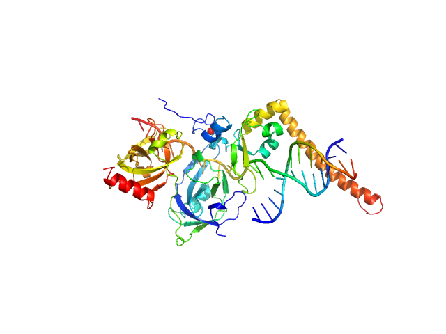
SASDJ82 – DNA repair protein XRCC1 - DNA Ligase IIIα complex

DNA repair protein XRCC1DNA ligase 3 (DNA ligase III alpha) experimental SAS data
BILBOMD model
Sample: DNA repair protein XRCC1 , 69 kDa Homo sapiens protein
DNA ligase 3 (DNA ligase III alpha) , Homo sapiens protein
Buffer: 50 mM Tris-HCl, pH 7.5, 100 mM NaCl, 5 mM MgCl₂,  0.2 mM PMSF, 1 mM benzamidine, pH: 7.5
Experiment: SAXS data collected at 12.3.1 (SIBYLS), Advanced Light Source (ALS) on 2019 Aug 18
An atypical BRCT-BRCT interaction with the XRCC1 scaffold protein compacts human DNA Ligase IIIα within a flexible DNA repair complex. Nucleic Acids Res (2020)
Hammel M, Rashid I, Sverzhinsky A, Pourfarjam Y, Tsai MS, Ellenberger T, Pascal JM, Kim IK, Tainer JA, Tomkinson AE
RgGuinier 6.2 nm
Dmax 27.9 nm
VolumePorod 675 nm3

SASDK92 – ...acids 215-619

Interferon-activable protein 204 experimental SAS data
CHIMERA model
Sample: Interferon-activable protein 204 monomer, 47 kDa Mus musculus protein
Buffer: 20 mM HEPES, 100 mM KCl, pH: 7.4
Experiment: SAXS data collected at X9A, National Synchrotron Light Source (NSLS) on 2013 Mar 14
Structural mechanism of DNA recognition by the p204 HIN domain. Nucleic Acids Res (2021)
Fan X, Jiang J, Zhao D, Chen F, Ma H, Smith P, Unterholzner L, Xiao TS, Jin T
RgGuinier 3.1 nm
Dmax 9.5 nm
VolumePorod 28 nm3

SASDJB2 – Restriction endonuclease R.AgeI in apo form, monomeric

Type-2 restriction enzyme AgeI experimental SAS data
DAMMIN model
Sample: ...restriction enzyme AgeI monomer, 31 kDa Thalassobius gelatinovorus protein
Buffer: 10 mM Tris-HCl, pH 7.5, 150 mM NaCl, 5 mM CaCl₂, pH: 7.5
Experiment: SAXS data collected at EMBL P12, PETRA III on 2013 Nov 14
Restriction endonuclease AgeI is a monomer which dimerizes to cleave DNA. Nucleic Acids Res 45(6):3547-3558 (2017)
Tamulaitiene G, Jovaisaite V, Tamulaitis G, Songailiene I, Manakova E, Zaremba M, Grazulis S, Xu SY, Siksnys V
RgGuinier 2.2 nm
Dmax 6.7 nm
VolumePorod 53 nm3

SASDJC2 – Restriction endonuclease R.AgeI complex with cognate DNA

Type-2 restriction enzyme AgeICognate DNA oligoduplex with 5'-T overhang experimental SAS data
PDB (PROTEIN DATA BANK) model
Sample: ...restriction enzyme AgeI dimer, 61 kDa Thalassobius gelatinovorus protein
Cognate DNA oligoduplex with 5'-T overhang dimer, 8 kDa DNA
Buffer: 10 mM Tris-HCl, pH 7.5, 150 mM NaCl, 5 mM CaCl₂, pH: 7.5
Experiment: SAXS data collected at EMBL P12, PETRA III on 2013 Nov 14
Restriction endonuclease AgeI is a monomer which dimerizes to cleave DNA. Nucleic Acids Res 45(6):3547-3558 (2017)
Tamulaitiene G, Jovaisaite V, Tamulaitis G, Songailiene I, Manakova E, Zaremba M, Grazulis S, Xu SY, Siksnys V
RgGuinier 2.4 nm
Dmax 7.4 nm
VolumePorod 69 nm3

SASDNL2 – LincRNA-p21 Sense Alu Inverted Repeat

LincRNA-p21 AluSx1 Sense RNA experimental SAS data
DAMMIN model
Sample: LincRNA-p21 AluSx1 Sense RNA monomer, 100 kDa Homo sapiens RNA
Buffer: 50mM HEPES,150 mM NaCl, 15 mM MgCl2, 3% glycerol, pH: 7.4
Experiment: SAXS data collected at B21, Diamond Light Source on 2021 Jul 1
Biophysical characterisation of human LincRNA-p21 sense and antisense Alu inverted repeats. Nucleic Acids Res 50(10):5881-5898 (2022)
D'Souza MH, Mrozowich T, Badmalia MD, Geeraert M, Frederickson A, Henrickson A, Demeler B, Wolfinger MT, Patel TR
RgGuinier 6.1 nm
Dmax 18.5 nm
VolumePorod 225 nm3

SASDNM2 – LincRNA-p21 Antisense Alu Inverted Repeat

LincRNA-p21 AluSx1 Antisense RNA experimental SAS data
DAMMIN model
Sample: LincRNA-p21 AluSx1 Antisense RNA monomer, 90 kDa Homo sapiens RNA
Buffer: 50mM HEPES,150 mM NaCl, 15 mM MgCl2, 3% glycerol, pH: 7.4
Experiment: SAXS data collected at B21, Diamond Light Source on 2021 Jul 1
Biophysical characterisation of human LincRNA-p21 sense and antisense Alu inverted repeats. Nucleic Acids Res 50(10):5881-5898 (2022)
D'Souza MH, Mrozowich T, Badmalia MD, Geeraert M, Frederickson A, Henrickson A, Demeler B, Wolfinger MT, Patel TR
RgGuinier 5.9 nm
Dmax 18.1 nm
VolumePorod 375 nm3

SASDXM2 – RAR/RXR heterodimer : BMS493 : DR0 element

Retinoic acid receptor alphaDirect Repeat binding site with zero-base-pair spacingBMS493 (RAR inverse agonist ligand)Retinoid X receptor alpha experimental SAS data
Retinoic acid receptor alpha Direct Repeat binding site with zero-base-pair spacing BMS493 (RAR inverse agonist ligand) Retinoid X receptor alpha Kratky plot
Sample: Retinoic acid receptor alpha monomer, 39 kDa Mus musculus protein
Direct Repeat binding site with zero-base-pair spacing monomer, 10 kDa DNA
BMS493 (RAR inverse agonist ligand) monomer, 0 kDa
Retinoid X receptor alpha monomer, 38 kDa Homo sapiens protein
Buffer: 20 mM Tris HCl, 75 mM NaCl, 75 mM KCl, 2 mM CHAPS, 4 mM MgCl₂, 12 mM TCEP, pH: 7.5
Experiment: SAXS data collected at SWING, SOLEIL on 2024 Apr 18
New structural insights into the control of the retinoic acid receptors RAR/RXR by DNA, ligands, and transcriptional coregulators. Nucleic Acids Res 53(18) (2025)
Tambones I, Sagar A, Vankova P, Loginov D, Carivenc C, Rochel N, Bourguet W, Man P, Bernadó P, le Maire A
RgGuinier 3.6 nm
Dmax 13.1 nm
VolumePorod 121 nm3

SASDXN2 – RAR/RXR heterodimer : BMS493 : DR0L element

Retinoic acid receptor alphaBMS493 (RAR inverse agonist ligand)Direct Repeat binding site with zero-base-pair spacing (long sequence)Retinoid X receptor alpha experimental SAS data
Retinoic acid receptor alpha BMS493 (RAR inverse agonist ligand) Direct Repeat binding site with zero-base-pair spacing (long sequence) Retinoid X receptor alpha Kratky plot
Sample: Retinoic acid receptor alpha monomer, 39 kDa Mus musculus protein
BMS493 (RAR inverse agonist ligand) monomer, 0 kDa
Direct Repeat binding site with zero-base-pair spacing (long sequence) monomer, 19 kDa DNA
Retinoid X receptor alpha monomer, 38 kDa Homo sapiens protein
Buffer: 20 mM Tris HCl, 75 mM NaCl, 75 mM KCl, 2 mM CHAPS, 4 mM MgCl₂, 12 mM TCEP, pH: 7.5
Experiment: SAXS data collected at SWING, SOLEIL on 2022 Dec 12
New structural insights into the control of the retinoic acid receptors RAR/RXR by DNA, ligands, and transcriptional coregulators. Nucleic Acids Res 53(18) (2025)
Tambones I, Sagar A, Vankova P, Loginov D, Carivenc C, Rochel N, Bourguet W, Man P, Bernadó P, le Maire A
RgGuinier 4.1 nm
Dmax 14.8 nm
VolumePorod 152 nm3

SASDXP2 – RAR/RXR heterodimer : BMS493 : DR1 element

Retinoic acid receptor alphaBMS493 (RAR inverse agonist ligand)Retinoid X receptor alphaDirect Repeat binding site with one-base-pair spacing experimental SAS data
Retinoic acid receptor alpha BMS493 (RAR inverse agonist ligand) Retinoid X receptor alpha Direct Repeat binding site with one-base-pair spacing Kratky plot
Sample: Retinoic acid receptor alpha monomer, 39 kDa Mus musculus protein
BMS493 (RAR inverse agonist ligand) monomer, 0 kDa
Retinoid X receptor alpha monomer, 38 kDa Homo sapiens protein
Direct Repeat binding site with one-base-pair spacing monomer, 11 kDa DNA
Buffer: 20 mM Tris HCl, 75 mM NaCl, 75 mM KCl, 2 mM CHAPS, 4 mM MgCl₂, 12 mM TCEP, pH: 7.5
Experiment: SAXS data collected at SWING, SOLEIL on 2022 Apr 20
New structural insights into the control of the retinoic acid receptors RAR/RXR by DNA, ligands, and transcriptional coregulators. Nucleic Acids Res 53(18) (2025)
Tambones I, Sagar A, Vankova P, Loginov D, Carivenc C, Rochel N, Bourguet W, Man P, Bernadó P, le Maire A
RgGuinier 3.8 nm
Dmax 13.9 nm
VolumePorod 120 nm3

SASDYP2 – Protein La 1 - full length

La protein 1 experimental SAS data
Protein La 1 - full length Rg histogram
Sample: La protein 1 monomer, 48 kDa Arabidopsis thaliana protein
Buffer: 100 mM Tris-HCl, 500 mM NaCl, pH: 8
Experiment: SAXS data collected at Rigaku BioSAXS-2000, CEITEC on 2025 Jul 31
La protein binding to telomerase RNA supports an evolutionary relationship between plant and ciliate telomerase pathways. Nucleic Acids Res 54(5) (2026)
Jenner L, Pruchkouski D, Štefanovie B, Nováková O, Kubíčková M, Fajkus P, Brázdová M, Paleček J, Sýkorová E
RgGuinier 5.0 nm
Dmax 13.3 nm
VolumePorod 111 nm3

SASDXQ2 – RAR/RXR heterodimer : BMS493 : DR5 element

Retinoic acid receptor alphaBMS493 (RAR inverse agonist ligand)Retinoid X receptor alphaDirect Repeat binding site with five-base-pair spacing experimental SAS data
Retinoic acid receptor alpha BMS493 (RAR inverse agonist ligand) Retinoid X receptor alpha Direct Repeat binding site with five-base-pair spacing Kratky plot
Sample: Retinoic acid receptor alpha monomer, 39 kDa Mus musculus protein
BMS493 (RAR inverse agonist ligand) monomer, 0 kDa
Retinoid X receptor alpha monomer, 38 kDa Homo sapiens protein
Direct Repeat binding site with five-base-pair spacing monomer, 13 kDa DNA
Buffer: 20 mM Tris HCl, 75 mM NaCl, 75 mM KCl, 2 mM CHAPS, 4 mM MgCl₂, 12 mM TCEP, pH: 7.5
Experiment: SAXS data collected at SWING, SOLEIL on 2022 Sep 12
New structural insights into the control of the retinoic acid receptors RAR/RXR by DNA, ligands, and transcriptional coregulators. Nucleic Acids Res 53(18) (2025)
Tambones I, Sagar A, Vankova P, Loginov D, Carivenc C, Rochel N, Bourguet W, Man P, Bernadó P, le Maire A
RgGuinier 3.9 nm
Dmax 14.3 nm
VolumePorod 130 nm3

SASDYQ2 – C-terminal domain of La protein 1

C-terminal domain of La protein 1 experimental SAS data
C-terminal domain of La protein 1 Rg histogram
Sample: C-terminal domain of La protein 1 monomer, 22 kDa Arabidopsis thaliana protein
Buffer: 100 mM Tris-HCl, 500 mM NaCl, pH: 8
Experiment: SAXS data collected at Rigaku BioSAXS-2000, CEITEC on 2025 Oct 9
La protein binding to telomerase RNA supports an evolutionary relationship between plant and ciliate telomerase pathways. Nucleic Acids Res 54(5) (2026)
Jenner L, Pruchkouski D, Štefanovie B, Nováková O, Kubíčková M, Fajkus P, Brázdová M, Paleček J, Sýkorová E
RgGuinier 2.6 nm
Dmax 10.7 nm
VolumePorod 24 nm3

SASDXR2 – RAR/RXR heterodimer : BMS493 : IR0 element

Retinoic acid receptor alphaBMS493 (RAR inverse agonist ligand)Retinoid X receptor alphaInverted Repeat binding site with zero-base-pair spacing experimental SAS data
Retinoic acid receptor alpha BMS493 (RAR inverse agonist ligand) Retinoid X receptor alpha Inverted Repeat binding site with zero-base-pair spacing Kratky plot
Sample: Retinoic acid receptor alpha monomer, 39 kDa Mus musculus protein
BMS493 (RAR inverse agonist ligand) monomer, 0 kDa
Retinoid X receptor alpha monomer, 38 kDa Homo sapiens protein
Inverted Repeat binding site with zero-base-pair spacing monomer, 11 kDa DNA
Buffer: 20 mM Tris HCl, 75 mM NaCl, 75 mM KCl, 2 mM CHAPS, 4 mM MgCl₂, 12 mM TCEP, pH: 7.5
Experiment: SAXS data collected at SWING, SOLEIL on 2022 Apr 20
New structural insights into the control of the retinoic acid receptors RAR/RXR by DNA, ligands, and transcriptional coregulators. Nucleic Acids Res 53(18) (2025)
Tambones I, Sagar A, Vankova P, Loginov D, Carivenc C, Rochel N, Bourguet W, Man P, Bernadó P, le Maire A
RgGuinier 3.9 nm
Dmax 12.9 nm
VolumePorod 121 nm3

SASDXS2 – RAR/RXR heterodimer : BMS493 : NCoR NID : DR0L element

Retinoic acid receptor alphaBMS493 (RAR inverse agonist ligand)Direct Repeat binding site with zero-base-pair spacing (long sequence)Retinoid X receptor alphaNuclear receptor corepressor 1 experimental SAS data
Retinoic acid receptor alpha BMS493 (RAR inverse agonist ligand) Direct Repeat binding site with zero-base-pair spacing (long sequence) Retinoid X receptor alpha Nuclear receptor corepressor 1 Kratky plot
Sample: Retinoic acid receptor alpha monomer, 39 kDa Mus musculus protein
BMS493 (RAR inverse agonist ligand) monomer, 0 kDa
Direct Repeat binding site with zero-base-pair spacing (long sequence) monomer, 19 kDa DNA
Retinoid X receptor alpha monomer, 38 kDa Homo sapiens protein
...ressor 1 monomer, 29 kDa Mus musculus protein
Buffer: 20 mM Tris HCl, 75 mM NaCl, 75 mM KCl, 2 mM CHAPS, 4 mM MgCl₂, 12 mM TCEP, pH: 7.5
Experiment: SAXS data collected at SWING, SOLEIL on 2022 Dec 12
New structural insights into the control of the retinoic acid receptors RAR/RXR by DNA, ligands, and transcriptional coregulators. Nucleic Acids Res 53(18) (2025)
Tambones I, Sagar A, Vankova P, Loginov D, Carivenc C, Rochel N, Bourguet W, Man P, Bernadó P, le Maire A
RgGuinier 5.4 nm
Dmax 20.1 nm
VolumePorod 363 nm3

SASDXT2 – RAR/RXR heterodimer : BMS493 : NCoR NID : DR1 element

Retinoic acid receptor alphaBMS493 (RAR inverse agonist ligand)Retinoid X receptor alphaDirect Repeat binding site with one-base-pair spacingNuclear receptor corepressor 1 experimental SAS data
Retinoic acid receptor alpha BMS493 (RAR inverse agonist ligand) Retinoid X receptor alpha Direct Repeat binding site with one-base-pair spacing Nuclear receptor corepressor 1 Kratky plot
Sample: Retinoic acid receptor alpha monomer, 39 kDa Mus musculus protein
BMS493 (RAR inverse agonist ligand) monomer, 0 kDa
Retinoid X receptor alpha monomer, 38 kDa Homo sapiens protein
Direct Repeat binding site with one-base-pair spacing monomer, 11 kDa DNA
...ressor 1 monomer, 29 kDa Mus musculus protein
Buffer: 20 mM Tris HCl, 75 mM NaCl, 75 mM KCl, 2 mM CHAPS, 4 mM MgCl₂, 12 mM TCEP, pH: 7.5
Experiment: SAXS data collected at SWING, SOLEIL on 2022 Apr 20
New structural insights into the control of the retinoic acid receptors RAR/RXR by DNA, ligands, and transcriptional coregulators. Nucleic Acids Res 53(18) (2025)
Tambones I, Sagar A, Vankova P, Loginov D, Carivenc C, Rochel N, Bourguet W, Man P, Bernadó P, le Maire A
RgGuinier 5.6 nm
Dmax 21.8 nm
VolumePorod 330 nm3

SASDXU2 – RAR/RXR heterodimer : BMS493 : NCoR NID : DR5 element

Retinoic acid receptor alphaBMS493 (RAR inverse agonist ligand)Retinoid X receptor alphaDirect Repeat binding site with five-base-pair spacingNuclear receptor corepressor 1 experimental SAS data
Retinoic acid receptor alpha BMS493 (RAR inverse agonist ligand) Retinoid X receptor alpha Direct Repeat binding site with five-base-pair spacing Nuclear receptor corepressor 1 Kratky plot
Sample: Retinoic acid receptor alpha monomer, 39 kDa Mus musculus protein
BMS493 (RAR inverse agonist ligand) monomer, 0 kDa
Retinoid X receptor alpha monomer, 38 kDa Homo sapiens protein
Direct Repeat binding site with five-base-pair spacing monomer, 13 kDa DNA
...ressor 1 monomer, 29 kDa Mus musculus protein
Buffer: 20 mM Tris HCl, 75 mM NaCl, 75 mM KCl, 2 mM CHAPS, 4 mM MgCl₂, 12 mM TCEP, pH: 7.5
Experiment: SAXS data collected at SWING, SOLEIL on 2022 Dec 12
New structural insights into the control of the retinoic acid receptors RAR/RXR by DNA, ligands, and transcriptional coregulators. Nucleic Acids Res 53(18) (2025)
Tambones I, Sagar A, Vankova P, Loginov D, Carivenc C, Rochel N, Bourguet W, Man P, Bernadó P, le Maire A
RgGuinier 5.7 nm
Dmax 21.8 nm
VolumePorod 363 nm3

SASDXV2 – RAR/RXR heterodimer : BMS493 : NCoR NID : IR0 element

Retinoic acid receptor alphaBMS493 (RAR inverse agonist ligand)Retinoid X receptor alphaInverted Repeat binding site with zero-base-pair spacingNuclear receptor corepressor 1 experimental SAS data
Retinoic acid receptor alpha BMS493 (RAR inverse agonist ligand) Retinoid X receptor alpha Inverted Repeat binding site with zero-base-pair spacing Nuclear receptor corepressor 1 Kratky plot
Sample: Retinoic acid receptor alpha monomer, 39 kDa Mus musculus protein
BMS493 (RAR inverse agonist ligand) monomer, 0 kDa
Retinoid X receptor alpha monomer, 38 kDa Homo sapiens protein
Inverted Repeat binding site with zero-base-pair spacing monomer, 11 kDa DNA
...ressor 1 monomer, 29 kDa Mus musculus protein
Buffer: 20 mM Tris HCl, 75 mM NaCl, 75 mM KCl, 2 mM CHAPS, 4 mM MgCl₂, 12 mM TCEP, pH: 7.5
Experiment: SAXS data collected at SWING, SOLEIL on 2022 Apr 20
New structural insights into the control of the retinoic acid receptors RAR/RXR by DNA, ligands, and transcriptional coregulators. Nucleic Acids Res 53(18) (2025)
Tambones I, Sagar A, Vankova P, Loginov D, Carivenc C, Rochel N, Bourguet W, Man P, Bernadó P, le Maire A
RgGuinier 5.5 nm
Dmax 22.9 nm
VolumePorod 257 nm3

SASDJW2 – ...restriction endonuclease

5-methylcytosine-specific restriction enzyme A experimental SAS data
PDB (PROTEIN DATA BANK) model
Sample: ...restriction enzyme A dimer, 65 kDa Escherichia coli protein
Buffer: 20 mM Tris–HCl pH 7.5, 200 mM KCl, 0.1 mM EDTA, 0.01% (w/v) sodium azide and 1 mM DTT, pH: 7.5
Experiment: SAXS data collected at EMBL P12, PETRA III on 2013 May 24
Activity and structure of EcoKMcrA. Nucleic Acids Res 46(18):9829-9841 (2018)
Czapinska H, Kowalska M, Zagorskaite E, Manakova E, Slyvka A, Xu SY, Siksnys V, Sasnauskas G, Bochtler M
RgGuinier 3.7 nm
Dmax 13.0 nm
VolumePorod 95 nm3

SASDJX2 – ...restriction endonuclease in complex with cognate hemimethylated 12bp oligoduplex

cognate hemimethylated 12-bp oligoduplex5-methylcytosine-specific restriction enzyme A experimental SAS data
SASREF model
Sample: cognate hemimethylated 12-bp oligoduplex dimer, 15 kDa DNA
...restriction enzyme A dimer, 65 kDa Escherichia coli protein
Buffer: 20 mM Tris–HCl pH 7.5, 200 mM KCl, 0.1 mM EDTA, 0.01% (w/v) sodium azide and 1 mM DTT, pH: 7.5
Experiment: SAXS data collected at EMBL P12, PETRA III on 2013 May 24
Activity and structure of EcoKMcrA. Nucleic Acids Res 46(18):9829-9841 (2018)
Czapinska H, Kowalska M, Zagorskaite E, Manakova E, Slyvka A, Xu SY, Siksnys V, Sasnauskas G, Bochtler M
RgGuinier 3.9 nm
Dmax 13.5 nm
VolumePorod 69 nm3

SASDJY2 – ...restriction endonuclease

5-methylcytosine-specific restriction enzyme A (N-terminal domain) experimental SAS data
OTHER model
Sample: ...restriction enzyme A (N-terminal domain) monomer, 21 kDa Escherichia coli protein
Buffer: 20 mM Tris–HCl pH 7.5, 200 mM KCl, 0.1 mM EDTA, 0.01% (w/v) sodium azide, 1 mM DTT, pH: 7.5
Experiment: SAXS data collected at EMBL P12, PETRA III on 2013 May 24
Activity and structure of EcoKMcrA. Nucleic Acids Res 46(18):9829-9841 (2018)
Czapinska H, Kowalska M, Zagorskaite E, Manakova E, Slyvka A, Xu SY, Siksnys V, Sasnauskas G, Bochtler M
RgGuinier 2.1 nm
Dmax 7.5 nm
VolumePorod 29 nm3

SASDJZ2 – ...restriction endonuclease in complex with cognate hemimethylated 12bp oligoduplex

5-methylcytosine-specific restriction enzyme A (N-terminal domain)cognate hemimethylated 12-bp oligoduplex experimental SAS data
OTHER model
Sample: ...restriction enzyme A (N-terminal domain) monomer, 21 kDa Escherichia coli protein
cognate hemimethylated 12-bp oligoduplex monomer, 7 kDa DNA
Buffer: 20 mM Tris–HCl pH 7.5, 200 mM KCl, 0.1 mM EDTA, 0.01% (w/v) sodium azide and 1 mM DTT, pH: 7.5
Experiment: SAXS data collected at EMBL P12, PETRA III on 2013 May 24
Activity and structure of EcoKMcrA. Nucleic Acids Res 46(18):9829-9841 (2018)
Czapinska H, Kowalska M, Zagorskaite E, Manakova E, Slyvka A, Xu SY, Siksnys V, Sasnauskas G, Bochtler M
RgGuinier 2.1 nm
Dmax 7.5 nm
VolumePorod 29 nm3

SASDY63 – human telomeric g-quadruplex (Tel50)

human telomeric g-quadruplex experimental SAS data
human telomeric g-quadruplex Kratky plot
Sample: human telomeric g-quadruplex , 16 kDa DNA
Buffer: 5 mM KH2PO4/K2HPO4, pH: 7
Experiment: SAXS data collected at BM29, ESRF on 2024 Jul 12
...reshape the compactness, stability, and topology of telomeric G-quadruplex dimers. Nucleic Acids Res 54(8) (2026)
Bertini L, Libera V, Arciuolo V, Trapella M, Marzano S, Mostarac D, Schirò G, Petrillo C, Giancola C, De Michele C, Amato J, Comez L, Pagano B, Paciaroni A
RgGuinier 2.1 nm

SASDY73 – human telomeric g-quadruplex (Tel26)

human telomeric g-quadruplex experimental SAS data
human telomeric g-quadruplex Kratky plot
Sample: human telomeric g-quadruplex monomer, 8 kDa DNA
Buffer: 5 mM KH2PO4/K2HPO4, pH: 7
Experiment: SAXS data collected at BM29, ESRF on 2024 Jul 12
...reshape the compactness, stability, and topology of telomeric G-quadruplex dimers. Nucleic Acids Res 54(8) (2026)
Bertini L, Libera V, Arciuolo V, Trapella M, Marzano S, Mostarac D, Schirò G, Petrillo C, Giancola C, De Michele C, Amato J, Comez L, Pagano B, Paciaroni A
RgGuinier 1.3 nm

SASDY83 – human telomeric g-quadruplex Tel50+BRACO-19 (1:2)

human telomeric g-quadruplex experimental SAS data
human telomeric g-quadruplex Kratky plot
Sample: human telomeric g-quadruplex , 16 kDa DNA
Buffer: 5 mM KH2PO4/K2HPO4, pH: 7
Experiment: SAXS data collected at BM29, ESRF on 2024 Jul 12
...reshape the compactness, stability, and topology of telomeric G-quadruplex dimers. Nucleic Acids Res 54(8) (2026)
Bertini L, Libera V, Arciuolo V, Trapella M, Marzano S, Mostarac D, Schirò G, Petrillo C, Giancola C, De Michele C, Amato J, Comez L, Pagano B, Paciaroni A
RgGuinier 2.0 nm

SASDY93 – human telomeric g-quadruplex Tel50+BRACO-19 (1:3)

human telomeric g-quadruplex experimental SAS data
human telomeric g-quadruplex Kratky plot
Sample: human telomeric g-quadruplex , 16 kDa DNA
Buffer: 5 mM KH2PO4/K2HPO4, pH: 7
Experiment: SAXS data collected at BM29, ESRF on 2024 Jul 12
...reshape the compactness, stability, and topology of telomeric G-quadruplex dimers. Nucleic Acids Res 54(8) (2026)
Bertini L, Libera V, Arciuolo V, Trapella M, Marzano S, Mostarac D, Schirò G, Petrillo C, Giancola C, De Michele C, Amato J, Comez L, Pagano B, Paciaroni A
RgGuinier 2.0 nm

SASDYA3 – human telomeric g-quadruplex Tel50+BRACO-19 (1:4)

human telomeric g-quadruplex experimental SAS data
human telomeric g-quadruplex Kratky plot
Sample: human telomeric g-quadruplex , 16 kDa DNA
Buffer: 5 mM KH2PO4/K2HPO4, pH: 7
Experiment: SAXS data collected at BM29, ESRF on 2024 Jul 12
...reshape the compactness, stability, and topology of telomeric G-quadruplex dimers. Nucleic Acids Res 54(8) (2026)
Bertini L, Libera V, Arciuolo V, Trapella M, Marzano S, Mostarac D, Schirò G, Petrillo C, Giancola C, De Michele C, Amato J, Comez L, Pagano B, Paciaroni A
RgGuinier 2.0 nm

SASDYB3 – human telomeric g-quadruplex Tel50+PhenDC3 (1:2)

human telomeric g-quadruplex experimental SAS data
human telomeric g-quadruplex Kratky plot
Sample: human telomeric g-quadruplex , 16 kDa DNA
Buffer: 5 mM KH2PO4/K2HPO4, pH: 7
Experiment: SAXS data collected at BM29, ESRF on 2024 Jul 12
...reshape the compactness, stability, and topology of telomeric G-quadruplex dimers. Nucleic Acids Res 54(8) (2026)
Bertini L, Libera V, Arciuolo V, Trapella M, Marzano S, Mostarac D, Schirò G, Petrillo C, Giancola C, De Michele C, Amato J, Comez L, Pagano B, Paciaroni A
RgGuinier 2.0 nm

SASDYC3 – human telomeric g-quadruplex Tel50+PhenDC3 (1:3)

human telomeric g-quadruplex experimental SAS data
human telomeric g-quadruplex Kratky plot
Sample: human telomeric g-quadruplex , 16 kDa DNA
Buffer: 5 mM KH2PO4/K2HPO4, pH: 7
Experiment: SAXS data collected at BM29, ESRF on 2024 Jul 12
...reshape the compactness, stability, and topology of telomeric G-quadruplex dimers. Nucleic Acids Res 54(8) (2026)
Bertini L, Libera V, Arciuolo V, Trapella M, Marzano S, Mostarac D, Schirò G, Petrillo C, Giancola C, De Michele C, Amato J, Comez L, Pagano B, Paciaroni A
RgGuinier 2.0 nm

SASDYD3 – human telomeric g-quadruplex Tel50+PhenDC3 (1:4)

human telomeric g-quadruplex experimental SAS data
human telomeric g-quadruplex Kratky plot
Sample: human telomeric g-quadruplex , 16 kDa DNA
Buffer: 5 mM KH2PO4/K2HPO4, pH: 7
Experiment: SAXS data collected at BM29, ESRF on 2024 Jul 12
...reshape the compactness, stability, and topology of telomeric G-quadruplex dimers. Nucleic Acids Res 54(8) (2026)
Bertini L, Libera V, Arciuolo V, Trapella M, Marzano S, Mostarac D, Schirò G, Petrillo C, Giancola C, De Michele C, Amato J, Comez L, Pagano B, Paciaroni A
RgGuinier 2.0 nm

SASDYE3 – human telomeric G-quadruplex Tel50+RHPS4 (1:2)

human telomeric g-quadruplex experimental SAS data
human telomeric g-quadruplex Kratky plot
Sample: human telomeric g-quadruplex , 16 kDa DNA
Buffer: 5 mM KH2PO4/K2HPO4, pH: 7
Experiment: SAXS data collected at BM29, ESRF on 2024 Jul 12
...reshape the compactness, stability, and topology of telomeric G-quadruplex dimers. Nucleic Acids Res 54(8) (2026)
Bertini L, Libera V, Arciuolo V, Trapella M, Marzano S, Mostarac D, Schirò G, Petrillo C, Giancola C, De Michele C, Amato J, Comez L, Pagano B, Paciaroni A
RgGuinier 2.0 nm

SASDKF3 – Human telomere monomer G4 hybrid-2 form (2JSL)

Human telomere G-quadruplex hybrid-2 form experimental SAS data
DAMMIN model
Sample: Human telomere G-quadruplex hybrid-2 form monomer, 8 kDa synthetic construct DNA
Buffer: 6 mM Na2HPO4, 2 mM NaH2PO4, 1 mM Na2EDTA, 185 mM KCl, pH: 7.2
Experiment: SAXS data collected at BioCAT 18ID, Advanced Photon Source (APS), Argonne National Laboratory on 2019 Aug 14
...res of higher-order human telomere G-quadruplex multimers. Nucleic Acids Res (2021)
...res JB, Trent JO
RgGuinier 1.2 nm
Dmax 3.8 nm
VolumePorod 9 nm3

SASDYF3 – human telomeric g-quadruplex Tel50+RHPS4(1:3)

human telomeric g-quadruplex experimental SAS data
human telomeric g-quadruplex Kratky plot
Sample: human telomeric g-quadruplex , 16 kDa DNA
Buffer: 5 mM KH2PO4/K2HPO4, pH: 7
Experiment: SAXS data collected at BM29, ESRF on 2024 Jul 12
...reshape the compactness, stability, and topology of telomeric G-quadruplex dimers. Nucleic Acids Res 54(8) (2026)
Bertini L, Libera V, Arciuolo V, Trapella M, Marzano S, Mostarac D, Schirò G, Petrillo C, Giancola C, De Michele C, Amato J, Comez L, Pagano B, Paciaroni A
RgGuinier 2.0 nm

SASDKG3 – Human wild-type telomere 48-mer d(TTAGGG)8

Human Telomere Repeat (TTAGGG)8 experimental SAS data
DAMMIN model
Sample: Human Telomere Repeat (TTAGGG)8 monomer, 15 kDa synthetic construct DNA
Buffer: 6 mM Na2HPO4, 2 mM NaH2PO4, 1 mM Na2EDTA, 185 mM KCl, pH: 7.2
Experiment: SAXS data collected at BioCAT 18ID, Advanced Photon Source (APS), Argonne National Laboratory on 2019 Aug 14
...res of higher-order human telomere G-quadruplex multimers. Nucleic Acids Res (2021)
...res JB, Trent JO
RgGuinier 1.9 nm
Dmax 6.4 nm
VolumePorod 16 nm3

SASDYG3 – human telomeric g-quadruplex Tel50+RHPS4(1:4)

human telomeric g-quadruplex experimental SAS data
human telomeric g-quadruplex Kratky plot
Sample: human telomeric g-quadruplex , 16 kDa DNA
Buffer: 5 mM KH2PO4/K2HPO4, pH: 7
Experiment: SAXS data collected at BM29, ESRF on 2024 Jul 12
...reshape the compactness, stability, and topology of telomeric G-quadruplex dimers. Nucleic Acids Res 54(8) (2026)
Bertini L, Libera V, Arciuolo V, Trapella M, Marzano S, Mostarac D, Schirò G, Petrillo C, Giancola C, De Michele C, Amato J, Comez L, Pagano B, Paciaroni A
RgGuinier 2.0 nm

SASDKH3 – Human wild-type telomere 72mer d(TTAGGG)12

Human Telomere Repeat (TTAGGG)12 experimental SAS data
DAMMIF model
Sample: Human Telomere Repeat (TTAGGG)12 monomer, 23 kDa synthetic construct DNA
Buffer: 6 mM Na2HPO4, 2 mM NaH2PO4, 1 mM Na2EDTA, 185 mM KCl, pH: 7.2
Experiment: SAXS data collected at BioCAT 18ID, Advanced Photon Source (APS), Argonne National Laboratory on 2019 Aug 14
...res of higher-order human telomere G-quadruplex multimers. Nucleic Acids Res (2021)
...res JB, Trent JO
RgGuinier 2.5 nm
Dmax 8.7 nm
VolumePorod 26 nm3

SASDYH3 – human telomeric g-quadruplex Tel50+SYUIQ-5 (1:2)

human telomeric g-quadruplex experimental SAS data
human telomeric g-quadruplex Kratky plot
Sample: human telomeric g-quadruplex , 16 kDa DNA
Buffer: 5 mM KH2PO4/K2HPO4, pH: 7
Experiment: SAXS data collected at BM29, ESRF on 2024 Jul 12
...reshape the compactness, stability, and topology of telomeric G-quadruplex dimers. Nucleic Acids Res 54(8) (2026)
Bertini L, Libera V, Arciuolo V, Trapella M, Marzano S, Mostarac D, Schirò G, Petrillo C, Giancola C, De Michele C, Amato J, Comez L, Pagano B, Paciaroni A
RgGuinier 2.0 nm

SASDKJ3 – Human telomere DNA d(TTAGGG)96

Human Telomere 96mer experimental SAS data
DAMMIF model
Sample: Human Telomere 96mer monomer, 31 kDa synthetic construct DNA
Buffer: 6 mM Na2HPO4, 2 mM NaH2PO4, 1 mM Na2EDTA, 185 mM KCl, pH: 7.2
Experiment: SAXS data collected at BioCAT 18ID, Advanced Photon Source (APS), Argonne National Laboratory on 2020 Feb 19
...res of higher-order human telomere G-quadruplex multimers. Nucleic Acids Res (2021)
...res JB, Trent JO
RgGuinier 3.2 nm
Dmax 10.9 nm
VolumePorod 38 nm3

SASDYJ3 – human telomeric g-quadruplex Tel50+SYUIQ-5 (1:3)

human telomeric g-quadruplex experimental SAS data
human telomeric g-quadruplex Kratky plot
Sample: human telomeric g-quadruplex , 16 kDa DNA
Buffer: 5 mM KH2PO4/K2HPO4, pH: 7
Experiment: SAXS data collected at BM29, ESRF on 2024 Jul 12
...reshape the compactness, stability, and topology of telomeric G-quadruplex dimers. Nucleic Acids Res 54(8) (2026)
Bertini L, Libera V, Arciuolo V, Trapella M, Marzano S, Mostarac D, Schirò G, Petrillo C, Giancola C, De Michele C, Amato J, Comez L, Pagano B, Paciaroni A
RgGuinier 1.9 nm

SASDYK3 – human telomeric g-quadruplex Tel50+SYUIQ-5 (1:4)

human telomeric g-quadruplex experimental SAS data
human telomeric g-quadruplex Kratky plot
Sample: human telomeric g-quadruplex , 16 kDa DNA
Buffer: 5 mM KH2PO4/K2HPO4, pH: 7
Experiment: SAXS data collected at BM29, ESRF on 2024 Jul 12
...reshape the compactness, stability, and topology of telomeric G-quadruplex dimers. Nucleic Acids Res 54(8) (2026)
Bertini L, Libera V, Arciuolo V, Trapella M, Marzano S, Mostarac D, Schirò G, Petrillo C, Giancola C, De Michele C, Amato J, Comez L, Pagano B, Paciaroni A
RgGuinier 1.9 nm

SASDYL3 – human telomeric g-quadruplex Tel26+BRACO-19 (1:1)

human telomeric g-quadruplex experimental SAS data
human telomeric g-quadruplex Kratky plot
Sample: human telomeric g-quadruplex monomer, 8 kDa DNA
Buffer: 5 mM KH2PO4/K2HPO4, pH: 7
Experiment: SAXS data collected at BM29, ESRF on 2024 Jul 12
...reshape the compactness, stability, and topology of telomeric G-quadruplex dimers. Nucleic Acids Res 54(8) (2026)
Bertini L, Libera V, Arciuolo V, Trapella M, Marzano S, Mostarac D, Schirò G, Petrillo C, Giancola C, De Michele C, Amato J, Comez L, Pagano B, Paciaroni A
RgGuinier 1.3 nm

SASDYM3 – human telomeric g-quadruplex Tel26+BRACO-19 (1:2)

human telomeric g-quadruplex experimental SAS data
human telomeric g-quadruplex Kratky plot
Sample: human telomeric g-quadruplex monomer, 8 kDa DNA
Buffer: 5 mM KH2PO4/K2HPO4, pH: 7
Experiment: SAXS data collected at BM29, ESRF on 2024 Jul 12
...reshape the compactness, stability, and topology of telomeric G-quadruplex dimers. Nucleic Acids Res 54(8) (2026)
Bertini L, Libera V, Arciuolo V, Trapella M, Marzano S, Mostarac D, Schirò G, Petrillo C, Giancola C, De Michele C, Amato J, Comez L, Pagano B, Paciaroni A
RgGuinier 1.3 nm

SASDYN3 – human telomeric g-quadruplex Tel26+PhenDC3 (1:1)

human telomeric g-quadruplex experimental SAS data
human telomeric g-quadruplex Kratky plot
Sample: human telomeric g-quadruplex monomer, 8 kDa DNA
Buffer: 5 mM KH2PO4/K2HPO4, pH: 7
Experiment: SAXS data collected at BM29, ESRF on 2024 Jul 12
...reshape the compactness, stability, and topology of telomeric G-quadruplex dimers. Nucleic Acids Res 54(8) (2026)
Bertini L, Libera V, Arciuolo V, Trapella M, Marzano S, Mostarac D, Schirò G, Petrillo C, Giancola C, De Michele C, Amato J, Comez L, Pagano B, Paciaroni A
RgGuinier 1.3 nm

SASDYP3 – human telomeric g-quadruplex Tel26+RHPS4 (1:1)

human telomeric g-quadruplex experimental SAS data
human telomeric g-quadruplex Kratky plot
Sample: human telomeric g-quadruplex monomer, 8 kDa DNA
Buffer: 5 mM KH2PO4/K2HPO4, pH: 7
Experiment: SAXS data collected at BM29, ESRF on 2024 Jul 12
...reshape the compactness, stability, and topology of telomeric G-quadruplex dimers. Nucleic Acids Res 54(8) (2026)
Bertini L, Libera V, Arciuolo V, Trapella M, Marzano S, Mostarac D, Schirò G, Petrillo C, Giancola C, De Michele C, Amato J, Comez L, Pagano B, Paciaroni A
RgGuinier 1.3 nm

SASDYQ3 – human telomeric g-quadruplex Tel26+PhenDC3 (1:2)

human telomeric g-quadruplex experimental SAS data
human telomeric g-quadruplex Kratky plot
Sample: human telomeric g-quadruplex monomer, 8 kDa DNA
Buffer: 5 mM KH2PO4/K2HPO4, pH: 7
Experiment: SAXS data collected at BM29, ESRF on 2024 Jul 12
...reshape the compactness, stability, and topology of telomeric G-quadruplex dimers. Nucleic Acids Res 54(8) (2026)
Bertini L, Libera V, Arciuolo V, Trapella M, Marzano S, Mostarac D, Schirò G, Petrillo C, Giancola C, De Michele C, Amato J, Comez L, Pagano B, Paciaroni A
RgGuinier 1.3 nm

SASDDR3 – Yeast tRNA Nm34 methyltransferase Trm7-Trm734 complex from Sacharomyces cerevisiae

Trm7: tRNA (cytidine(32)/guanosine(34)-2'-O)-methyltransferaseTrm734: Regulator of Ty1 transposition protein 10 experimental SAS data
CORAL model
Sample: Trm7: tRNA (cytidine(32)/guanosine(34)-2'-O)-methyltransferase monomer, 36 kDa Saccharomyces cerevisiae protein
Trm734: Regulator of Ty1 transposition protein 10 monomer, 116 kDa Saccharomyces cerevisiae protein
Buffer: 50 mM HEPES, 200 mM KCl, 5% v/v Glycerol, 10mM β-mercaptoethanol, pH: 8
Experiment: SAXS data collected at BL-10C, ...Research Organization (KEK) on 2015 Dec 16
Structure of tRNA methyltransferase complex of Trm7 and Trm734 reveals a novel binding interface for tRNA recognition. Nucleic Acids Res (2019)
Hirata A, Okada K, Yoshii K, Shiraishi H, Saijo S, Yonezawa K, Shimizu N, Hori H
RgGuinier 3.8 nm
Dmax 13.0 nm
VolumePorod 218 nm3

SASDYR3 – human telomeric g-quadruplex Tel26+RHPS4 (1:2)

human telomeric g-quadruplex experimental SAS data
human telomeric g-quadruplex Kratky plot
Sample: human telomeric g-quadruplex monomer, 8 kDa DNA
Buffer: 5 mM KH2PO4/K2HPO4, pH: 7
Experiment: SAXS data collected at BM29, ESRF on 2024 Jul 12
...reshape the compactness, stability, and topology of telomeric G-quadruplex dimers. Nucleic Acids Res 54(8) (2026)
Bertini L, Libera V, Arciuolo V, Trapella M, Marzano S, Mostarac D, Schirò G, Petrillo C, Giancola C, De Michele C, Amato J, Comez L, Pagano B, Paciaroni A
RgGuinier 1.3 nm

SASDYS3 – human telomeric g-quadruplex Tel26+SYUIQ-5 (1:1)

human telomeric g-quadruplex experimental SAS data
human telomeric g-quadruplex Kratky plot
Sample: human telomeric g-quadruplex monomer, 8 kDa DNA
Buffer: 5 mM KH2PO4/K2HPO4, pH: 7
Experiment: SAXS data collected at BM29, ESRF on 2024 Jul 12
...reshape the compactness, stability, and topology of telomeric G-quadruplex dimers. Nucleic Acids Res 54(8) (2026)
Bertini L, Libera V, Arciuolo V, Trapella M, Marzano S, Mostarac D, Schirò G, Petrillo C, Giancola C, De Michele C, Amato J, Comez L, Pagano B, Paciaroni A
RgGuinier 1.3 nm

SASDYT3 – human telomeric g-quadruplex Tel26+SYUIQ-5 (1:2)

human telomeric g-quadruplex experimental SAS data
human telomeric g-quadruplex Kratky plot
Sample: human telomeric g-quadruplex monomer, 8 kDa DNA
Buffer: 5 mM KH2PO4/K2HPO4, pH: 7
Experiment: SAXS data collected at BM29, ESRF on 2024 Jul 12
...reshape the compactness, stability, and topology of telomeric G-quadruplex dimers. Nucleic Acids Res 54(8) (2026)
Bertini L, Libera V, Arciuolo V, Trapella M, Marzano S, Mostarac D, Schirò G, Petrillo C, Giancola C, De Michele C, Amato J, Comez L, Pagano B, Paciaroni A
RgGuinier 1.3 nm

SASDXU3 – Farnesoid X Receptor Protein

Isoform 3 of Bile acid receptor experimental SAS data
ALPHAFOLD model
Sample: Isoform 3 of Bile acid receptor monomer, 41 kDa Homo sapiens protein
Buffer: 50 mM HEPES, 150 mM NaCl, 5 % glycerol, 1 mM DTT, 20 µM CDCA, pH: 7.4
Experiment: SAXS data collected at Rigaku BioSAXS-2000, Pennsylvania State University on 2024 May 9
DNA induces non-canonical dimerization of the farnesoid X receptor. Nucleic Acids Res 54(4) (2026)
Khan SH, Yennawar N, Okafor CD
RgGuinier 3.5 nm
Dmax 11.6 nm
VolumePorod 75 nm3

SASDXV3 – Farnesoid X receptor homo-dimer bound to inverted repeat DNA

inverted repeat DNAIsoform 3 of Bile acid receptor experimental SAS data
ALPHAFOLD model
Sample: inverted repeat DNA monomer, 11 kDa DNA
Isoform 3 of Bile acid receptor dimer, 83 kDa Homo sapiens protein
Buffer: 50 mM HEPES, 150 mM NaCl, 5 % glycerol, 1 mM DTT, 20 µM CDCA, pH: 7.4
Experiment: SAXS data collected at Rigaku BioSAXS-2000, Pennsylvania State University on 2024 May 9
DNA induces non-canonical dimerization of the farnesoid X receptor. Nucleic Acids Res 54(4) (2026)
Khan SH, Yennawar N, Okafor CD
RgGuinier 5.1 nm
Dmax 23.2 nm
VolumePorod 120 nm3

SASDBX3 – Aureobox dsDNA

Aureobox dsDNA experimental SAS data
DAMFILT model
Sample: Aureobox dsDNA monomer, 13 kDa synthetic construct DNA
Buffer: 50 mM Tris 50 mM boric acid 1 mM EDTA, pH: 8
Experiment: SAXS data collected at BM29, ESRF on 2014 Nov 6
...responsive domains of diatom class I aureochromes. Nucleic Acids Res 44(12):5957-70 (2016)
Banerjee A, Herman E, Serif M, Maestre-Reyna M, Hepp S, Pokorny R, Kroth PG, Essen LO, Kottke T
RgGuinier 2.0 nm
Dmax 8.6 nm
VolumePorod 18 nm3

SASDBY3 – Aureochrome 1a bZIP-LOV module: PtAUREO1a bZIP-LOV (Light oxygen voltage) module (light state-TBE)

Aureochrome 1a bZIP-LOV module experimental SAS data
DAMFILT model
Sample: Aureochrome 1a bZIP-LOV module dimer, 57 kDa Phaeodactylum tricornutum protein
Buffer: 50 mM Tris 50 mM boric acid 1 mM EDTA, pH: 8
Experiment: SAXS data collected at BM29, ESRF on 2014 Nov 6
...responsive domains of diatom class I aureochromes. Nucleic Acids Res 44(12):5957-70 (2016)
Banerjee A, Herman E, Serif M, Maestre-Reyna M, Hepp S, Pokorny R, Kroth PG, Essen LO, Kottke T
RgGuinier 3.4 nm
Dmax 12.6 nm
VolumePorod 115 nm3

SASDBZ3 – Aureochrome 1a bZIP-LOV module: PtAUREO1a bZIP-LOV (Light oxygen voltage)/DNA complex (light state)

Aureobox dsDNAAureochrome 1a bZIP-LOV module experimental SAS data
DAMFILT model
Sample: Aureobox dsDNA monomer, 13 kDa synthetic construct DNA
Aureochrome 1a bZIP-LOV module dimer, 57 kDa Phaeodactylum tricornutum protein
Buffer: 50 mM Tris 50 mM boric acid 1 mM EDTA, pH: 8
Experiment: SAXS data collected at BM29, ESRF on 2014 Nov 6
...responsive domains of diatom class I aureochromes. Nucleic Acids Res 44(12):5957-70 (2016)
Banerjee A, Herman E, Serif M, Maestre-Reyna M, Hepp S, Pokorny R, Kroth PG, Essen LO, Kottke T
RgGuinier 4.5 nm
Dmax 16.7 nm
VolumePorod 97 nm3

SASDB24 – Aureochrome 1a bZIP-LOV module: PtAUREO1a bZIP-LOV (Light oxygen voltage) module (light state, Tris)

Aureochrome 1a bZIP-LOV module experimental SAS data
Aureochrome 1a bZIP-LOV module Kratky plot
Sample: Aureochrome 1a bZIP-LOV module dimer, 57 kDa Phaeodactylum tricornutum protein
Buffer: 10 mM Tris 300 mM NaCl, pH: 8
Experiment: SAXS data collected at BM29, ESRF on 2014 Nov 6
...responsive domains of diatom class I aureochromes. Nucleic Acids Res 44(12):5957-70 (2016)
Banerjee A, Herman E, Serif M, Maestre-Reyna M, Hepp S, Pokorny R, Kroth PG, Essen LO, Kottke T
RgGuinier 3.9 nm
Dmax 12.5 nm
VolumePorod 121 nm3

SASDB34 – Aureochrome 1a bZIP-LOV module: PtAUREO1a bZIP-LOV (Light oxygen voltage) module (dark state, Tris)

Aureochrome 1a bZIP-LOV module experimental SAS data
Aureochrome 1a bZIP-LOV module Kratky plot
Sample: Aureochrome 1a bZIP-LOV module dimer, 57 kDa Phaeodactylum tricornutum protein
Buffer: 10 mM Tris 300 mM NaCl, pH: 8
Experiment: SAXS data collected at BM29, ESRF on 2014 Nov 6
...responsive domains of diatom class I aureochromes. Nucleic Acids Res 44(12):5957-70 (2016)
Banerjee A, Herman E, Serif M, Maestre-Reyna M, Hepp S, Pokorny R, Kroth PG, Essen LO, Kottke T
RgGuinier 3.8 nm
Dmax 12.5 nm
VolumePorod 117 nm3

SASDH44 – 3' Complex of XPA-DBD and RPA70AB

DNA repair protein complementing XP-A cellsReplication protein A 70 kDa DNA-binding subunit3-prime  Nucleotide Excision Repair Junction Model Substrate experimental SAS data
HADDOCK model
Sample: DNA repair protein complementing XP-A cells monomer, 17 kDa Homo sapiens protein
Replication protein A 70 kDa DNA-binding subunit monomer, 27 kDa Homo sapiens protein
3-prime Nucleotide Excision Repair Junction Model Substrate monomer, 11 kDa DNA
Buffer: 20 mM Tris, 150 mM NaCl, 2% glycerol, 1 mM DTT, pH: 7.5
Experiment: SAXS data collected at 12.3.1 (SIBYLS), Advanced Light Source (ALS) on 17 Nov 2
A key interaction with RPA orients XPA in NER complexes. Nucleic Acids Res (2020)
Topolska-Woś AM, Sugitani N, Cordoba JJ, Le Meur KV, Le Meur RA, Kim HS, Yeo JE, Rosenberg D, Hammel M, Schärer OD, Chazin WJ
RgGuinier 3.1 nm
Dmax 9.7 nm
VolumePorod 103 nm3

SASDH54 – 5' Complex of XPA-DBD with RPA70AB

DNA repair protein complementing XP-A cellsReplication protein A 70 kDa DNA-binding subunit5-prime Nucleotide Excision Repair Junction Model Substrate experimental SAS data
HADDOCK model
Sample: DNA repair protein complementing XP-A cells monomer, 17 kDa Homo sapiens protein
Replication protein A 70 kDa DNA-binding subunit monomer, 27 kDa Homo sapiens protein
5-prime Nucleotide Excision Repair Junction Model Substrate monomer, 11 kDa DNA
Buffer: 20 mM Tris, 150 mM NaCl, 2% glycerol, 1 mM DTT, pH: 7.5
Experiment: SAXS data collected at 12.3.1 (SIBYLS), Advanced Light Source (ALS) on 2019 Jun 4
A key interaction with RPA orients XPA in NER complexes. Nucleic Acids Res (2020)
Topolska-Woś AM, Sugitani N, Cordoba JJ, Le Meur KV, Le Meur RA, Kim HS, Yeo JE, Rosenberg D, Hammel M, Schärer OD, Chazin WJ
RgGuinier 2.9 nm
Dmax 97.0 nm
VolumePorod 87 nm3

SASDC74 – ...resolved 0ms

Truncated P5abc subdomain from tetrahymena ribozyme experimental SAS data
Truncated P5abc subdomain from tetrahymena ribozyme Kratky plot
Sample: Truncated P5abc subdomain from tetrahymena ribozyme monomer, 18 kDa RNA
Buffer: 10mM KMOPS 20mM KCl 1mM MgCl2 20uM EDTA, pH: 7
Experiment: SAXS data collected at G1, Cornell High Energy Synchrotron Source (CHESS) on 2016 Dec 3
Revealing the distinct folding phases of an RNA three-helix junction. Nucleic Acids Res 46(14):7354-7365 (2018)
Plumridge A, Katz AM, Calvey GD, Elber R, Kirmizialtin S, Pollack L
RgGuinier 2.5 nm
Dmax 8.0 nm

SASDK74 – Streptococcus agalactiae transcription factor BusR - RCK_C domain

Transcriptional repressor BusR RCK_C domain experimental SAS data
OTHER model
Sample: ...ressor BusR RCK_C domain dimer, 22 kDa Streptococcus agalactiae serotype … protein
Buffer: 100 mM NaCl, 30mM Hepes, pH: 7.5
Experiment: SAXS data collected at EMBL P12, PETRA III on 2019 Jul 2
BusR senses bipartite DNA binding motifs by a unique molecular ruler architecture. Nucleic Acids Res (2021)
Bandera AM, Bartho J, Lammens K, Drexler DJ, Kleinschwärzer J, Hopfner KP, Witte G
RgGuinier 1.9 nm
Dmax 6.4 nm
VolumePorod 44 nm3

SASDK84 – Streptococcus agalactiae transcription factor BusR

Transcriptional repressor BusR experimental SAS data
Transcriptional repressor BusR Kratky plot
Sample: ...ressor BusR tetramer, 95 kDa Streptococcus agalactiae protein
Buffer: 20mM HEPES, pH6.5, 100mM NaCl, 3% glycerol (v/v), pH: 6.5
Experiment: SAXS data collected at EMBL P12, PETRA III on 2019 Jul 2
BusR senses bipartite DNA binding motifs by a unique molecular ruler architecture. Nucleic Acids Res (2021)
Bandera AM, Bartho J, Lammens K, Drexler DJ, Kleinschwärzer J, Hopfner KP, Witte G
RgGuinier 4.4 nm
Dmax 13.9 nm
VolumePorod 168 nm3

SASDC94 – ...resolved 10ms

Truncated P5abc subdomain from tetrahymena ribozyme experimental SAS data
Truncated P5abc subdomain from tetrahymena ribozyme Kratky plot
Sample: Truncated P5abc subdomain from tetrahymena ribozyme monomer, 18 kDa RNA
Buffer: 10mM KMOPS 20mM KCl 1mM MgCl2 20uM EDTA, pH: 7
Experiment: SAXS data collected at G1, Cornell High Energy Synchrotron Source (CHESS) on 2016 Dec 3
Revealing the distinct folding phases of an RNA three-helix junction. Nucleic Acids Res 46(14):7354-7365 (2018)
Plumridge A, Katz AM, Calvey GD, Elber R, Kirmizialtin S, Pollack L
RgGuinier 2.3 nm
Dmax 7.2 nm

SASDK94 – Streptococcus agalactiae transcription factor BusR dsDNA complex

Transcriptional repressor BusRBusR Recognition sequence experimental SAS data
OTHER model
Sample: ...ressor BusR tetramer, 95 kDa Streptococcus agalactiae protein
BusR Recognition sequence monomer, 28 kDa synthetic construct DNA
Buffer: 20mM HEPES, pH6.5, 100mM NaCl, 3% glycerol (v/v), pH: 6.5
Experiment: SAXS data collected at EMBL P12, PETRA III on 2019 Jul 2
BusR senses bipartite DNA binding motifs by a unique molecular ruler architecture. Nucleic Acids Res (2021)
Bandera AM, Bartho J, Lammens K, Drexler DJ, Kleinschwärzer J, Hopfner KP, Witte G
RgGuinier 4.3 nm
Dmax 14.2 nm
VolumePorod 210 nm3

SASDCA4 – ...resolved 30ms

Truncated P5abc subdomain from tetrahymena ribozyme experimental SAS data
Truncated P5abc subdomain from tetrahymena ribozyme Kratky plot
Sample: Truncated P5abc subdomain from tetrahymena ribozyme monomer, 18 kDa RNA
Buffer: 10mM KMOPS 20mM KCl 1mM MgCl2 20uM EDTA, pH: 7
Experiment: SAXS data collected at G1, Cornell High Energy Synchrotron Source (CHESS) on 2016 Dec 3
Revealing the distinct folding phases of an RNA three-helix junction. Nucleic Acids Res 46(14):7354-7365 (2018)
Plumridge A, Katz AM, Calvey GD, Elber R, Kirmizialtin S, Pollack L
RgGuinier 2.2 nm
Dmax 7.0 nm

SASDCB4 – ...resolved 100ms

Truncated P5abc subdomain from tetrahymena ribozyme experimental SAS data
Truncated P5abc subdomain from tetrahymena ribozyme Kratky plot
Sample: Truncated P5abc subdomain from tetrahymena ribozyme monomer, 18 kDa RNA
Buffer: 10mM KMOPS 20mM KCl 1mM MgCl2 20uM EDTA, pH: 7
Experiment: SAXS data collected at G1, Cornell High Energy Synchrotron Source (CHESS) on 2016 Dec 3
Revealing the distinct folding phases of an RNA three-helix junction. Nucleic Acids Res 46(14):7354-7365 (2018)
Plumridge A, Katz AM, Calvey GD, Elber R, Kirmizialtin S, Pollack L
RgGuinier 2.3 nm
Dmax 7.1 nm

SASDCC4 – ...resolved 300ms

Truncated P5abc subdomain from tetrahymena ribozyme experimental SAS data
Truncated P5abc subdomain from tetrahymena ribozyme Kratky plot
Sample: Truncated P5abc subdomain from tetrahymena ribozyme monomer, 18 kDa RNA
Buffer: 10mM KMOPS 20mM KCl 1mM MgCl2 20uM EDTA, pH: 7
Experiment: SAXS data collected at G1, Cornell High Energy Synchrotron Source (CHESS) on 2016 Dec 3
Revealing the distinct folding phases of an RNA three-helix junction. Nucleic Acids Res 46(14):7354-7365 (2018)
Plumridge A, Katz AM, Calvey GD, Elber R, Kirmizialtin S, Pollack L
RgGuinier 2.2 nm
Dmax 6.8 nm

SASDCD4 – ...resolved 1000ms

Truncated P5abc subdomain from tetrahymena ribozyme experimental SAS data
Truncated P5abc subdomain from tetrahymena ribozyme Kratky plot
Sample: Truncated P5abc subdomain from tetrahymena ribozyme monomer, 18 kDa RNA
Buffer: 10mM KMOPS 20mM KCl 1mM MgCl2 20uM EDTA, pH: 7
Experiment: SAXS data collected at G1, Cornell High Energy Synchrotron Source (CHESS) on 2016 Dec 3
Revealing the distinct folding phases of an RNA three-helix junction. Nucleic Acids Res 46(14):7354-7365 (2018)
Plumridge A, Katz AM, Calvey GD, Elber R, Kirmizialtin S, Pollack L
RgGuinier 2.2 nm
Dmax 6.8 nm

SASDGD4 – Mixed lineage leukemia protein-1 complex, MLL1-WDR5-ASH2L-RBBP5(2-381)

Retinoblastoma-binding protein 5Histone-lysine N-methyltransferase 2AWD repeat-containing protein 5Set1/Ash2 histone methyltransferase complex subunit ASH2 experimental SAS data
Retinoblastoma-binding protein 5 Histone-lysine N-methyltransferase 2A WD repeat-containing protein 5 Set1/Ash2 histone methyltransferase complex subunit ASH2 Kratky plot
Sample: Retinoblastoma-binding protein 5 monomer, 42 kDa Homo sapiens protein
Histone-lysine N-methyltransferase 2A monomer, 25 kDa Homo sapiens protein
WD repeat-containing protein 5 monomer, 34 kDa Homo sapiens protein
Set1/Ash2 histone methyltransferase complex subunit ASH2 monomer, 60 kDa Homo sapiens protein
Buffer: 300 mM NaCl, 25mM Tris-HCl, 4% glycerol, 1 mM TCEP, pH: 8
Experiment: SAXS data collected at BL19U2, Shanghai Synchrotron Radiation Facility (SSRF) on 2019 Jun 22
The internal interaction in RBBP5 regulates assembly and activity of MLL1 methyltransferase complex. Nucleic Acids Res (2019)
Han J, Li T, Li Y, Li M, Wang X, Peng C, Su C, Li N, Li Y, Xu Y, Chen Y
RgGuinier 5.7 nm
Dmax 18.6 nm
VolumePorod 360 nm3

SASDCE4 – ...resolved 3000ms

Truncated P5abc subdomain from tetrahymena ribozyme experimental SAS data
Truncated P5abc subdomain from tetrahymena ribozyme Kratky plot
Sample: Truncated P5abc subdomain from tetrahymena ribozyme monomer, 18 kDa RNA
Buffer: 10mM KMOPS 20mM KCl 1mM MgCl2 20uM EDTA, pH: 7
Experiment: SAXS data collected at G1, Cornell High Energy Synchrotron Source (CHESS) on 2016 Dec 3
Revealing the distinct folding phases of an RNA three-helix junction. Nucleic Acids Res 46(14):7354-7365 (2018)
Plumridge A, Katz AM, Calvey GD, Elber R, Kirmizialtin S, Pollack L
RgGuinier 2.2 nm
Dmax 6.7 nm

SASDGE4 – Mixed lineage leukemia protein-1 complex, MLL1-WDR5-ASH2L-RBBP5(2-480)

Histone-lysine N-methyltransferase 2AWD repeat-containing protein 5Set1/Ash2 histone methyltransferase complex subunit ASH2Retinoblastoma-binding protein 5 experimental SAS data
Histone-lysine N-methyltransferase 2A WD repeat-containing protein 5 Set1/Ash2 histone methyltransferase complex subunit ASH2 Retinoblastoma-binding protein 5 Kratky plot
Sample: Histone-lysine N-methyltransferase 2A monomer, 25 kDa Homo sapiens protein
WD repeat-containing protein 5 monomer, 34 kDa Homo sapiens protein
Set1/Ash2 histone methyltransferase complex subunit ASH2 monomer, 60 kDa Homo sapiens protein
Retinoblastoma-binding protein 5 monomer, 53 kDa Homo sapiens protein
Buffer: 300 mM NaCl, 25mM Tris-HCl, 4% glycerol, 1 mM TCEP, pH: 8
Experiment: SAXS data collected at BL19U2, Shanghai Synchrotron Radiation Facility (SSRF) on 2019 Jun 22
The internal interaction in RBBP5 regulates assembly and activity of MLL1 methyltransferase complex. Nucleic Acids Res (2019)
Han J, Li T, Li Y, Li M, Wang X, Peng C, Su C, Li N, Li Y, Xu Y, Chen Y
RgGuinier 5.0 nm
Dmax 15.3 nm
VolumePorod 256 nm3

SASDCF4 – Truncated P5abc subdomain from tetrahymena ribozyme: Static 20mM KCl

Truncated P5abc subdomain from tetrahymena ribozyme experimental SAS data
Truncated P5abc subdomain from tetrahymena ribozyme Kratky plot
Sample: Truncated P5abc subdomain from tetrahymena ribozyme monomer, 18 kDa RNA
Buffer: 20mM KCl 10mM KMOPS 20uM EDTA, pH: 7
Experiment: SAXS data collected at G1, Cornell High Energy Synchrotron Source (CHESS) on 2016 Jun 17
Revealing the distinct folding phases of an RNA three-helix junction. Nucleic Acids Res 46(14):7354-7365 (2018)
Plumridge A, Katz AM, Calvey GD, Elber R, Kirmizialtin S, Pollack L
RgGuinier 2.6 nm
Dmax 8.5 nm

SASDGF4 – Mixed lineage leukemia protein-1 complex, MLL1-WDR5-ASH2L-RBBP5(2-480)L399A/L400A/I457A/L459A

Histone-lysine N-methyltransferase 2AWD repeat-containing protein 5Set1/Ash2 histone methyltransferase complex subunit ASH2Retinoblastoma-binding protein 5 experimental SAS data
Histone-lysine N-methyltransferase 2A WD repeat-containing protein 5 Set1/Ash2 histone methyltransferase complex subunit ASH2 Retinoblastoma-binding protein 5 Kratky plot
Sample: Histone-lysine N-methyltransferase 2A monomer, 25 kDa Homo sapiens protein
WD repeat-containing protein 5 monomer, 34 kDa Homo sapiens protein
Set1/Ash2 histone methyltransferase complex subunit ASH2 monomer, 60 kDa Homo sapiens protein
Retinoblastoma-binding protein 5 monomer, 53 kDa Homo sapiens protein
Buffer: 300 mM NaCl, 25mM Tris-HCl, 4% glycerol, 1 mM TCEP, pH: 8
Experiment: SAXS data collected at BL19U2, Shanghai Synchrotron Radiation Facility (SSRF) on 2019 Jun 22
The internal interaction in RBBP5 regulates assembly and activity of MLL1 methyltransferase complex. Nucleic Acids Res (2019)
Han J, Li T, Li Y, Li M, Wang X, Peng C, Su C, Li N, Li Y, Xu Y, Chen Y
RgGuinier 5.3 nm
Dmax 17.2 nm
VolumePorod 313 nm3

SASDCG4 – Truncated P5abc subdomain from tetrahymena ribozyme: Static 100mM KCl

Truncated P5abc subdomain from tetrahymena ribozyme experimental SAS data
Truncated P5abc subdomain from tetrahymena ribozyme Kratky plot
Sample: Truncated P5abc subdomain from tetrahymena ribozyme monomer, 18 kDa RNA
Buffer: 100mM KCl 10mM KMOPS 20uM EDTA, pH: 7
Experiment: SAXS data collected at G1, Cornell High Energy Synchrotron Source (CHESS) on 2016 Jun 17
Revealing the distinct folding phases of an RNA three-helix junction. Nucleic Acids Res 46(14):7354-7365 (2018)
Plumridge A, Katz AM, Calvey GD, Elber R, Kirmizialtin S, Pollack L
RgGuinier 2.5 nm
Dmax 8.0 nm

SASDGG4 – Mixed lineage leukemia protein-1 complex, MLL1-WDR5-ASH2L-RBBP5(2-538)

Histone-lysine N-methyltransferase 2AWD repeat-containing protein 5Set1/Ash2 histone methyltransferase complex subunit ASH2Retinoblastoma-binding protein 5 experimental SAS data
Histone-lysine N-methyltransferase 2A WD repeat-containing protein 5 Set1/Ash2 histone methyltransferase complex subunit ASH2 Retinoblastoma-binding protein 5 Kratky plot
Sample: Histone-lysine N-methyltransferase 2A monomer, 25 kDa Homo sapiens protein
WD repeat-containing protein 5 monomer, 34 kDa Homo sapiens protein
Set1/Ash2 histone methyltransferase complex subunit ASH2 monomer, 60 kDa Homo sapiens protein
Retinoblastoma-binding protein 5 monomer, 59 kDa Homo sapiens protein
Buffer: 300 mM NaCl, 25mM Tris-HCl, 4% glycerol, 1 mM TCEP, pH: 8
Experiment: SAXS data collected at BL19U2, Shanghai Synchrotron Radiation Facility (SSRF) on 2019 Jun 22
The internal interaction in RBBP5 regulates assembly and activity of MLL1 methyltransferase complex. Nucleic Acids Res (2019)
Han J, Li T, Li Y, Li M, Wang X, Peng C, Su C, Li N, Li Y, Xu Y, Chen Y
RgGuinier 5.0 nm
Dmax 15.5 nm
VolumePorod 282 nm3

SASDCH4 – Truncated P5abc subdomain from tetrahymena ribozyme: Static 200mm KCl

Truncated P5abc subdomain from tetrahymena ribozyme experimental SAS data
Truncated P5abc subdomain from tetrahymena ribozyme Kratky plot
Sample: Truncated P5abc subdomain from tetrahymena ribozyme monomer, 18 kDa RNA
Buffer: 200mM KCl 10mM KMOPS 20uM EDTA, pH: 7
Experiment: SAXS data collected at G1, Cornell High Energy Synchrotron Source (CHESS) on 2016 Jun 17
Revealing the distinct folding phases of an RNA three-helix junction. Nucleic Acids Res 46(14):7354-7365 (2018)
Plumridge A, Katz AM, Calvey GD, Elber R, Kirmizialtin S, Pollack L
RgGuinier 2.5 nm
Dmax 8.0 nm

SASDCJ4 – Truncated P5abc subdomain from tetrahymena ribozyme: Static 400mM KCl

Truncated P5abc subdomain from tetrahymena ribozyme experimental SAS data
Truncated P5abc subdomain from tetrahymena ribozyme Kratky plot
Sample: Truncated P5abc subdomain from tetrahymena ribozyme monomer, 18 kDa RNA
Buffer: 400mM KCl 10mM KMOPS 20uM EDTA, pH: 7
Experiment: SAXS data collected at G1, Cornell High Energy Synchrotron Source (CHESS) on 2016 Jun 17
Revealing the distinct folding phases of an RNA three-helix junction. Nucleic Acids Res 46(14):7354-7365 (2018)
Plumridge A, Katz AM, Calvey GD, Elber R, Kirmizialtin S, Pollack L
RgGuinier 2.4 nm
Dmax 8.0 nm

SASDBK4 – The 1:1:3:1 crRNA:Cas6f:Cas7fv:Cas5fv CRISPR/Cas Type I-F short Cascade complex

short-crRNA: CRISPR/Cas Type I-F Cascade componentCas6f: CRISPR/Cas Type I-F Cascade component (CRISPR-associated protein, Csy4 family)Trimeric Cas7fv: CRISPR/Cas Type I-F Cascade component (Uncharacterized protein, Sputcn32_1821)Cas5fv: CRISPR/Cas Type I-F Cascade component (Uncharacterized protein, Sputcn32_1822) experimental SAS data
DAMMIF model
Sample: short-crRNA: CRISPR/Cas Type I-F Cascade component monomer, 14 kDa Shewanella putrefaciens RNA
Cas6f: CRISPR/Cas Type I-F Cascade component (CRISPR-associated protein, Csy4 family) monomer, 21 kDa Shewanella putrefaciens protein
Trimeric Cas7fv: CRISPR/Cas Type I-F Cascade component (Uncharacterized protein, Sputcn32_1821) trimer, 112 kDa Shewanella putrefaciens protein
Cas5fv: CRISPR/Cas Type I-F Cascade component (Uncharacterized protein, Sputcn32_1822) monomer, 38 kDa Shewanella putrefaciens protein
Buffer: 50 mM HEPES 150 mM NaCl 1mM DTT 1mM EDTA, pH: 7
Experiment: SAXS data collected at BM29, ESRF on 2015 Jun 27
Modulating the Cascade architecture of a minimal Type I-F CRISPR-Cas system. Nucleic Acids Res 44(12):5872-82 (2016)
Gleditzsch D, Müller-Esparza H, Pausch P, Sharma K, Dwarakanath S, Urlaub H, Bange G, Randau L
RgGuinier 4.1 nm
Dmax 14.2 nm

SASDCK4 – Truncated P5abc subdomain from tetrahymena ribozyme: Static 0.25mM MgCl2

Truncated P5abc subdomain from tetrahymena ribozyme experimental SAS data
Truncated P5abc subdomain from tetrahymena ribozyme Kratky plot
Sample: Truncated P5abc subdomain from tetrahymena ribozyme monomer, 18 kDa RNA
Buffer: 20mM KCl 0.25mM MgCl2 10mM KMOPS 20uM EDTA, pH: 7
Experiment: SAXS data collected at G1, Cornell High Energy Synchrotron Source (CHESS) on 2016 Jun 17
Revealing the distinct folding phases of an RNA three-helix junction. Nucleic Acids Res 46(14):7354-7365 (2018)
Plumridge A, Katz AM, Calvey GD, Elber R, Kirmizialtin S, Pollack L
RgGuinier 2.4 nm
Dmax 7.6 nm

SASDBL4 – The 1:1:6:1 crRNA:Cas6f:Cas7fv:Cas5fv CRISPR/Cas Type I-F wild-type Cascade complex

Cas6f: CRISPR/Cas Type I-F Cascade component (CRISPR-associated protein, Csy4 family)Cas5fv: CRISPR/Cas Type I-F Cascade component (Uncharacterized protein, Sputcn32_1822)Hexameric Cas7fv: CRISPR/Cas Type I-F Cascade component (Uncharacterized protein, Sputcn32_1821)wildtype-crRNA: CRISPR/Cas Type I-F Cascade component experimental SAS data
DAMMIF model
Sample: Cas6f: CRISPR/Cas Type I-F Cascade component (CRISPR-associated protein, Csy4 family) monomer, 21 kDa Shewanella putrefaciens protein
Cas5fv: CRISPR/Cas Type I-F Cascade component (Uncharacterized protein, Sputcn32_1822) monomer, 38 kDa Shewanella putrefaciens protein
Hexameric Cas7fv: CRISPR/Cas Type I-F Cascade component (Uncharacterized protein, Sputcn32_1821) hexamer, 223 kDa Shewanella putrefaciens protein
wildtype-crRNA: CRISPR/Cas Type I-F Cascade component monomer, 19 kDa RNA
Buffer: 50 mM HEPES 150 mM NaCl 1mM DTT 1mM EDTA, pH: 7
Experiment: SAXS data collected at BM29, ESRF on 2015 Jun 27
Modulating the Cascade architecture of a minimal Type I-F CRISPR-Cas system. Nucleic Acids Res 44(12):5872-82 (2016)
Gleditzsch D, Müller-Esparza H, Pausch P, Sharma K, Dwarakanath S, Urlaub H, Bange G, Randau L
RgGuinier 5.4 nm
Dmax 18.4 nm

SASDCL4 – Truncated P5abc subdomain from tetrahymena ribozyme: Static 0.50mM MgCl2

Truncated P5abc subdomain from tetrahymena ribozyme experimental SAS data
Truncated P5abc subdomain from tetrahymena ribozyme Kratky plot
Sample: Truncated P5abc subdomain from tetrahymena ribozyme monomer, 18 kDa RNA
Buffer: 0.5mM MgCl2 20mM KCl 10mM KMOPS 20uM EDTA, pH: 7
Experiment: SAXS data collected at G1, Cornell High Energy Synchrotron Source (CHESS) on 2016 Jun 17
Revealing the distinct folding phases of an RNA three-helix junction. Nucleic Acids Res 46(14):7354-7365 (2018)
Plumridge A, Katz AM, Calvey GD, Elber R, Kirmizialtin S, Pollack L
RgGuinier 2.3 nm
Dmax 7.2 nm

SASDBM4 – The 1:1:9:1 crRNA:Cas6f:Cas7fv:Cas5fv CRISPR/Cas Type I-F long Cascade complex

Cas6f: CRISPR/Cas Type I-F Cascade component (CRISPR-associated protein, Csy4 family)Cas5fv: CRISPR/Cas Type I-F Cascade component (Uncharacterized protein, Sputcn32_1822)Nonameric Cas7fv: CRISPR/Cas Type I-F Cascade component (Uncharacterized protein, Sputcn32_1821)long-crRNA: CRISPR/Cas Type I-F Cascade component experimental SAS data
DAMMIF model
Sample: Cas6f: CRISPR/Cas Type I-F Cascade component (CRISPR-associated protein, Csy4 family) monomer, 21 kDa Shewanella putrefaciens protein
Cas5fv: CRISPR/Cas Type I-F Cascade component (Uncharacterized protein, Sputcn32_1822) monomer, 38 kDa Shewanella putrefaciens protein
Nonameric Cas7fv: CRISPR/Cas Type I-F Cascade component (Uncharacterized protein, Sputcn32_1821) nonamer, 335 kDa Shewanella putrefaciens protein
long-crRNA: CRISPR/Cas Type I-F Cascade component monomer, 25 kDa RNA
Buffer: 50 mM HEPES 150 mM NaCl 1mM DTT 1mM EDTA, pH: 7
Experiment: SAXS data collected at BM29, ESRF on 2016 Jan 30
Modulating the Cascade architecture of a minimal Type I-F CRISPR-Cas system. Nucleic Acids Res 44(12):5872-82 (2016)
Gleditzsch D, Müller-Esparza H, Pausch P, Sharma K, Dwarakanath S, Urlaub H, Bange G, Randau L
RgGuinier 6.5 nm
Dmax 21.6 nm

SASDCM4 – Truncated P5abc subdomain from tetrahymena ribozyme: Static 1mM MgCl2

Truncated P5abc subdomain from tetrahymena ribozyme experimental SAS data
Truncated P5abc subdomain from tetrahymena ribozyme Kratky plot
Sample: Truncated P5abc subdomain from tetrahymena ribozyme monomer, 18 kDa RNA
Buffer: 1mM MgCl2 20mM KCl 10mM KMOPS 20uM EDTA, pH: 7
Experiment: SAXS data collected at G1, Cornell High Energy Synchrotron Source (CHESS) on 2016 Jun 17
Revealing the distinct folding phases of an RNA three-helix junction. Nucleic Acids Res 46(14):7354-7365 (2018)
Plumridge A, Katz AM, Calvey GD, Elber R, Kirmizialtin S, Pollack L
RgGuinier 2.2 nm
Dmax 7.2 nm

SASDUA5 – Double-stranded RNA-binding domains of Interleukin enhancer-binding factor 3 (395-592)

Interleukin enhancer-binding factor 3 experimental SAS data
GASBOR model
Sample: Interleukin enhancer-binding factor 3 monomer, 21 kDa Mus musculus protein
Buffer: 20 mM HEPES, 150 mM NaCl, 1 mM DTT, pH: 7.5
Experiment: SAXS data collected at B21, Diamond Light Source on 2017 Oct 23
Integrative structural analysis of NF45-NF90 heterodimers reveals architectural rearrangements and oligomerization on binding dsRNA. Nucleic Acids Res 53(6) (2025)
Winterbourne S, Jayachandran U, Zou J, Rappsilber J, Granneman S, Cook AG
RgGuinier 3.5 nm
Dmax 12.6 nm
VolumePorod 39 nm3

SASDUB5 – Heterodimer complex of domain-associated zinc finger domains of Interleukin enhancer-binding factor 2 (29-390) and Interleukin enhancer-binding factor 3 (1-381)

Interleukin enhancer-binding factor 3Interleukin enhancer-binding factor 2 experimental SAS data
GASBOR model
Sample: Interleukin enhancer-binding factor 3 monomer, 42 kDa Mus musculus protein
Interleukin enhancer-binding factor 2 monomer, 40 kDa Mus musculus protein
Buffer: 20 mM HEPES, 150 mM NaCl, 1 mM DTT, pH: 7.5
Experiment: SAXS data collected at B21, Diamond Light Source on 2017 Oct 23
Integrative structural analysis of NF45-NF90 heterodimers reveals architectural rearrangements and oligomerization on binding dsRNA. Nucleic Acids Res 53(6) (2025)
Winterbourne S, Jayachandran U, Zou J, Rappsilber J, Granneman S, Cook AG
RgGuinier 3.5 nm
Dmax 12.6 nm
VolumePorod 127 nm3

SASDUC5 – Interleukin enhancer-binding factor 3 (1-591) and Interleukin enhancer-binding factor 2 (1-390) heterodimer complex (SEC-SAXS 1)

Interleukin enhancer-binding factor 2Interleukin enhancer-binding factor 3 experimental SAS data
DAMMIN model
Sample: Interleukin enhancer-binding factor 2 monomer, 44 kDa Homo sapiens protein
Interleukin enhancer-binding factor 3 monomer, 66 kDa Mus musculus protein
Buffer: 20 mM HEPES, 150 mM NaCl, 1 mM DTT, pH: 7.5
Experiment: SAXS data collected at B21, Diamond Light Source on 2023 May 9
Integrative structural analysis of NF45-NF90 heterodimers reveals architectural rearrangements and oligomerization on binding dsRNA. Nucleic Acids Res 53(6) (2025)
Winterbourne S, Jayachandran U, Zou J, Rappsilber J, Granneman S, Cook AG
RgGuinier 4.7 nm
Dmax 17.1 nm
VolumePorod 210 nm3

SASDUD5 – Interleukin enhancer-binding factor 3 (1-591) and Interleukin enhancer-binding factor 2 (1-390) heterodimer complex (SEC-SAXS 2)

Interleukin enhancer-binding factor 3Interleukin enhancer-binding factor 2 experimental SAS data
GASBOR model
Sample: Interleukin enhancer-binding factor 3 monomer, 66 kDa Mus musculus protein
Interleukin enhancer-binding factor 2 monomer, 44 kDa Homo sapiens protein
Buffer: 20 mM HEPES, 150 mM NaCl, 1 mM DTT, pH: 7.5
Experiment: SAXS data collected at B21, Diamond Light Source on 2017 Oct 23
Integrative structural analysis of NF45-NF90 heterodimers reveals architectural rearrangements and oligomerization on binding dsRNA. Nucleic Acids Res 53(6) (2025)
Winterbourne S, Jayachandran U, Zou J, Rappsilber J, Granneman S, Cook AG
RgGuinier 4.7 nm
Dmax 17.1 nm
VolumePorod 210 nm3

SASDUE5 – Interleukin enhancer-binding factor 3 (1-591) and Interleukin enhancer-binding factor 2 (1-390) heterodimer complex oligomerised along 25mer of dsRNA in a 2:1 ratio

Interleukin enhancer-binding factor 2Interleukin enhancer-binding factor 325-mer dsRNAInterleukin enhancer-binding factor 2Interleukin enhancer-binding factor 3 experimental SAS data
DAMMIN model
Sample: Interleukin enhancer-binding factor 2 monomer, 44 kDa Homo sapiens protein
Interleukin enhancer-binding factor 3 monomer, 66 kDa Mus musculus protein
25-mer dsRNA monomer, 16 kDa RNA
Interleukin enhancer-binding factor 2 monomer, 44 kDa Homo sapiens protein
Interleukin enhancer-binding factor 3 monomer, 66 kDa Mus musculus protein
Buffer: 20 mM HEPES, 150 mM NaCl, 1 mM DTT, pH: 7.5
Experiment: SAXS data collected at B21, Diamond Light Source on 2023 May 9
Integrative structural analysis of NF45-NF90 heterodimers reveals architectural rearrangements and oligomerization on binding dsRNA. Nucleic Acids Res 53(6) (2025)
Winterbourne S, Jayachandran U, Zou J, Rappsilber J, Granneman S, Cook AG
RgGuinier 5.7 nm
Dmax 19.9 nm
VolumePorod 453 nm3

SASDUF5 – Interleukin enhancer-binding factor 3 (1-591) and Interleukin enhancer-binding factor 2 (1-390) heterodimer complex oligomerised along 25mer of dsRNA in a 4:1 ratio

Interleukin enhancer-binding factor 2Interleukin enhancer-binding factor 325-mer dsRNAInterleukin enhancer-binding factor 2Interleukin enhancer-binding factor 3 experimental SAS data
DAMMIN model
Sample: Interleukin enhancer-binding factor 2 monomer, 44 kDa Homo sapiens protein
Interleukin enhancer-binding factor 3 monomer, 66 kDa Mus musculus protein
25-mer dsRNA monomer, 16 kDa RNA
Interleukin enhancer-binding factor 2 monomer, 44 kDa Homo sapiens protein
Interleukin enhancer-binding factor 3 monomer, 66 kDa Mus musculus protein
Buffer: 20 mM HEPES, 150 mM NaCl, 1 mM DTT, pH: 7.5
Experiment: SAXS data collected at B21, Diamond Light Source on 2023 May 9
Integrative structural analysis of NF45-NF90 heterodimers reveals architectural rearrangements and oligomerization on binding dsRNA. Nucleic Acids Res 53(6) (2025)
Winterbourne S, Jayachandran U, Zou J, Rappsilber J, Granneman S, Cook AG
RgGuinier 5.7 nm
Dmax 20.5 nm
VolumePorod 431 nm3

SASDUG5 – Interleukin enhancer-binding factor 3 (1-591) and Interleukin enhancer-binding factor 2 (1-390) heterodimer complex oligomerised along 36mer of dsRNA in a 2:1 ratio

Interleukin enhancer-binding factor 2Interleukin enhancer-binding factor 3Interleukin enhancer-binding factor 2Interleukin enhancer-binding factor 336-mer dsRNAInterleukin enhancer-binding factor 2Interleukin enhancer-binding factor 3 experimental SAS data
DAMMIN model
Sample: Interleukin enhancer-binding factor 2 monomer, 44 kDa Homo sapiens protein
Interleukin enhancer-binding factor 3 monomer, 66 kDa Mus musculus protein
Interleukin enhancer-binding factor 2 monomer, 44 kDa Homo sapiens protein
Interleukin enhancer-binding factor 3 monomer, 66 kDa Mus musculus protein
36-mer dsRNA monomer, 23 kDa RNA
Interleukin enhancer-binding factor 2 monomer, 44 kDa Homo sapiens protein
Interleukin enhancer-binding factor 3 monomer, 66 kDa Mus musculus protein
Buffer: 20 mM HEPES, 150 mM NaCl, 1 mM DTT, pH: 7.5
Experiment: SAXS data collected at B21, Diamond Light Source on 2023 May 9
Integrative structural analysis of NF45-NF90 heterodimers reveals architectural rearrangements and oligomerization on binding dsRNA. Nucleic Acids Res 53(6) (2025)
Winterbourne S, Jayachandran U, Zou J, Rappsilber J, Granneman S, Cook AG
RgGuinier 6.1 nm
Dmax 21.2 nm
VolumePorod 587 nm3

SASDUH5 – Interleukin enhancer-binding factor 3 (1-591) and Interleukin enhancer-binding factor 2 (1-390) heterodimer complex oligomerised along 36mer of dsRNA in a 4:1 ratio

Interleukin enhancer-binding factor 2Interleukin enhancer-binding factor 3Interleukin enhancer-binding factor 2Interleukin enhancer-binding factor 336-mer dsRNAInterleukin enhancer-binding factor 2Interleukin enhancer-binding factor 3 experimental SAS data
DAMMIN model
Sample: Interleukin enhancer-binding factor 2 monomer, 44 kDa Homo sapiens protein
Interleukin enhancer-binding factor 3 monomer, 66 kDa Mus musculus protein
Interleukin enhancer-binding factor 2 monomer, 44 kDa Homo sapiens protein
Interleukin enhancer-binding factor 3 monomer, 66 kDa Mus musculus protein
36-mer dsRNA monomer, 23 kDa RNA
Interleukin enhancer-binding factor 2 monomer, 44 kDa Homo sapiens protein
Interleukin enhancer-binding factor 3 monomer, 66 kDa Mus musculus protein
Buffer: 20 mM HEPES, 150 mM NaCl, 1 mM DTT, pH: 7.5
Experiment: SAXS data collected at B21, Diamond Light Source on 2023 May 9
Integrative structural analysis of NF45-NF90 heterodimers reveals architectural rearrangements and oligomerization on binding dsRNA. Nucleic Acids Res 53(6) (2025)
Winterbourne S, Jayachandran U, Zou J, Rappsilber J, Granneman S, Cook AG
RgGuinier 6.4 nm
Dmax 22.1 nm
VolumePorod 626 nm3

SASDUJ5 – Interleukin enhancer-binding factor 3 (1-591) and Interleukin enhancer-binding factor 2 (1-390) heterodimer complex oligomerised along 54mer of dsRNA in a 2:1 ratio

Interleukin enhancer-binding factor 2Interleukin enhancer-binding factor 3Interleukin enhancer-binding factor 2Interleukin enhancer-binding factor 3Interleukin enhancer-binding factor 2Interleukin enhancer-binding factor 354-mer dsRNA experimental SAS data
DAMMIN model
Sample: Interleukin enhancer-binding factor 2 monomer, 44 kDa Homo sapiens protein
Interleukin enhancer-binding factor 3 monomer, 66 kDa Mus musculus protein
Interleukin enhancer-binding factor 2 monomer, 44 kDa Homo sapiens protein
Interleukin enhancer-binding factor 3 monomer, 66 kDa Mus musculus protein
Interleukin enhancer-binding factor 2 monomer, 44 kDa Homo sapiens protein
Interleukin enhancer-binding factor 3 monomer, 66 kDa Mus musculus protein
54-mer dsRNA monomer, 35 kDa RNA
Buffer: 20 mM HEPES, 150 mM NaCl, 1 mM DTT, pH: 7.5
Experiment: SAXS data collected at B21, Diamond Light Source on 2023 May 9
Integrative structural analysis of NF45-NF90 heterodimers reveals architectural rearrangements and oligomerization on binding dsRNA. Nucleic Acids Res 53(6) (2025)
Winterbourne S, Jayachandran U, Zou J, Rappsilber J, Granneman S, Cook AG
RgGuinier 7.1 nm
Dmax 25.0 nm
VolumePorod 797 nm3

SASDUK5 – Interleukin enhancer-binding factor 3 (1-591) and Interleukin enhancer-binding factor 2 (1-390) heterodimer complex oligomerised along 54mer of dsRNA in a 4:1 ratio

Interleukin enhancer-binding factor 2Interleukin enhancer-binding factor 3Interleukin enhancer-binding factor 2Interleukin enhancer-binding factor 3Interleukin enhancer-binding factor 2Interleukin enhancer-binding factor 354-mer dsRNAInterleukin enhancer-binding factor 3Interleukin enhancer-binding factor 2 experimental SAS data
DAMMIN model
Sample: Interleukin enhancer-binding factor 2 monomer, 44 kDa Homo sapiens protein
Interleukin enhancer-binding factor 3 monomer, 66 kDa Mus musculus protein
Interleukin enhancer-binding factor 2 monomer, 44 kDa Homo sapiens protein
Interleukin enhancer-binding factor 3 monomer, 66 kDa Mus musculus protein
Interleukin enhancer-binding factor 2 monomer, 44 kDa Homo sapiens protein
Interleukin enhancer-binding factor 3 monomer, 66 kDa Mus musculus protein
54-mer dsRNA monomer, 35 kDa RNA
Interleukin enhancer-binding factor 3 monomer, 66 kDa Mus musculus protein
Interleukin enhancer-binding factor 2 monomer, 44 kDa Homo sapiens protein
Buffer: 20 mM HEPES, 150 mM NaCl, 1 mM DTT, pH: 7.5
Experiment: SAXS data collected at B21, Diamond Light Source on 2023 May 9
Integrative structural analysis of NF45-NF90 heterodimers reveals architectural rearrangements and oligomerization on binding dsRNA. Nucleic Acids Res 53(6) (2025)
Winterbourne S, Jayachandran U, Zou J, Rappsilber J, Granneman S, Cook AG
RgGuinier 7.7 nm
Dmax 26.5 nm
VolumePorod 1075 nm3

SASDUL5 – Interleukin enhancer-binding factor 3 (1-591) and Interleukin enhancer-binding factor 2 (1-390) heterodimer complex oligomerised along 54mer of dsRNA in a 6:1 ratio

Interleukin enhancer-binding factor 2Interleukin enhancer-binding factor 3Interleukin enhancer-binding factor 2Interleukin enhancer-binding factor 3Interleukin enhancer-binding factor 2Interleukin enhancer-binding factor 354-mer dsRNAInterleukin enhancer-binding factor 3Interleukin enhancer-binding factor 2 experimental SAS data
DAMMIN model
Sample: Interleukin enhancer-binding factor 2 monomer, 44 kDa Homo sapiens protein
Interleukin enhancer-binding factor 3 monomer, 66 kDa Mus musculus protein
Interleukin enhancer-binding factor 2 monomer, 44 kDa Homo sapiens protein
Interleukin enhancer-binding factor 3 monomer, 66 kDa Mus musculus protein
Interleukin enhancer-binding factor 2 monomer, 44 kDa Homo sapiens protein
Interleukin enhancer-binding factor 3 monomer, 66 kDa Mus musculus protein
54-mer dsRNA monomer, 35 kDa RNA
Interleukin enhancer-binding factor 3 monomer, 66 kDa Mus musculus protein
Interleukin enhancer-binding factor 2 monomer, 44 kDa Homo sapiens protein
Buffer: 20 mM HEPES, 150 mM NaCl, 1 mM DTT, pH: 7.5
Experiment: SAXS data collected at B21, Diamond Light Source on 2023 May 9
Integrative structural analysis of NF45-NF90 heterodimers reveals architectural rearrangements and oligomerization on binding dsRNA. Nucleic Acids Res 53(6) (2025)
Winterbourne S, Jayachandran U, Zou J, Rappsilber J, Granneman S, Cook AG
RgGuinier 7.8 nm
Dmax 26.1 nm
VolumePorod 1050 nm3

SASDWL5 – I-RNA, an A-to-I hyper-edited 20mer dsRNA

I-RNA, Top-strandI-RNA, Bottom-strand experimental SAS data
I-RNA, Top-strand I-RNA, Bottom-strand Kratky plot
Sample: I-RNA, Top-strand monomer, 7 kDa RNA
I-RNA, Bottom-strand monomer, 7 kDa RNA
Buffer: 25 mM sodium phosphate 25 mM sodium chloride, pH: 6.4
Experiment: SAXS data collected at BM29, ESRF on 2022 Nov 9
Unique conformational dynamics and protein recognition of A-to-I hyper-edited dsRNA. Nucleic Acids Res 53(12) (2025)
Müller-Hermes C, Piomponi V, Hilber S, Asami S, Kreutz C, Bussi G, Sattler M
RgGuinier 2.0 nm
Dmax 6.9 nm
VolumePorod 17 nm3

SASDWM5 – A-RNA, non-edited 20mer dsRNA

A-RNA, Top-StrandA-RNA, Bottom-Strand experimental SAS data
A-RNA, Top-Strand A-RNA, Bottom-Strand Kratky plot
Sample: A-RNA, Top-Strand monomer, 6 kDa RNA
A-RNA, Bottom-Strand monomer, 6 kDa RNA
Buffer: 25 mM sodium phosphate 25 mM sodium chloride, pH: 6.4
Experiment: SAXS data collected at BM29, ESRF on 2022 Nov 9
Unique conformational dynamics and protein recognition of A-to-I hyper-edited dsRNA. Nucleic Acids Res 53(12) (2025)
Müller-Hermes C, Piomponi V, Hilber S, Asami S, Kreutz C, Bussi G, Sattler M
RgGuinier 1.8 nm
Dmax 6.2 nm
VolumePorod 17 nm3

SASDGS5 – MvaT (low salt data set)

MvaT(mutant) experimental SAS data
OTHER [STATIC IMAGE] model
Sample: MvaT(mutant) dimer, 28 kDa Pseudomonas aeruginosa protein
Buffer: 20 mM Bis-Tris 50 mM KCl, pH: 6
Experiment: SAXS data collected at BM29, ESRF on 2018 May 11
Structural basis for osmotic regulation of the DNA binding properties of H-NS proteins. Nucleic Acids Res (2020)
Qin L, Bdira FB, Sterckx YGJ, Volkov AN, Vreede J, Giachin G, van Schaik P, Ubbink M, Dame RT
RgGuinier 3.6 nm
Dmax 14.7 nm
VolumePorod 47 nm3

SASDGT5 – MvaT (high salt data set)

MvaT(mutant) experimental SAS data
OTHER [STATIC IMAGE] model
Sample: MvaT(mutant) dimer, 28 kDa Pseudomonas aeruginosa protein
Buffer: 20 mM Bis-Tris 300 mM KCl, pH: 6
Experiment: SAXS data collected at BM29, ESRF on 2018 May 11
Structural basis for osmotic regulation of the DNA binding properties of H-NS proteins. Nucleic Acids Res (2020)
Qin L, Bdira FB, Sterckx YGJ, Volkov AN, Vreede J, Giachin G, van Schaik P, Ubbink M, Dame RT
RgGuinier 3.8 nm
Dmax 15.8 nm
VolumePorod 50 nm3